Hemoflagellates1 Assis Prof Dr Suhad Faisal Hatem Flagellate


: It is skin lesion may appear 210 years after")


 -found in central & south America. -infect")

- Slides: 14
Hemoflagellates-1 Assis. Prof. Dr. Suhad Faisal Hatem
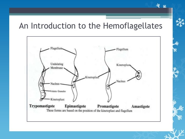
Flagellate of blood and tissue This parasite include two genus Leishmania spp. and Trypansoma spp. They are pathogenic to man and may exist in two or more form in life cycle (Promastigote, Epimastigote, Trypomastigote, Amastigote). Leishmania spp Include three important spp. 1. Leishmania donovani (visceral Leishmaniasis) 2. Leishmania tropica: (cutaneous Leishmaniasis) 3. Leishmania braziliensis: It causes (muco-cutaneous Leishmaniasis). 1 -transmitted to human by female sand fly genous Phlebotomus 2 -life cycle pass in two hosts vertebrate & invertebrate 3 -in invertebrate host the parasite are extracellular in the gut and transmission via the mouth parts during blood feeding. or Espondia
Leishmania donovani -Obligate, intracellular arasite of reticuloendothelial cells, predominat in liver , spleen, bone marrow and lymph node of man and other vertebrate host were occurs in amastigote form. Morphologically, found in two forms 1 - Amastigote : Size: 5 by 3µm, Shape: oval to round, Nucleus One, eccentric. Kinetoplast: Present, Consisting of dot-like blepharoplast, with small axoneme and prabasal body. Flagellum absent 2 -promastigote: Size: 9 -15µm, Shape: long and slender, Nucleus one, central. Kinetoplast: Anterior end of the organism, no undulating membrane. Flagellum: Single, anterior free flagellum. -It causes a disease called Kala-azar or Dum-Dum fever or visceral Leishmaniasis or black fever.
Life cycle
Pathogenesis 1 -L. donovani is the causative agent of visceral leishmaniasis, traditionally known as kala-azar ("black fever", particularly in India), because of its characteristic symptoms. 2 -The disease is highly lethal if not treated properly. The incubation period generally ranges from 3 to 6 months, and in some cases may be over 2 year. In Indian leishmaniasis, incubation can be as short as 10 days. 3 -The target cells are those of mononuclear phagocyte system. The two main tissues of infection are spleen and liver leading to enlargement of spleen, hepato -splenomegaly) and lymph node with fever, malaise, headache, emaciation and anaemia. 4 - Heavy skin pigmentation which darkens the physical appearance (the reason for naming "black fever").
-Post kala-azar dermal Leishmaniasis (PKDL): It is skin lesion may appear 210 years after successful therapy for visceral Leishmaniasis caused by L. donovani in india. It is caused by the reversal of L. donovani from viscero-tropic to dermatotropic. These lesions are soft, painless, granulomatous of varying size. Lab diagnosis 1 -Parasiyological diagnosis: -Blood film by thick smear to see the amastigote form by using Gimsa stain -Needle biopsy/aspiration for lymph node, liver, spleen -Culture of blood or needle biopsy/aspiration to see the promastigote form. NNN agar. 2 -Non Specific Lab. Test -Blood Count: normally, total leucosyte count is 3000/µl, during disease the count reach to 1000/ µl or less also decrease in erythrocytes number reveals pancytopenia. -Haemoglobin estimation - Serum protein estimation: It will be raised reveal to albumin: globulin ratio (Ig. G ) high.
• 3 -Molecular method: using DNA probes, PCR • 4 -Immunological test using ELISA , Monoclonal antibody • • 5 -Animal inoculation • 6 -Leishmania (Montengro) test:
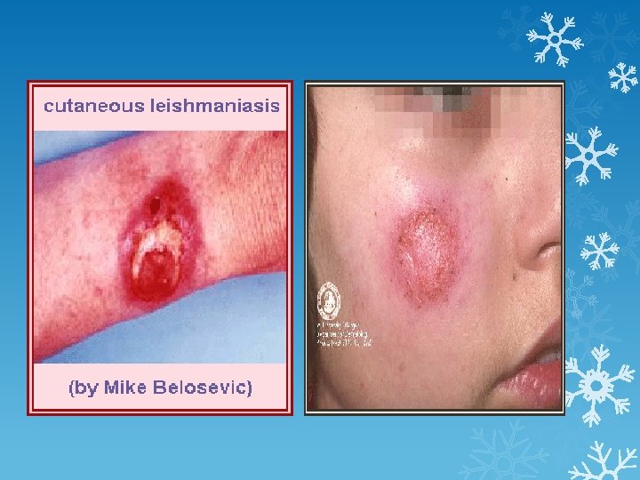
Leishmania tropica It causes a disease called Dry or urban cutaneous Leishmaniasis or oriental sore or Baghdad boil or Old world cutaneous Leishmaniasis or tropica sore. Morphological and life cycle resemble to Leishmania donovani has two form amastigote in man and the promastigote in sandfly called (P. sergenti). It is have 3 varieties: urban: cased by L. tropica, Miner , dry type(dog reservoir) rural: cased by L. tropica , major , wet type (rodent reservoir ) diffuse form (Ethiopia)
Pathogenesis üLesion will breakdown with purulent exudates üCutaneous lesions develop causing chronic infective granuloma with fibrosis. üPyrogenic complication cause local ulcer (sore on face and extremities), septicemia, and fever. üAfter proliferation of amastigote , it will infilterate the lymphocytes and plasma cells causing delay hypersensitivity skin reaction. üUncomplicated cases will heal at self limiting
Lab. Diagnosis -Puncture of the sore edge then stained with Gimsa will allow to see the amastigotes inside macrophage. -aspiration from the ulcer can culture it in NNN media -Leishmania skin test: injection (killed promastigotes of Leishmania tropica in 0. 5% phenol salin) show delayed hypersensitive response. Treatment 1 -Pentavalent antimonials , sodium antimony gluconate(pentostam), meglumine antimonate in many lesions 2 Cryotherapy for single lesion
3 -L. Brazilionsis cause mucocutaneous leishmaniasis(espondia) -found in central & south America. -infect mucous membrane of mouth, nasopharynx, nasal septa -lesions , nodules, ulcer the same with oriental sore but its differ by type of lesion& course of the disease. -Secondary infection lead to losse mucosal tissue, oedema, necrosis extensive destruction, also deep erosion then spread to lung.