posture Assis Prof Neveen Abdel Latif Objectives At


posture Assis. Prof. Neveen Abdel Latif

Objectives At the end of this lecture you should be able to; 1. Identify causes of poor posture 2. Investigate Characteristics of poor posture 3. Define common postural deficiencies 4. Identify postural assessment. 5. Investigate spinal deformities 6. Investigate assessment & ttt of Kyphosis. 7. Investigate assessment & ttt of scoliosis

Causes of poor posture Structural causes -Due to permanent anatomical deformities -Not corrected by conservative treatments Positional causes (flexable) -Due to poor posture

Positional causes of poor posture include • • • Poor postural habit, the individual does not maintain a correct posture. Psychological factors especially selfesteem. Normal developmental and degenerative processes. Pain leading to muscle guarding and avoidance postures. Muscle imbalance, spasm, or contracture.

• • • General weakness. Working at a desk with poorly designed seats. Poor sleep pattern. Excessive weight (obesity). Foot problems/improper shoes. Careless sitting, standing, sleeping habits.

Characteristics of poor posture • Increase discomfort and pain – -forward-head position result in headaches and pain in the shoulders, arms, hands and around the eyes. -Rounded shoulders can trigger the headaches at the base of skull where the shoulder muscles attach. • Create pain in the jaw - a forward-head position can lead to jaw pain. • Decrease lung capacity - reducing the amount of oxygen in body can decrease the space in chest cavity, restricting efficient functioning of lungs as in scoliosis. • Cause low back pain • Cause nerve interference - spine is the basis of posture. If posture is bad, spine can be misaligned which may cause interference in nerve function

Postural examinations consist of three parts: 1. Examination of alignment in standing. 2. Tests for flexibility& muscle length. 3. Tests for muscle strength.

POSTURE ASSESSMENT 1. After the initial examination, prior to treatment; 2. During performance of a treatment technique; 3. Immediately after the application of each treatment technique; 4. At the end of a treatment session; 5. At the beginning of the next session; 6. Retrospectively, after a number of treatments; 7. At the completion of treatment.

Common Postural Deficiencies
. -Head Forward, C-Spine slightly extended. -Thoracic Spine:")
Flat-Back Posture: (Flattening of the lumbar vertebrae). -Head Forward, C-Spine slightly extended. -Thoracic Spine: Upper part is flexed while lower part is straight. -Lumbar Spine: Flexed (straight). -Pelvis: posterior tilting. -Hip joints: Extended. -Knee Joints: Extended. -Ankle Joints: slightly plantar flexed.

Causes -Weakness of the hip flexor & lower back ms &stretched posterior longitudinal ligaments. -Tightness of hip extensor ms & Abd ms Corrective Posture Exercises -strengthening exercises for Hip Flexors & lower back ms -stretching exercises for Abdominal & hip extensor muscles.

strengthening exercises of hip flexor muscles

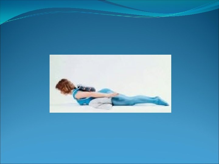
Stretching of hip extensor muscles.

Back strengthening exercises
. Upper trunk")
Sway Back. Flattening of the lumbar vertebrae (the pelvis is displaced forward). Upper trunk backward. Causes -Thoracic Kyphosis. -Posterior pelvic tilt. -Hips Hyperextended. -Compression of vertebrae anteriorly. - Weakness of posterior longitudinal ligaments, back extensors, and hip flexor muscles& anterior hip ligament
 • arched")
Military posture: • head pulled back • shoulder blades tightly (pulled back) • arched lower back • knees locked (straight) • minimizes the spinal column's ability to be a shock absorber for the body
 -Arched lower back (Lordosis)")
Slouched Posture: -Head forward, shoulders rounded -Rounded upper back (kyphosis) -Arched lower back (Lordosis) -Protruding buttocks Corrective Posture exercises: • Strengthening of cervical extensors • Isometric Neck Retraction • Stretching of cervical vertebral column flexors

Spinal Deformities of the spine alter the shape of the whole back and occur • in the sagital plane as Kyphosis and Lordosis and • in the coronal plane as Scoliosis.
")
Kyphosis. Increased posterior convexity of the vertebrae( excessive forward bending in the thoracic area) In the thoracic level, it exaggerates the normal curve. In the lumbar and cervical levels, it obliterates the normal lordosis Kyphosis occurs in older& adults, particularly women with osteoporosis and osteoarthritis. Kyphosis is sometime accompanied with other posterior problems such as posterior or anterior pelvic tilt.

Causes -Compression of intervertebral discs anteriorly. -lengthening thoracic extensors and middle and lower trapezius muscles and posterior ligaments. -Tightness of anterior longitudinal ligament, upper abdominal, and anterior chest muscles.

Types of Kyphosis 1 -Postural Kyphosis Postural kyphosis-sometimes called "round back"-is the result of poor posture. most common in adolescents and young adults. Postural kyphosis is often accompanied by hyperlordosis of the lumbar (lower) spine due to its compensation in the inward direction.
 kyphosis corrects itself when lying down on a flat surface, or when")
A postural(flexable) kyphosis corrects itself when lying down on a flat surface, or when the spine is hyper-extended. There are no noticeable vertebral abnormalities on X-rays. Postural kyphosis is easily corrected with education about proper posture, including some retraining on how to sit and stand correctly. Corrective exercises include strengthening exercises for the back ms &stretching exercises for the anterior chest muscles.

2 -Scheuermann's Kyphosis It affects the shape of the vertebral bodies in the mid back. The affected bones appear wedge-shaped, producing forward rounding (kyphosis) in the thoracic spine. Causes: genetics or osteoporosis that runs in families.

3 -Congenital Kyphosis Congenital kyphosis means a person is born with some sort defect, such as incomplete formation of the spine. This can lead to a severe abnormal kyphosis. Extreme (severe) kyphosis is the most common cause of paralysis in the lower part of the body. Treatment: usually surgically not Conservative treatment

Physical Examination >Observation of the posture from lateral view reveal round-back or gibbous deformity. Balance of the head and trunk over the pelvis viewing from the side can be assessed. >Adam's Forward Bending Test requires the patient to bend forward at the waist. This may reveal a thoracolumbar kyphosis.

>Palpation determines spinal abnormalities by feel. Often the paraspinal musculature is tender. >Range of Motion measures the degree to which a patient can perform movements of flexion, extension, lateral bending, and spinal rotation. The deformity is palpated during range of motion to assess flexibility or rigidity of the curve.

Treatment. -Postural exercises include -Strengthening of thoracic vertebral column extensors -Stretching of thoracic flexors and -bracing may be necessary. -Severe deformities with symptoms will require surgical correction.

Lordosis is a posterior curvature of the spine. It occurs commonly in the lumbar spine, most often as compensation for a kyphosis above or a flexion deformity at the hip join Lordosis is found in all age groups. It primarily affects the lumbar spine, but can occur in the neck (cervical).
 may")
Causes • Discitis is inflammation of intervertebral disc space. • Kyphosis (eg 'humpback') may force the low back to compensate • Obesity may cause some overweight people to lean backward to improve balance. • Osteoporosis is a bone density disease that may cause vertebrae to loose strength, compromising the spine's structural integrity. • Spondylolisthesis occurs when one vertebra slips forward in relation to an adjacent one. • Fashion of wearing high heels.
.")
Characteristics of lordosis • Pelvis is positioned forward and downward ( anterior pelvic tilt). • Hips are slightly flexed • Lumbar spine is excessively hyperextended. • Hip flexors, lower back extensor (erector spinae) &posterior longitudinal ligaments are short. • anterior longitudinal ligament and lower abdominal muscles & gluteus maximus muscles may be weak. Increased risk of lower back injury during standing or lying hip extension.

Corrective exercises: • www. exrx. net/Stretches/Hip. Flexors/Kne eling. Hip. Flexor. html &Lower Back Stretch exercises. • Strength exercise for abdominal muscles& gluteus maximus

Lower Back Stretch exercises. Hip Flexor Stretch

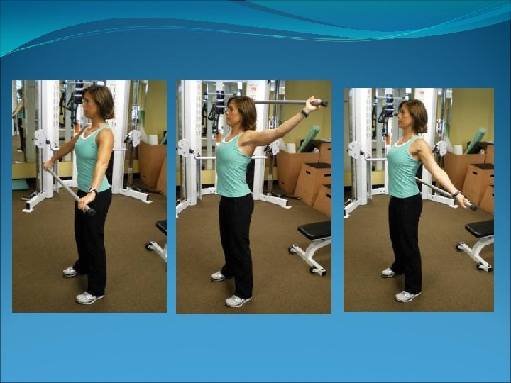
• abdominal muscles & gluteus maximus Strengthening exercise

Kyphosis-Lordosis posture: • Increases intervertebral disc pressure L-5/S-1. • Head is forward, • C-Spine is extended, • Scapulae are abducted • Thoracic spine increased flexion (Kyphosis) • Lumbar spine Hyperextended (Lordosis) • Hip joints flexed • Knee Hyperextended • Ankle joints slightly plantar flexed.

Scoliosis is lateral curvature of the spine.

Classification of scoliosis Structural scoliosis 1 - Idiopathic Scoliosis 2 -Neuromuscular Scoliosis 3 -Congenital Scoliosis 4 -Degenerative Scoliosis: Nonstructural scoliosis (Postural ) (functional) A) Infantile B) Juvenile C) Adolescent D) Adult
 Postural Scoliosis(mobile curve) • Postural scoliosis is common in the younger age group.")
I) Postural Scoliosis(mobile curve) • Postural scoliosis is common in the younger age group. • It does not show structural changes in the spine. • It corrects completely on lying down. • It shows no rotatory deformity on forward flexion.

Etiology of Nonstructural scoliosis • Leg length discrepancy: • Measurable difference because of a dislocated hip, asymmetric leg or foot postures. • Habitual asymmetric posture: -Sitting with weight shifted onto one hip or -standing with weight primarily supported on one leg results in asymmetric flexibility and tightness in soft tissue of the trunk and hips.
 Structural scoliosis(rigid curve). 1 - Idiopathic Scoliosis \"occurring without known cause. “ Idiopathic")
II) Structural scoliosis(rigid curve). 1 - Idiopathic Scoliosis "occurring without known cause. “ Idiopathic scoliosis is broken down into four categories: A) Infantile idiopathic scoliosis children under 3. B) Juvenile idiopathic scoliosis children ages 3 to 9. C) Adolescent idiopathic scoliosis children ages 10 to 18. D) Adult idiopathic scoliosis refers to people who've reached skeletal maturity. Over 80% of scoliosis cases are idiopathic, and of those cases, 80% are adolescent idiopathic scoliosis. Idiopathic scoliosis is most common in girls.

2 -Neuromuscular Scoliosis: Children who have a neurological system disorder-such as cerebral palsy, spina bifida 3 -Congenital Scoliosis: "present at birth. " Congenital scoliosis is the result of malformation of the spine which happens sometime in the 3 to 6 week of a pregnancy— that's when the spine starts to develop. The deformity becomes obvious in early childhood and tends to progress rapidly. These require aggressive surgical treatment

4 -Degenerative Scoliosis: becomes apparent in later life. It usually occurs when the disease went unnoticed or was not treated during childhood or due to Osteoporosis, disc degeneration.

-Characteristics of Nonstructural scoliosis: 1. A reversible lateral curve of the spine 2. There are no structural or rotational changes in the alignment of the vertebrae. 3. The curve also disappears when the patient is supine or prone, or while making forward or side bending. - Characteristics of Structural scoliosis: An irreversible lateral curvature of the spine with fixed rotation of the vertebrae

Biomechanics If any curved rod is bent in the lateral plane to form a second curve, it must twist in the long axis. In the case of the vertebral column, the lateral curvature causes rotation of the vertebral bodies. The bodies move towards the convex side and the spinous processes move towards the concave side.

Signs of Scoliosis • The head is not centered over the pelvis. • One shoulder may be higher than the other. on the convex side • One scapula (shoulder blade) may be higher or more prominent than the other. • With the arms hanging loosely at the side, there may be more space between the arm and the body on one side. • One hip may appear to be higher or more prominent than the other. on concave side

The direction of the curve is always identified by the convexity. The scoliosis is named according to the level and side to which the main convexity; of the curve is directed. For example, the term left dorsal scoliosis denotes that the convexity of the main curve is towards the left side and is at the dorsal level.

The primary curve has compensatory curves above and below called the secondary curves. By the upper compensatory curve, nature attempts to keep the shoulders level & the eyes are in a horizontal level. By the lower secondary curve, nature helps to maintain the pelvis level & the parallelism of the legs for normal locomotion.

Compensatory curve • A minor curve • less severe than primary curve. • develop in the opposite direction above and/or below a major curve. • Is often uncompensated leading to a high shoulder on the convex side of the curve and high pelvis on the concave side.

The common patterns of scoliosis 1. Thoracic scoliosis. 2. lumbar scoliosis. 3. Thoracolumbar scoliosis. 4. Cervico-Thoracic Scoliosis is depending upon the site of the primary

Classification of severity of the curvature: Mild scoliosis: Curves of less than 20 degrees. Moderate scoliosis: Curves from 20 to 40 or 50 degrees. Severe scoliosis: Curves of 40 to 50 degrees or greater.
 and diagnosis of scoliosis leads to early treatment")
Evaluation of scoliosis: Early recognition (Screening) and diagnosis of scoliosis leads to early treatment of this progressive spinal deformity. School screening programs using moiré topography have become more common in recent years

Postural assessment: • assessment must be done with the client standing barefoot on a level surface from posterior view. • The curve is assessed by palpation of the spinous processes of the vertebrae. • Plumb line test: This is a quick visual test to see if the spine is straight. In scoliosis, the plumb line will fall to the left or right of the spine instead of through the middle of the buttocks.

Test flexibility of the curve a. Lateral Bending Test b. Forward Bending Test: helps to identify an unusual curve & to differentiate between postural &structural scoliosis, but it can't the severity of the curve functional scoliosis: curve will disappear. (mobile) structural scoliosis : curve will persist with rib hump. (rigid)

To differentiate Functional and Structural • scoliosis. Make the patient lie in the lateral position on the concave side. Or put the patient in hanging position • The curvature is diminished in mobile cases.

Investigation • Scoliometer: , a used to measure the size of the rib hump if it is seen. It's a painless and non-invasive test. • X-ray: An x-ray can help the doctor to confirm scoliosis by measuring the Cobb's angle& showing : -site of scoliosis(lumbar, thoracic) -the extent of the curve. -degree of the curve (mild, moderate or severe The x-rays will capture pictures of the front, back, and sides of the spine.

Treatment of scoliosis • In mild cases: Physical Therapy Intervention • In moderate cases: Brace treatment & Physical Therapy Intervention • In severe cases: Surgical treatment(correction & fusion)

Physical Therapy Treatment • Postural correction exercises including 1 -positioning exercises to decrease stress on the joints of the body due to faulty posture. 2 -Stretching exercises for tight ms on concave side to restore ms flexibility. 3 -strengthening exercises for weak ms on convex side to restore ms strength. 4 - breathing exercises to restore lung function. • Educational exercises to prevent recurrence of deformity including; 1 -Relaxation exercises to relief pain &ms tension. 2 -Teaching pt to sleep on the side of concavity • home exercise program or sleep on the convex side using a small cushion under the apex of the primary curve.

Quiz Choose the most appropriate answer: 1 - Anterior pelvic tilting is a result of: A) Weakness of abdominal muscles B) Tightness of hip flexors C) Compression of vertebrae anteriorly D) A and B 2 - The Flat – Back posture caused by: A) Cervical spine slightly flexed B) Thoracic spine: upper part is extended while lower part is flexed C) Posterior tilting of pelvis D) Slight dorsiflexion of ankle joint
 Decrease Kyphotic curve in lumbar spine")
3 - Pregnancy affects on posture by: A) Decrease Kyphotic curve in lumbar spine B) Increase Lordotic curve (thoracic and lumbar) C) Increase Kyphotic curve in cervical spine D) Increase Lordotic curve (cervical and lumbar) 4 - Causes of lordosis include: A) Obesity B) Osteoporosis C) Discitis D) All of the above 5 - Causes of Hallus Valgus include all the following except: A) Joint dislocation B) Too tight shoes C) Elongation of abductor hallus muscle D) Tightness of the long toe flexors

Quiz answer 1 -D 2 -C 3 -D 4 -D 5 -D

Assis. Prof. Neveen Abdel Latif
- Slides: 63