Greater Manchester Cancer Diagnosis Staging Amit Kumar Kat


")


 Low grade (grade 1) leiomyosarcoma")


- Slides: 20
Greater Manchester Cancer Diagnosis & Staging Amit Kumar / Kat Boros NWOOC
Diagnosis & Staging • Suspected soft tissue sarcoma on clinical assessment and imaging • Require histopathological diagnosis – grade / cellular analysis • Staging – disease extent
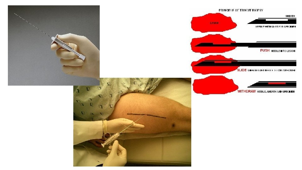
Principles of Biopsy • Treatment centre • Core biospy • Incisional or excisional; image guided • Don’t contaminate adjacent compartments; stick to involved compartment • In line with longitudinal incision • Haemostasis • Do not overly expose adjacent structures ie NV • Drains – in line with skin incision
Staging • Combines grade and histopath diagnosis • 1 – low grade, small, not spread • 2 – any grade, larger, not spread • 3 – high grade, not spread • 4 – any grade , spread
What next?
Histopathological assessment of soft tissue sarcomas
Key questions from the clinical point of view: What is the diagnosis? (diagnostic biopsies) and/or Is excision complete/adequate? (resection specimens) When will the result be ready?
The path of a specimen in the histopathology laboratory Specimen reception – • labeling, • sorting (subspecialty, BMS/consultant, high risk, urgent), • booking in same day/ next morning usually overnight fixation before cut-up, sometimes 48 hours (large specimens, high risk)
The path of a specimen in the histopathology laboratory Dissection – • “opening” • photos • macroscopic description • sampling calcified/bone samples – decalcification before processing
The path of a specimen in the histopathology laboratory • Processing - overnight • Embedding, cutting staining – next day(s) • Reporting – often further work including further stains, molecular tests
Reporting of soft tissue specimens Standards – RCPath guidelines/datasets ‘Layers of information’ – H&E stained slides - adequacy, cell/tissue types, benign/malignant features Immunohistochemistry – confirmation/further information on cell differentiation, proliferation index Special stains – matrix elements, infective organisms Molecular studies – FISH, RT-PCR, WGS
Dataset for histopathological reporting of soft tissue sarcomas Author: Prof. Cyril Fisher, 16 Aug 2017
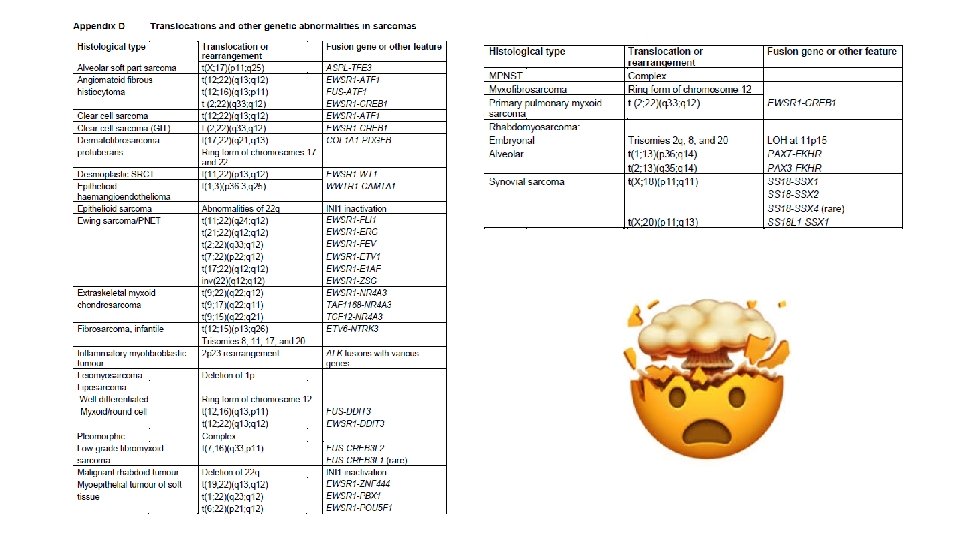
Clinical • Site Core data items • depth from surface Pathological • size of tumour • histological type and subtype • grade • tissue planes involved • relationship to margins • • • Adipocytic Fibroblastic/myofibroblastic Pericytic (perivascular) So-called fibrohistiocytic Smooth muscle Skeletal muscle Vascular Chondro-osseus Gastrointestinal stromal tumours Peripheral nerve sheath tumours Tumours of uncertain differentiation Undifferentiated/unclassified sarcoma • stage • cytogenetic and molecular genetic findings (for small round cell tumours)
Immunohistochemical staining H&E S 100
Clinical • Site Core data items • depth from surface Pathological • size of tumour • histological type and subtype • grade • tissue planes involved • relationship to margins • stage • cytogenetic and molecular genetic findings (for small round cell tumours)
Morphological features of tumours of different grades: Benign (leiomyoma) Low grade (grade 1) leiomyosarcoma High grade (grade 2 -3) leiomyosarcoma
An example of the use of molecular studies in the diagnosis of sarcomas: FISH for MDM 2 amplification in atypical lipomatous tumour/well differentiated liposarcoma Thway, Khin et al. “Fluorescence In Situ Hybridization for MDM 2 Amplification as a Routine Ancillary Diagnostic Tool for Suspected Well-Differentiated and Dedifferentiated Liposarcomas: Experience at a Tertiary Center. ” Sarcoma vol. 2015 (2015): 812089. doi: 10. 1155/2015/812089
Thank you