Grand Rounds Raafay Sophie MD 11182016 Department of


 Past Ocular Hx: Low Tension Glaucoma. – Hx of combined (Trab+Phaco) OU")


 • Unknown")


 -accumulation of blood in the space between the choroid and sclera")


- Slides: 35
Grand Rounds Raafay Sophie, MD 11/18/2016 Department of Ophthalmology and Visual Sciences
Patient Presentation CC Painful Red Right eye HPI 70 y White Female seen in the ED at night, with acute pain in right eye that stared around 8 hours ago – Had called her Ophthalmologist earlier and had been instructed to come to clinic the same day, but had initially deferred due to improvement in pain – Pain was suddenly exacerbated later in evening when she bent down to play with her dog.
Patient Presentation HPI – Some “flashes of light” when she blinked - Eye had been “watering” on and off - Was starting to have a headache with associated nausea and had vomited x 1 - Vision was “about the same” No new floaters No discharge No photophobia.
History (Hx) Past Ocular Hx: Low Tension Glaucoma. – Hx of combined (Trab+Phaco) OU 10 years ago – currently not on drops Past Medical Hx: HTN, Asthma, DM, Hypothyroidism Meds: ASA, Metformin, Synthroid, Lisinopril, Lipitor, Albuterol inhaler Allergies: PCN, Latex, Antihistamines Social Hx: no smoking or alcohol. Fam Hx: non-contributory ROS: No fever, palpitations, Shortness of breath
External Exam ODcc. N OScc. N VA 20/400 Pupils 4→ 3 mm, irregular IOP 7, 6 mm. Hg 11, 12 mm. Hg EOM full No r. APD CVF 4→ 2 mm
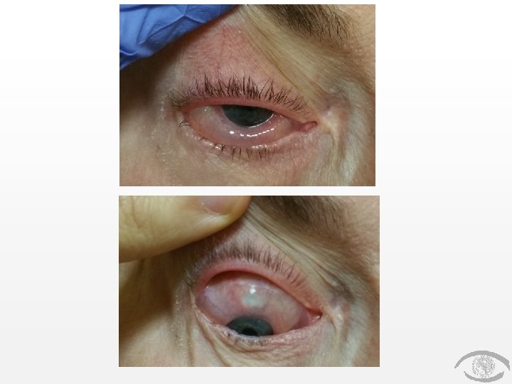
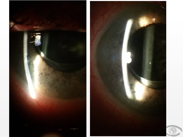
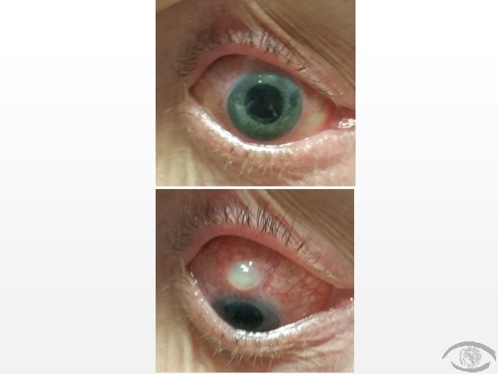
Anterior Segment Exam PLE or SLE OD External/Lids OS WNL Conj/Sclera Diffuse chemosis with +2 injection. white bleb superiorly Superior bleb Cornea Clear, Siedel negative Clear Ant Chamber Shallow, +1 cell and flare, <5% hyphema with trace diffuse RBCs Deep and Quiet Iris Bowed forward slightly but not touching cornea No NVI PI@12 Lens PCIOL
OS
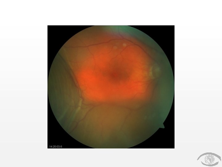
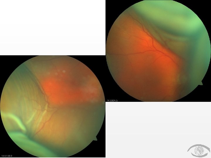
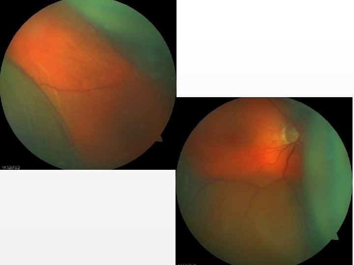
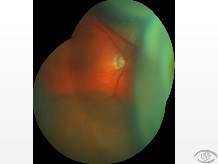
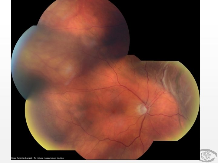
Posterior Segment Exam Fundus Optic Nerve OD C/D 0. 9, sharp border Macula WNL C/D 0. 8 WNL Vessels Periphery OS WNL Dome shaped dark choroidal elevation (no central retinal apposition) WNL
Assessment DDx: • Suprachoroidal Hemorrhage • Suprachoroidal effusion/serous choroidal detachment • Blebitis • Rhegmatogenous Retinal detachment
Plan • Started on – Pred Forte QID – Cyclopentolate 1% TID – Prophylactic Vigamox Q 2 h WA – PO Tylenol and Zofran PRN – Instructed not to touch eye and keep eye shield on at night – F/up next day in General Clinic and then referred to Retina Clinic
F/up • Retina Clinic: – Suprachoroidal hemorrhage (with no central retinal apposition) • Unknown etiology • VAcc 20/HM and 20/200 – Stopped Vigamox – Stopped ASA – PF QID + Atropine 1% BID – Subsequently monitored with bi-weekly checkups
Day 52 VAcc 20/200
Month 11 VAcc 20/200
Suprachoroidal Hemmorhage (SCH) -accumulation of blood in the space between the choroid and sclera (ie, suprachoroidal space) -is a distinct entity from a choroidal detachment in which serous fluid (rather than blood) pools in the suprachoroidal space. -First case report of choroidal hemorrhage in the setting of ophthalmic surgery was in 1760, by Baron de Wetzel. • Expulsive Suprachoroidal Hemorrhage: usually associated with substantial bleeding that leads to the eviction of intraocular contents through the surgical wound, usually with a poor visual prognosis. • Delayed Suprachoroidal Hemorrhage: Closed wound prevents the extrusion of intraocular contents. Have a better visual prognosis.
Discussion Proposed Mechanism: 1. engorgement of the choriocapillaris 2. serous effusion into the suprachoroidal space, occurring mainly in the posterior pole 3. stretching and tearing of the vessels and attachments at the base of the ciliary body as the effusion enlarges 4. resultant massive extravasation of blood arising from torn ciliary body vessels, which leads to SCH (and expulsion of intraocular contents through the surgical wound)
Discussion • Risk factors include – Systemic conditions: advanced age, atherosclerosis, hypertension, blood dyscrasias – Ocular conditions: including glaucoma, aphakia/pseudophakia, choroidal arteriolar sclerosis, myopia, choroiditis, recent intraocular surgery, SCH in fellow eye
Discussion – Perioperative conditions: • Valsalva maneuver, precipitous drop in IOP, retrobulbar anesthesia without epinephrine, vitreous loss, intraoperative systemic hypertension, • tube shunts vs trabeculectomy – Postoperative conditions: • hypotony, systemic thrombolytic agents, and postoperative trauma. • Glaucoma surgery: White race, post operative hypotony, anticoagulants and aphakia/anterior chamber lens
Discussion Clinical Presentation: • Severe ocular pain (from stretching ciliary nerves in the posterior segment) • ± Headache and Nausea/ Vomitting • Decreased vision • IOP may be low, normal or high
Discussion Clinical Presentation : • Shallowing of the anterior chamber • May have vitreous prolapse into the anterior chamber in aphakic and pseudophakic eyes • May have a loss of red reflex • Exhibit classically dark dome-shaped choroidal elevations that do not transillluminate well (can be confirmed by B-scan ultrasound).
Discussion Treatment: • Topical and oral steroids to control inflammation. • Analgesics (except aspirin and NSAIDs) and cycloplegic agents control the pain. • Manage IOP- Topical and oral antiglaucoma medications.
Discussion • Secondary surgical management remains controversial. • No consensus exists about whether the surgical drainage of the Suprachoroidal space is appropriate. Indications for surgery may include: – retinal detachment – central retinal apposition – vitreous incarceration into a surgical wound or a breakthrough vitreous hemorrhage – increased IOP – retained lens material during cataract surgery – intractable eye pain.
Discussion • SCH with associated retinal detachment – evidence suggests that patients may be followed for progression, because the majority may experience spontaneous regression. • Delayed SCH with central retinal apposition – Treatment strategies range from close follow up without intervention to surgical drainage and vitrectomy 10 to 14 days after the event
Conclusions • Suprachoroidal Hemorrhage is a rare but potentially devastating complication of ocular surgery. • Expulsive Suprachoroidal Hemorrhage vs Delayed Suprachoroidal Hemorrhage • Treatment goals: control IOP, inflammation and pain • Surgical management: no current consensus
References • Chu TG, Green RL. Suprachoroidal hemorrhage. Surv Ophthalmol. 1999; 43: 471 -486. • Chu TG, Cano MR, Green RL. Massive suprachoroidal hemorrhage with central retinal apposition: a clinical and echographic study. Arch Ophthalmol. 1991; 109: 1575 -1581. • Berrocal JA. Adhesion of the retina secondary to large choroidal detachment as a cause of failure in retinal detachment surgery. Mod Probl Ophthalmol. 1979; 20: 51 -52. • Reynolds MG, Haimovici R, Flynn HW Jr, et al. Suprachoroidal hemorrhage: clinical features and results of secondary surgical management. Ophthalmology. 1993; 100: 460 -465. • Scott IU, Flynn HW Jr, Schiffman J, et al. Visual acuity outcomes among patients with appositional suprachoroidal hemorrhage. Ophthalmology. 1997; 104: 2039 -2046 • Tuli SS 1, Wu. Dunn D, Ciulla TA, Cantor LB. Delayed suprachoroidal hemorrhage after glaucoma filtration procedures. Ophthalmology. 2001 Oct; 108(10): 1808 -11.