Electrocardiography Saeed Oraii MD Cardiologist Interventional Electrophysiologist Tehran

- Slides: 98
Electrocardiography Saeed Oraii MD, Cardiologist Interventional Electrophysiologist Tehran Arrhythmia Clinic Tehran Arrhythmia Center
Some slides have accompanied notes. To view them you can right click on the screen, choose ‘Screen’ and then ‘Speaker Notes’. Tehran Arrhythmia Center
ECG A graphic recording of electrical potentials generated by the heart A noninvasive, inexpensive and highly versatile test Tehran Arrhythmia Center
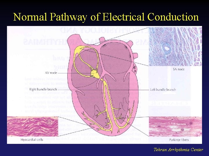
Normal Pathway of Electrical Conduction Tehran Arrhythmia Center
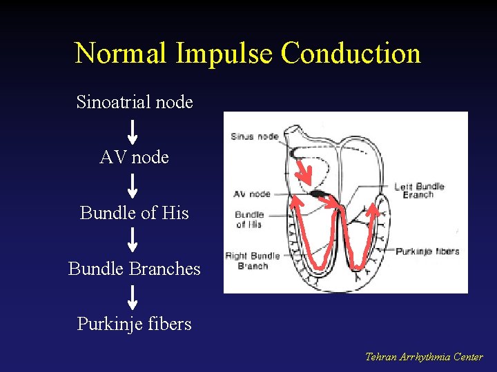
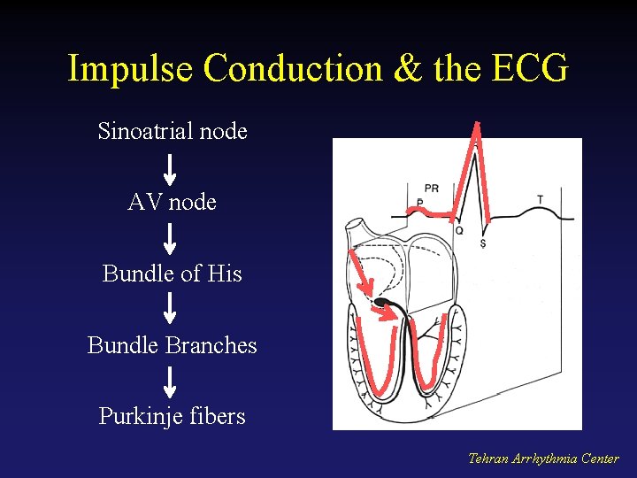
Normal Impulse Conduction Sinoatrial node AV node Bundle of His Bundle Branches Purkinje fibers Tehran Arrhythmia Center
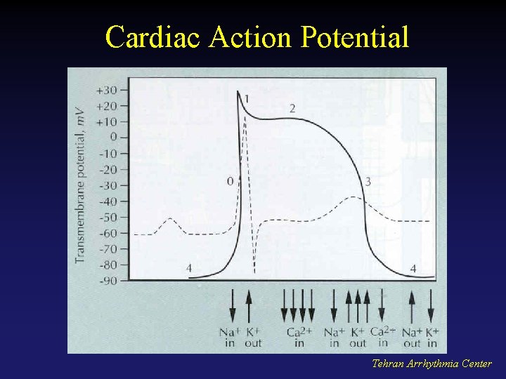
Cardiac Action Potential Tehran Arrhythmia Center
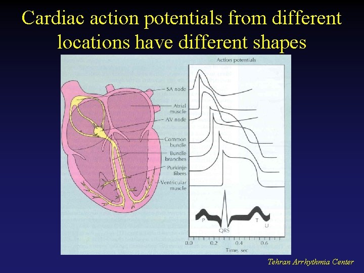
Cardiac action potentials from different locations have different shapes Tehran Arrhythmia Center
Electrophysiology • Electric currents that spread through the heart are produced by three components – Cardiac pacemaker cells – Specialized conduction tissue – The heart muscle • ECG only records the depolarization and repolarization potentials generated by atrial and ventricular myocardium. Tehran Arrhythmia Center
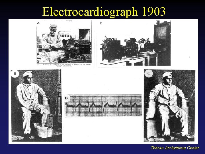
Electrocardiograph 1903 Tehran Arrhythmia Center
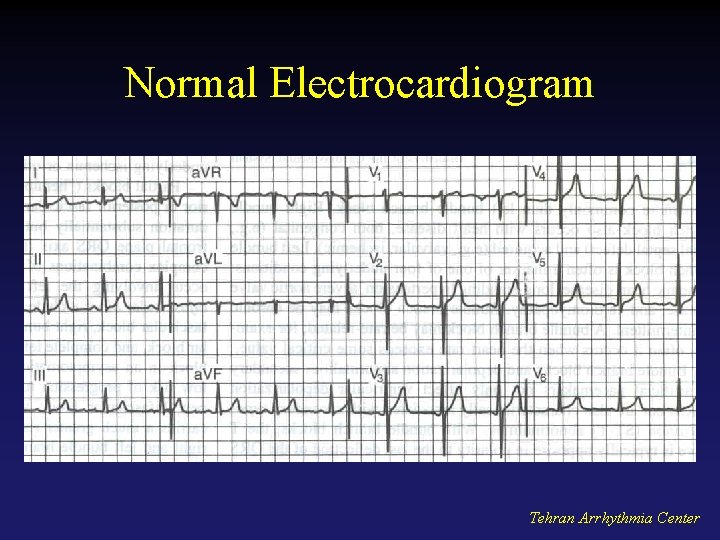
Normal Electrocardiogram Tehran Arrhythmia Center
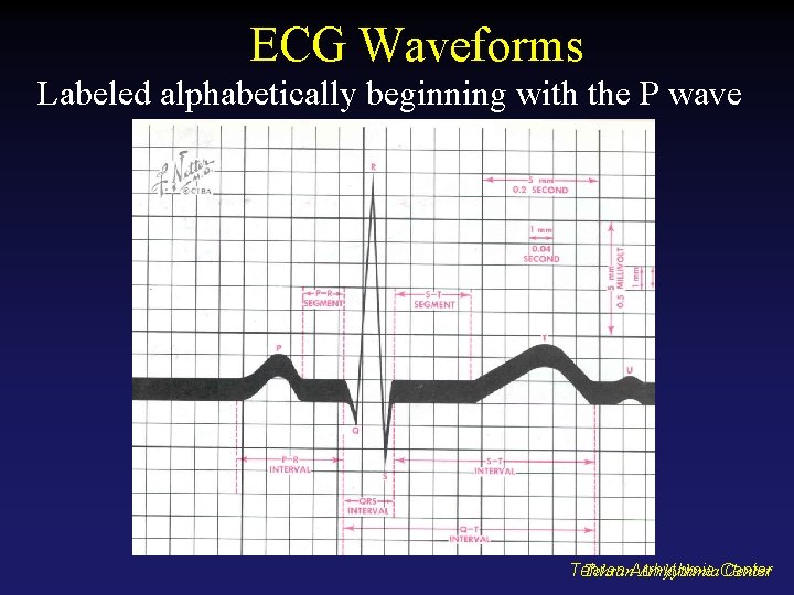
ECG Waveforms Labeled alphabetically beginning with the P wave Tehran. Arrhythmia. Center Tehran
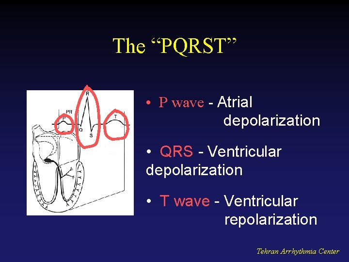
The “PQRST” • P wave - Atrial depolarization • QRS - Ventricular depolarization • T wave - Ventricular repolarization Tehran Arrhythmia Center
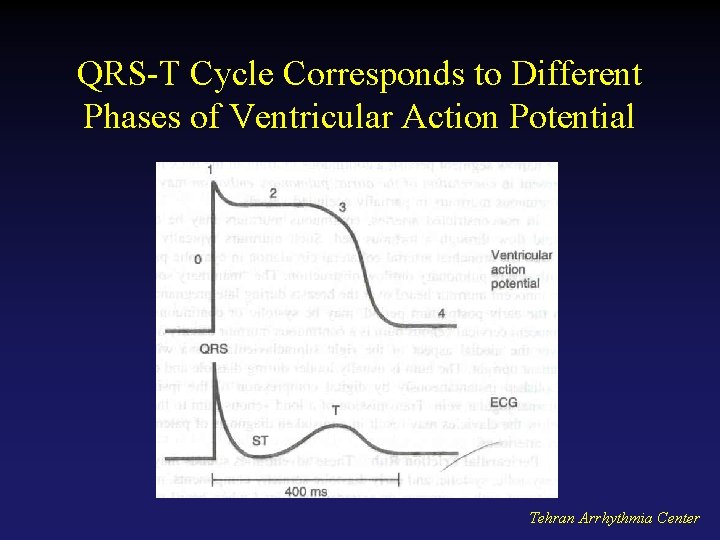
QRS-T Cycle Corresponds to Different Phases of Ventricular Action Potential Tehran Arrhythmia Center
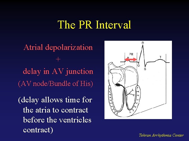
The PR Interval Atrial depolarization + delay in AV junction (AV node/Bundle of His) (delay allows time for the atria to contract before the ventricles contract) Tehran Arrhythmia Center
Impulse Conduction & the ECG Sinoatrial node AV node Bundle of His Bundle Branches Purkinje fibers Tehran Arrhythmia Center
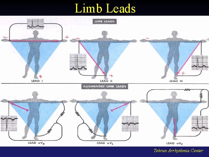
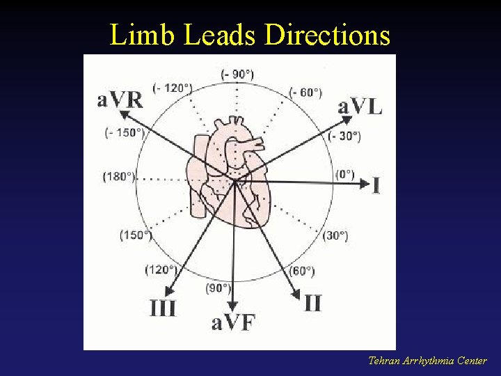
Limb Leads Tehran Arrhythmia Center
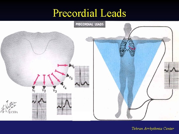
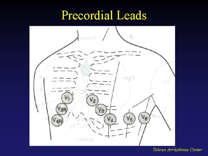
Precordial Leads Tehran Arrhythmia Center
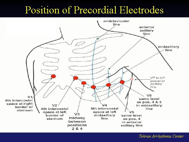
Position of Precordial Electrodes Tehran Arrhythmia Center
Precordial Leads Tehran Arrhythmia Center
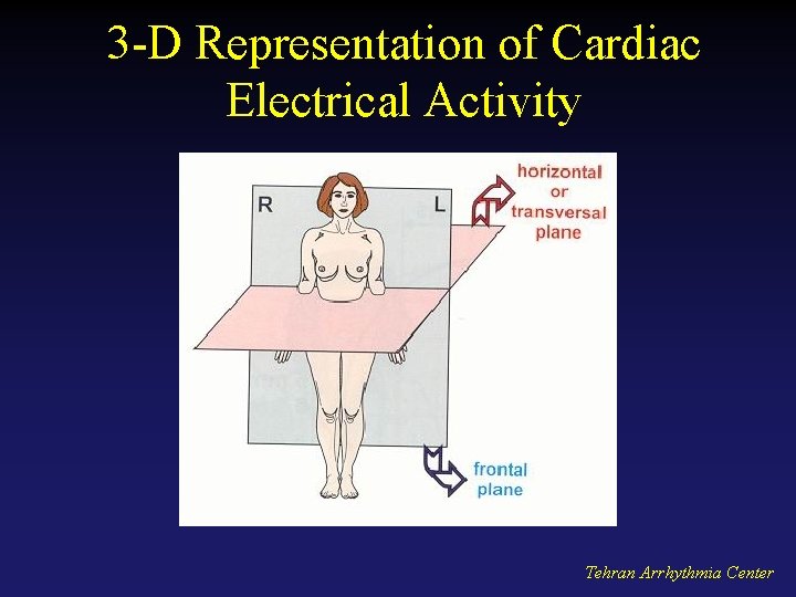
3 -D Representation of Cardiac Electrical Activity Tehran Arrhythmia Center
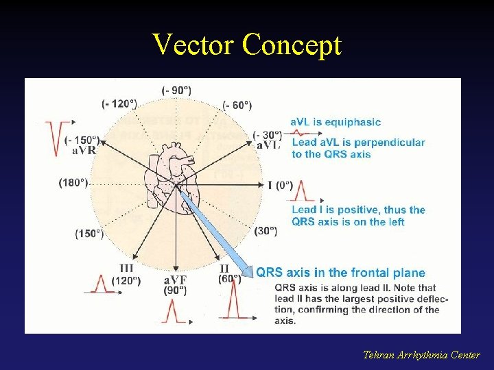
Vector Concept • Cardiac depolarization and repolarization waves have direction and magnitude. • They can, therefore, be represented by vectors. • ECG records the complex spatial and temporal summation of electrical potentials from multiple myocardial fibers conducted to the surface of the body. Tehran Arrhythmia Center
Limb Leads Directions Tehran Arrhythmia Center
Vector Concept Tehran Arrhythmia Center
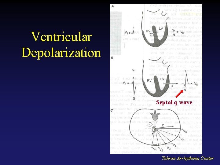
Ventricular Depolarization Septal q wave Tehran Arrhythmia Center
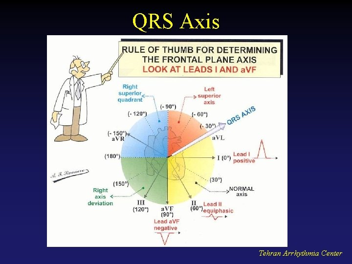
QRS Axis Tehran Arrhythmia Center
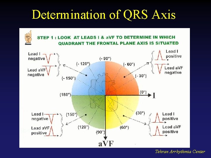
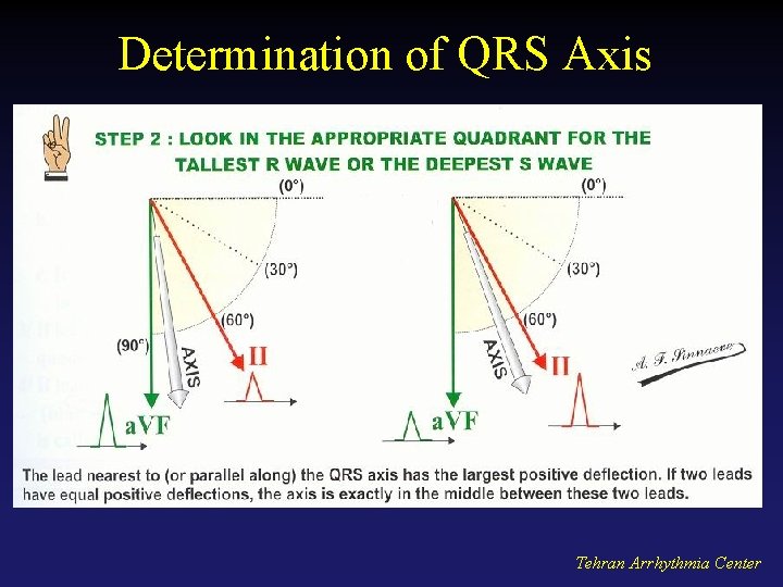
Determination of QRS Axis Tehran Arrhythmia Center
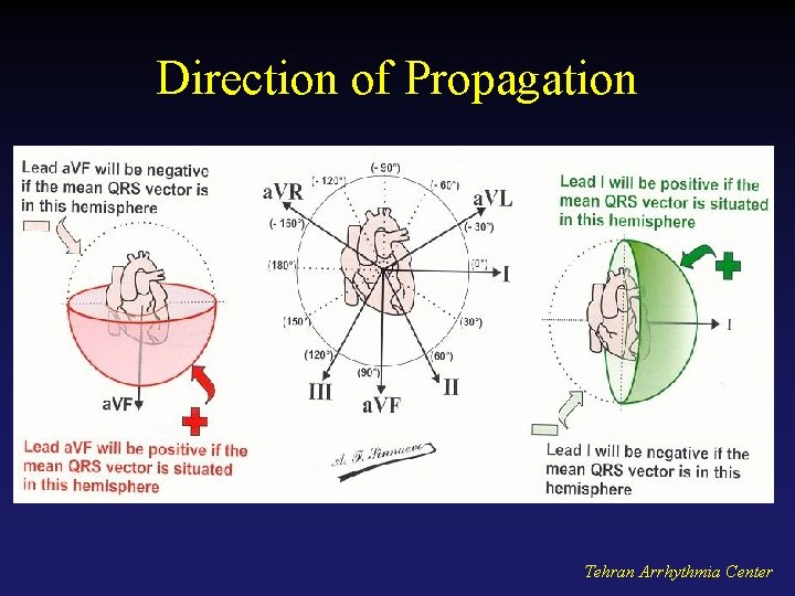
Direction of Propagation Tehran Arrhythmia Center
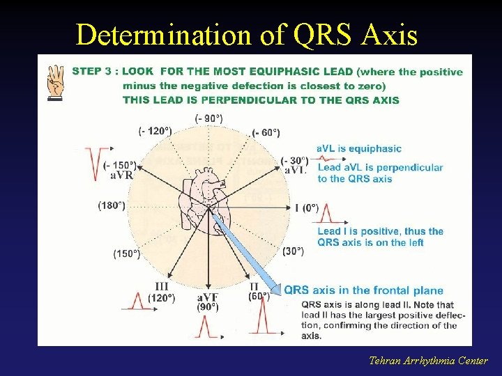
Determination of QRS Axis Tehran Arrhythmia Center
Determination of QRS Axis Tehran Arrhythmia Center
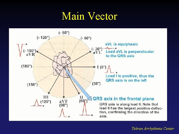
Main Vector Tehran Arrhythmia Center
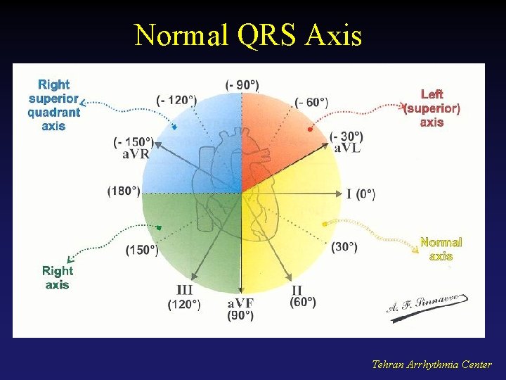
Normal QRS Axis Tehran Arrhythmia Center
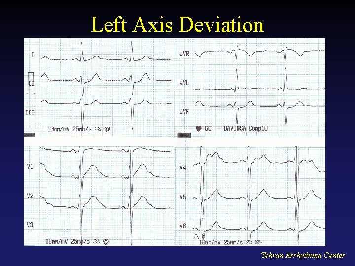
Left Axis Deviation Tehran Arrhythmia Center
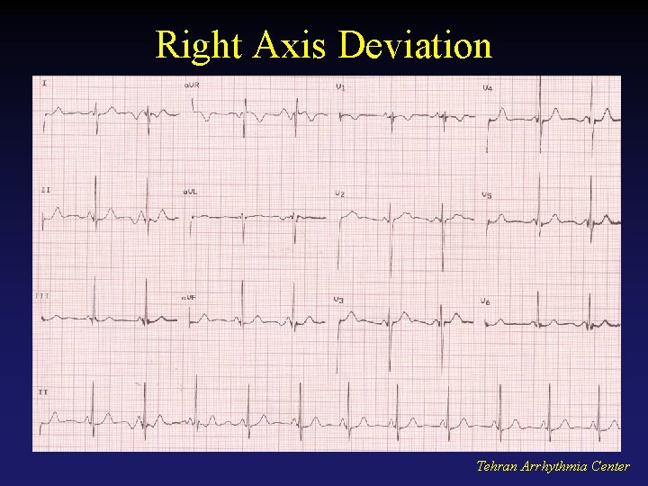
Right Axis Deviation Tehran Arrhythmia Center
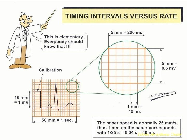
Timing Intervals Tehran Arrhythmia Center
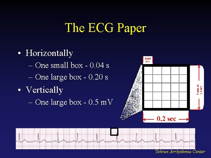
The ECG Paper • Horizontally – One small box - 0. 04 s – One large box - 0. 20 s • Vertically – One large box - 0. 5 m. V Tehran Arrhythmia Center
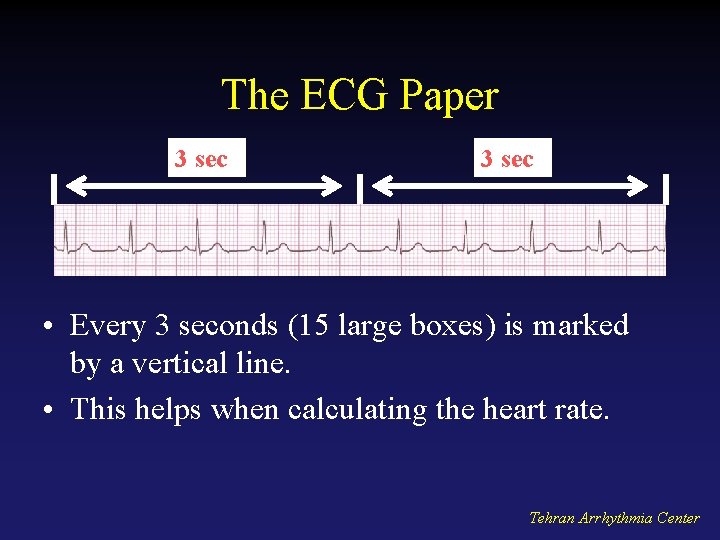
The ECG Paper 3 sec • Every 3 seconds (15 large boxes) is marked by a vertical line. • This helps when calculating the heart rate. Tehran Arrhythmia Center
Major ECG Abnormalities Tehran. Arrhythmia. Center Tehran
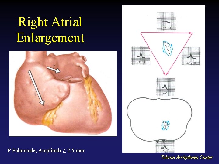
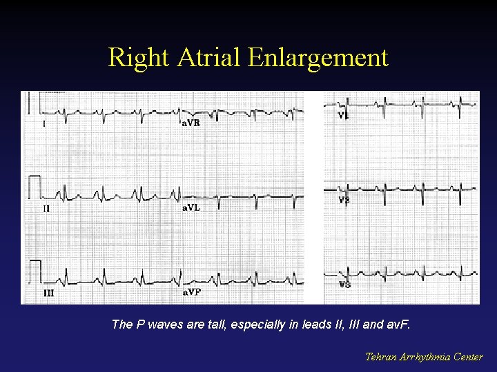
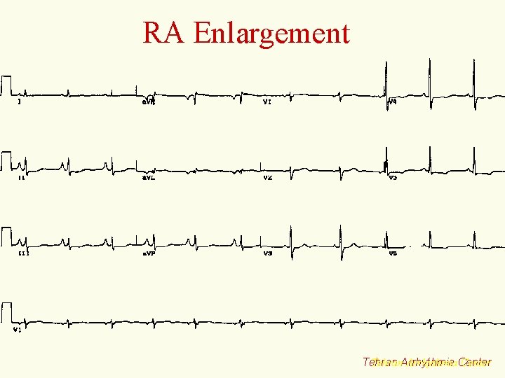
Right Atrial Enlargement P Pulmonale, Amplitude ≥ 2. 5 mm Tehran Arrhythmia Center
Right Atrial Enlargement The P waves are tall, especially in leads II, III and av. F. Tehran Arrhythmia Center
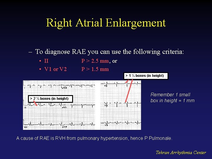
Right Atrial Enlargement – To diagnose RAE you can use the following criteria: • II • V 1 or V 2 P > 2. 5 mm, or P > 1. 5 mm > 1 ½ boxes (in height) > 2 ½ boxes (in height) Remember 1 small box in height = 1 mm A cause of RAE is RVH from pulmonary hypertension, hence P Pulmonale. Tehran Arrhythmia Center
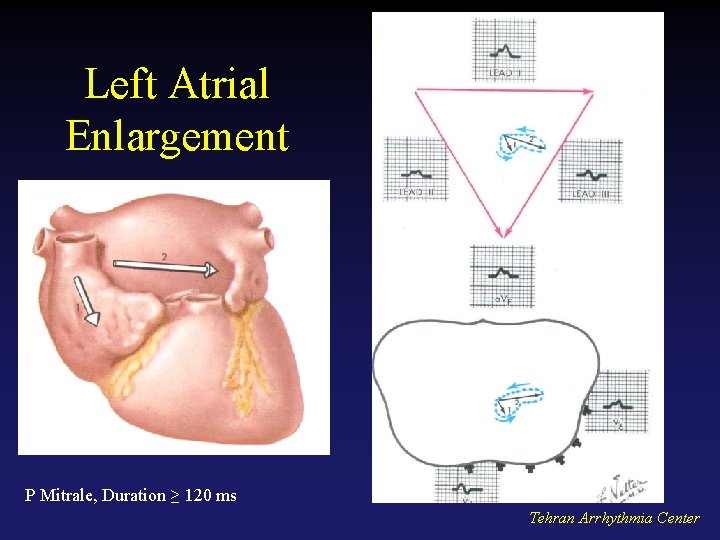
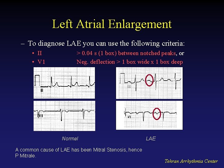
Left Atrial Enlargement P Mitrale, Duration ≥ 120 ms Tehran Arrhythmia Center
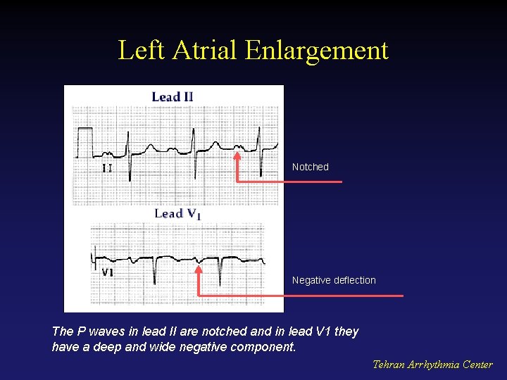
Left Atrial Enlargement Notched Negative deflection The P waves in lead II are notched and in lead V 1 they have a deep and wide negative component. Tehran Arrhythmia Center
Left Atrial Enlargement – To diagnose LAE you can use the following criteria: • II • V 1 > 0. 04 s (1 box) between notched peaks, or Neg. deflection > 1 box wide x 1 box deep Normal A common cause of LAE has been Mitral Stenosis, hence P Mitrale. LAE Tehran Arrhythmia Center
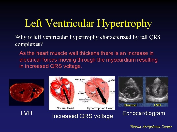
Left Ventricular Hypertrophy Why is left ventricular hypertrophy characterized by tall QRS complexes? As the heart muscle wall thickens there is an increase in electrical forces moving through the myocardium resulting in increased QRS voltage. LVH Increased QRS voltage Echocardiogram Tehran Arrhythmia Center
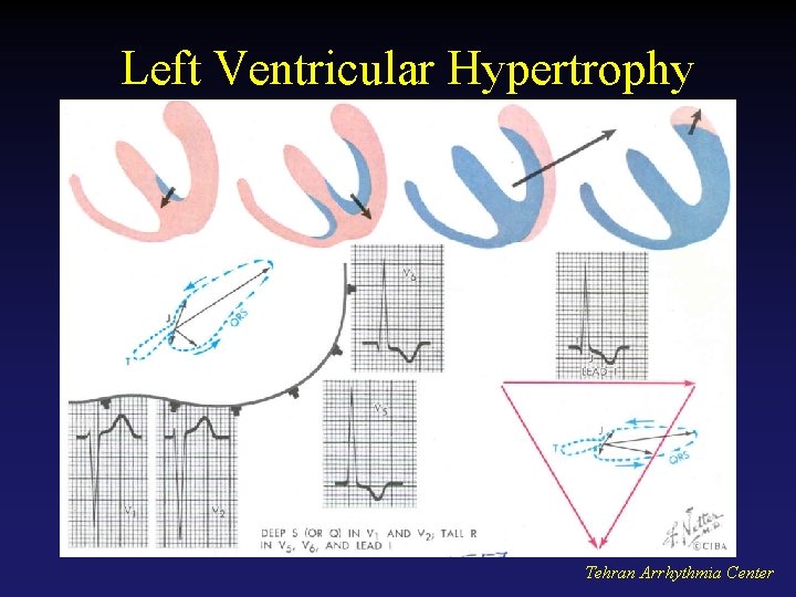
Left Ventricular Hypertrophy Tehran Arrhythmia Center
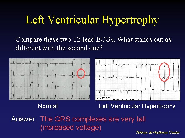
Left Ventricular Hypertrophy Compare these two 12 -lead ECGs. What stands out as different with the second one? Normal Left Ventricular Hypertrophy Answer: The QRS complexes are very tall (increased voltage) Tehran Arrhythmia Center
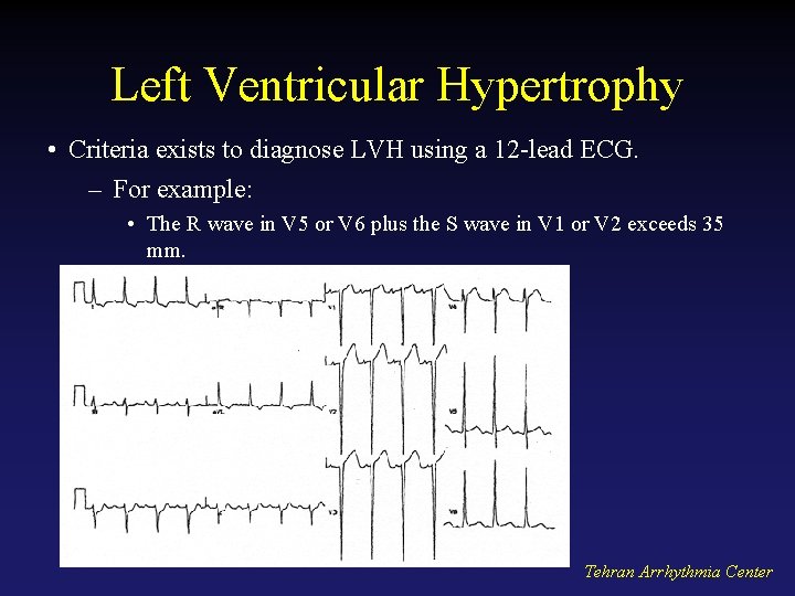
Left Ventricular Hypertrophy • Criteria exists to diagnose LVH using a 12 -lead ECG. – For example: • The R wave in V 5 or V 6 plus the S wave in V 1 or V 2 exceeds 35 mm. Tehran Arrhythmia Center
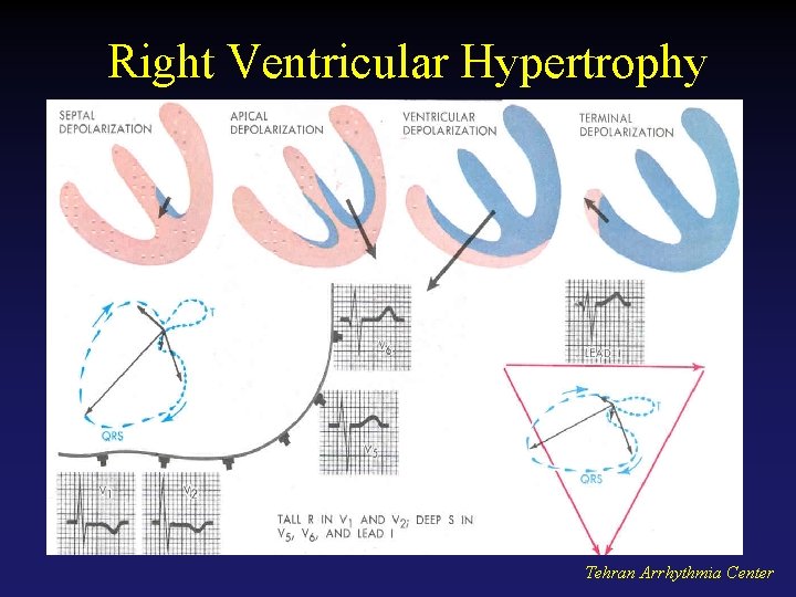
Right Ventricular Hypertrophy Tehran Arrhythmia Center
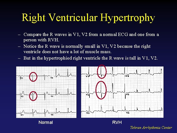
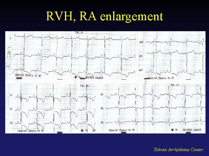
Right Ventricular Hypertrophy – Compare the R waves in V 1, V 2 from a normal ECG and one from a person with RVH. – Notice the R wave is normally small in V 1, V 2 because the right ventricle does not have a lot of muscle mass. – But in the hypertrophied right ventricle the R wave is tall in V 1, V 2. Normal RVH Tehran Arrhythmia Center
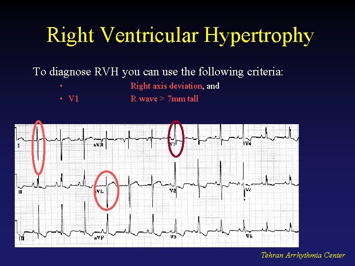
Right Ventricular Hypertrophy To diagnose RVH you can use the following criteria: • • V 1 Right axis deviation, and R wave > 7 mm tall Tehran Arrhythmia Center
RVH, RA enlargement Tehran Arrhythmia Center
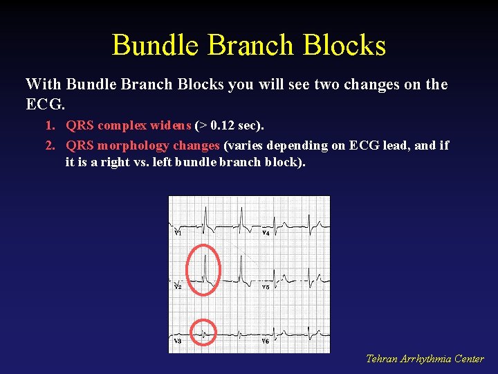
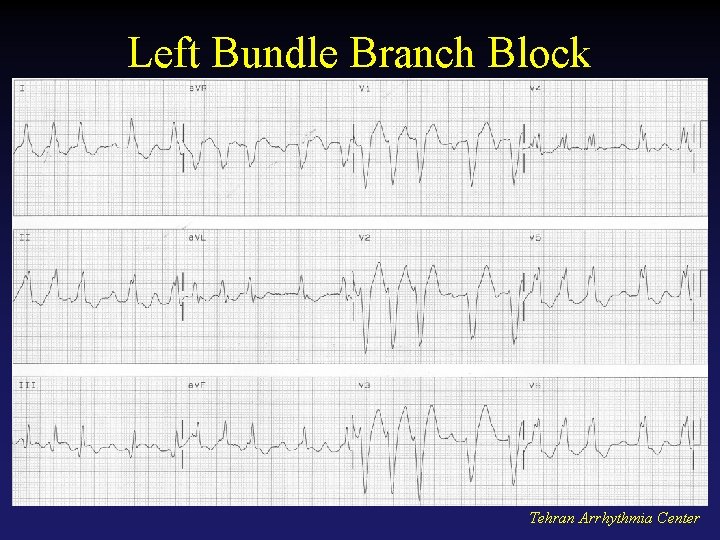
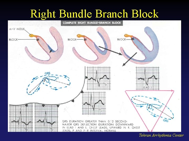
Bundle Branch Blocks With Bundle Branch Blocks you will see two changes on the ECG. 1. QRS complex widens (> 0. 12 sec). 2. QRS morphology changes (varies depending on ECG lead, and if it is a right vs. left bundle branch block). Tehran Arrhythmia Center
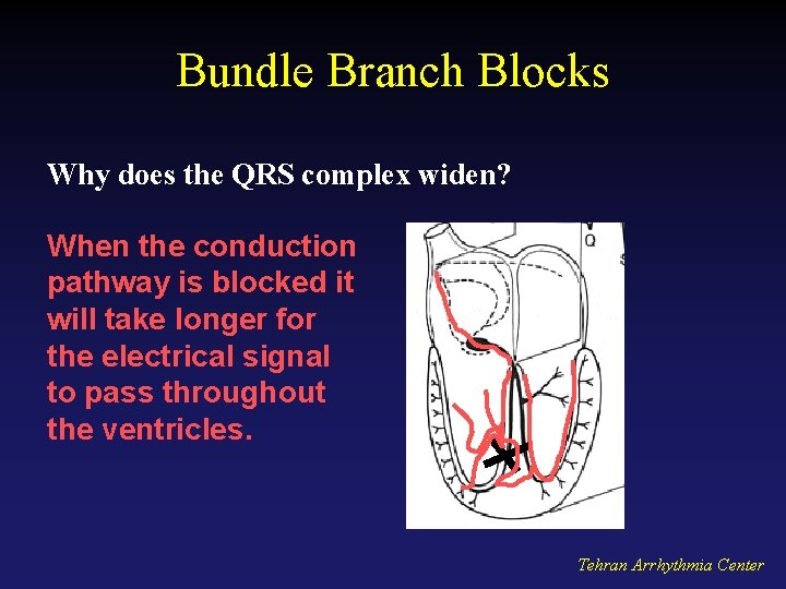
Bundle Branch Blocks Why does the QRS complex widen? When the conduction pathway is blocked it will take longer for the electrical signal to pass throughout the ventricles. Tehran Arrhythmia Center
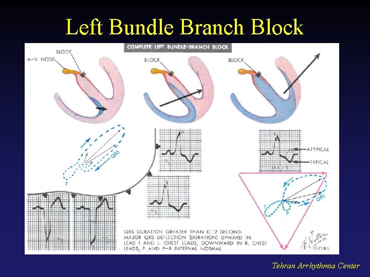
Left Bundle Branch Block Tehran Arrhythmia Center
Left Bundle Branch Block Tehran Arrhythmia Center
Right Bundle Branch Block Tehran Arrhythmia Center
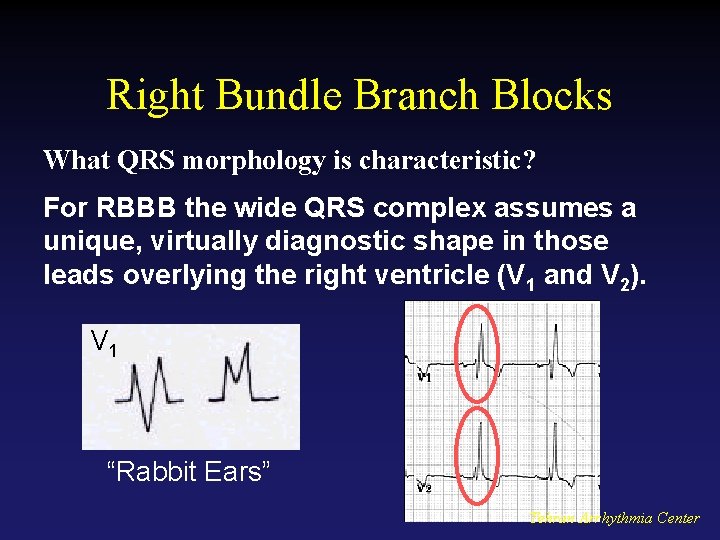
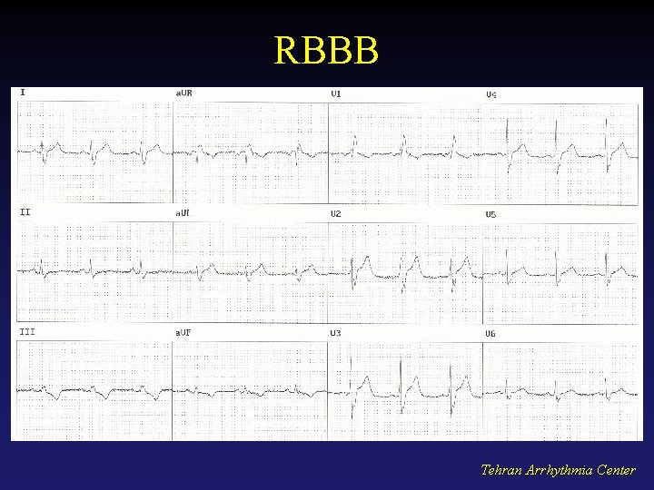
Right Bundle Branch Blocks What QRS morphology is characteristic? For RBBB the wide QRS complex assumes a unique, virtually diagnostic shape in those leads overlying the right ventricle (V 1 and V 2). V 1 “Rabbit Ears” Tehran Arrhythmia Center
RBBB Tehran Arrhythmia Center
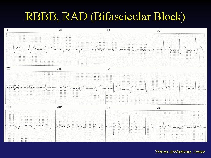
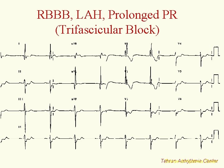
RBBB, RAD (Bifascicular Block) Tehran Arrhythmia Center
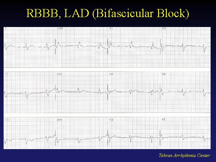
RBBB, LAD (Bifascicular Block) Tehran Arrhythmia Center
Tehran Arrhythmia Center
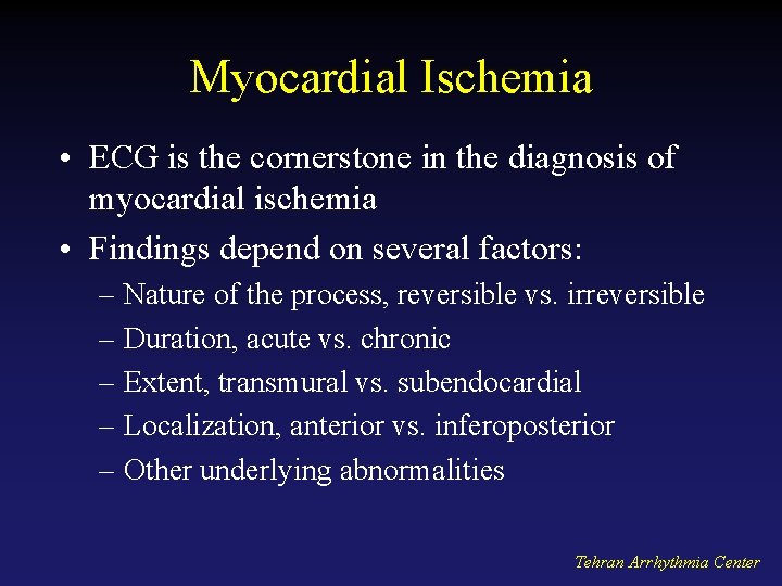
Myocardial Ischemia • ECG is the cornerstone in the diagnosis of myocardial ischemia • Findings depend on several factors: – Nature of the process, reversible vs. irreversible – Duration, acute vs. chronic – Extent, transmural vs. subendocardial – Localization, anterior vs. inferoposterior – Other underlying abnormalities Tehran Arrhythmia Center
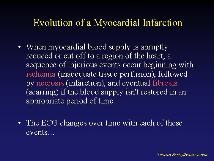
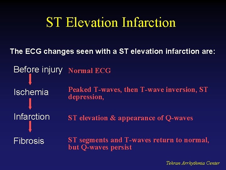
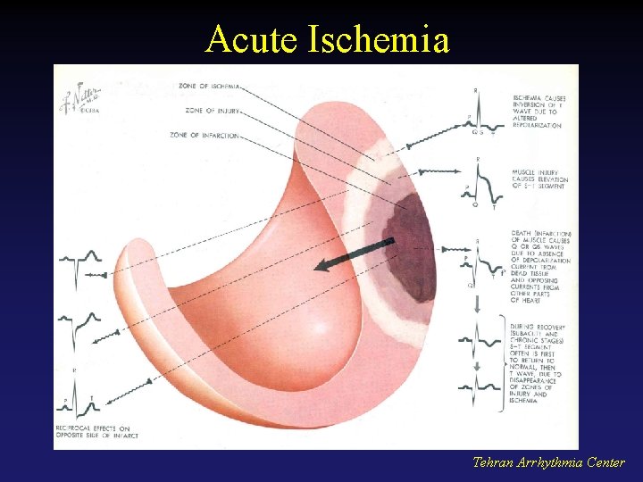
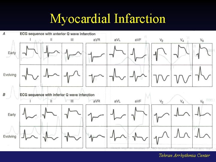
Evolution of a Myocardial Infarction • When myocardial blood supply is abruptly reduced or cut off to a region of the heart, a sequence of injurious events occur beginning with ischemia (inadequate tissue perfusion), followed by necrosis (infarction), and eventual fibrosis (scarring) if the blood supply isn't restored in an appropriate period of time. • The ECG changes over time with each of these events… Tehran Arrhythmia Center
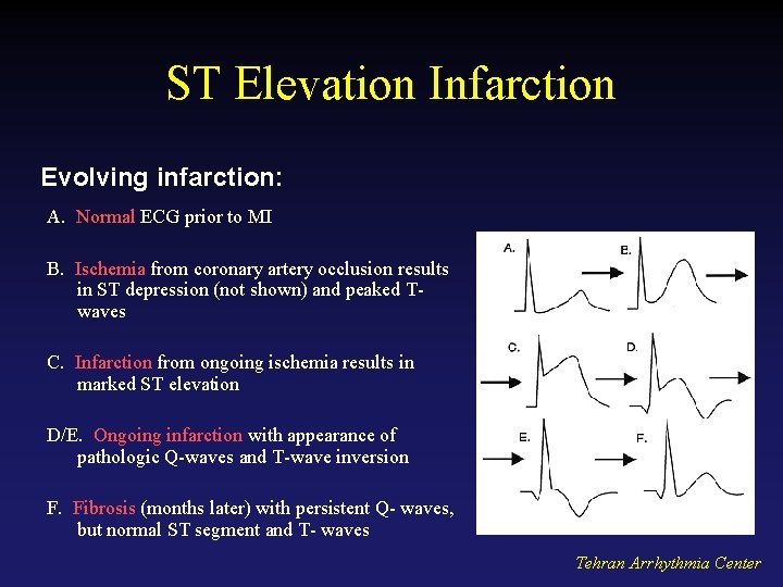
ST Elevation Infarction The ECG changes seen with a ST elevation infarction are: Before injury Normal ECG Ischemia Peaked T-waves, then T-wave inversion, ST depression, Infarction ST elevation & appearance of Q-waves Fibrosis ST segments and T-waves return to normal, but Q-waves persist Tehran Arrhythmia Center
Acute Ischemia Tehran Arrhythmia Center
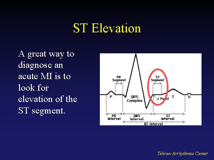
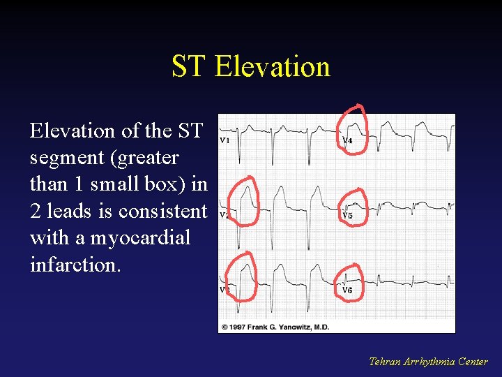
ST Elevation A great way to diagnose an acute MI is to look for elevation of the ST segment. Tehran Arrhythmia Center
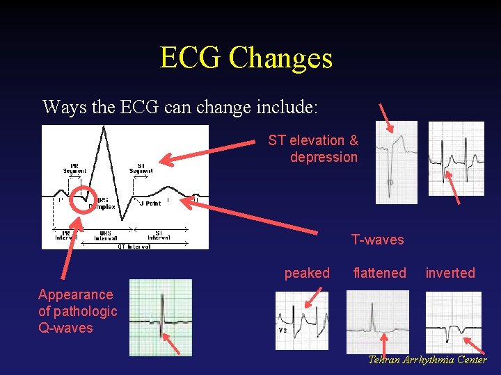
ECG Changes Ways the ECG can change include: ST elevation & depression T-waves peaked flattened inverted Appearance of pathologic Q-waves Tehran Arrhythmia Center
ST Elevation of the ST segment (greater than 1 small box) in 2 leads is consistent with a myocardial infarction. Tehran Arrhythmia Center
ST Elevation Infarction Evolving infarction: A. Normal ECG prior to MI B. Ischemia from coronary artery occlusion results in ST depression (not shown) and peaked Twaves C. Infarction from ongoing ischemia results in marked ST elevation D/E. Ongoing infarction with appearance of pathologic Q-waves and T-wave inversion F. Fibrosis (months later) with persistent Q- waves, but normal ST segment and T- waves Tehran Arrhythmia Center
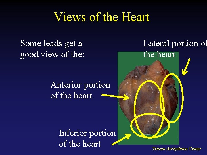
Views of the Heart Some leads get a good view of the: Lateral portion of the heart Anterior portion of the heart Inferior portion of the heart Tehran Arrhythmia Center
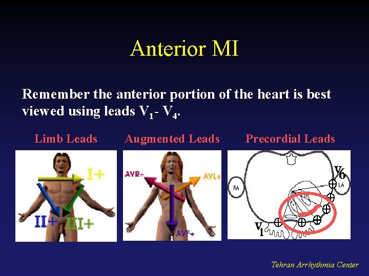
Anterior MI Remember the anterior portion of the heart is best viewed using leads V 1 - V 4. Limb Leads Augmented Leads Precordial Leads Tehran Arrhythmia Center
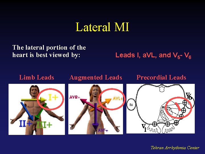
Lateral MI The lateral portion of the heart is best viewed by: Limb Leads I, a. VL, and V 5 - V 6 Augmented Leads Precordial Leads Tehran Arrhythmia Center
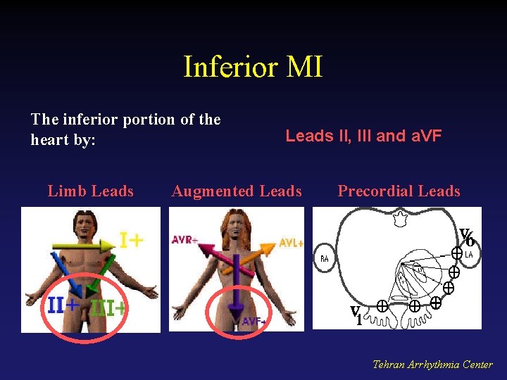
Inferior MI The inferior portion of the heart by: Limb Leads II, III and a. VF Augmented Leads Precordial Leads Tehran Arrhythmia Center
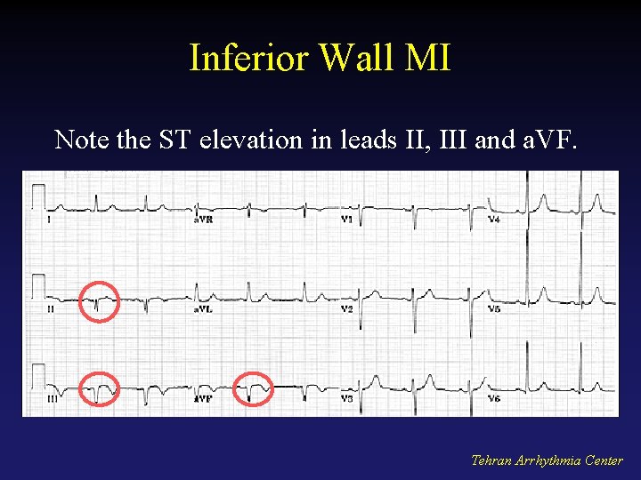
Inferior Wall MI Note the ST elevation in leads II, III and a. VF. Tehran Arrhythmia Center
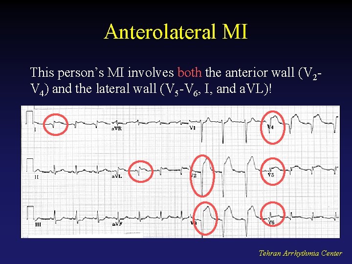
Anterolateral MI This person’s MI involves both the anterior wall (V 2 V 4) and the lateral wall (V 5 -V 6, I, and a. VL)! Tehran Arrhythmia Center
Myocardial Infarction Tehran Arrhythmia Center
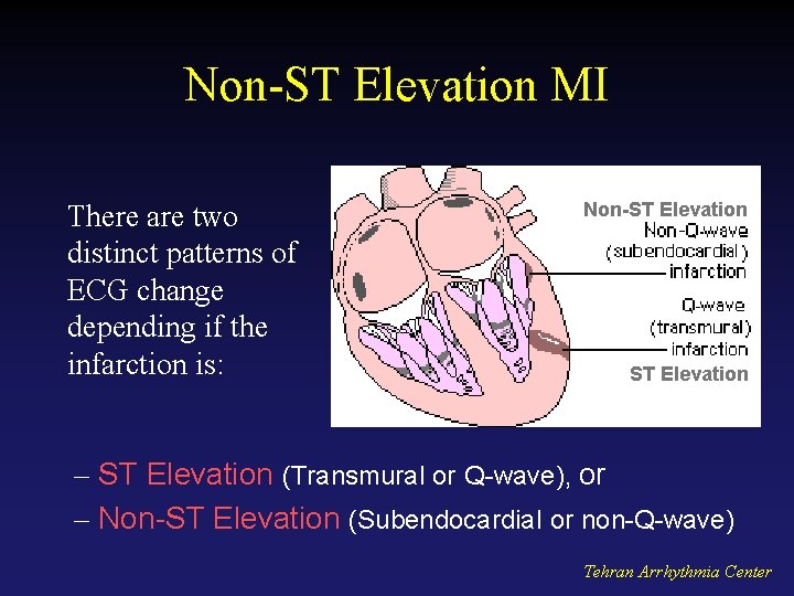
Non-ST Elevation MI There are two distinct patterns of ECG change depending if the infarction is: Non-ST Elevation – ST Elevation (Transmural or Q-wave), or – Non-ST Elevation (Subendocardial or non-Q-wave) Tehran Arrhythmia Center
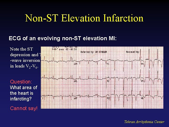
Non-ST Elevation Infarction ECG of an evolving non-ST elevation MI: Note the ST depression and T -wave inversion in leads V 2 -V 6. Question: What area of the heart is infarcting? Cannot say! Tehran Arrhythmia Center
Acute Pericarditis Tehran Arrhythmia Center
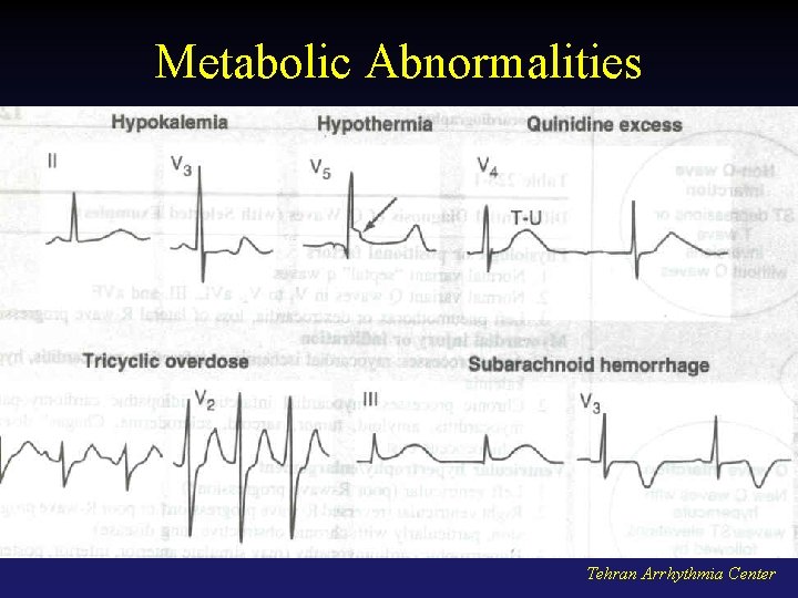
Metabolic Abnormalities Tehran Arrhythmia Center
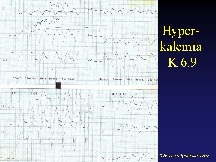
Hyperkalemia K 6. 9 Tehran Arrhythmia Center
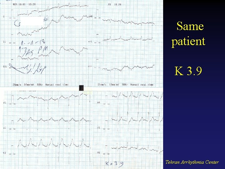
Same patient K 3. 9 Tehran Arrhythmia Center
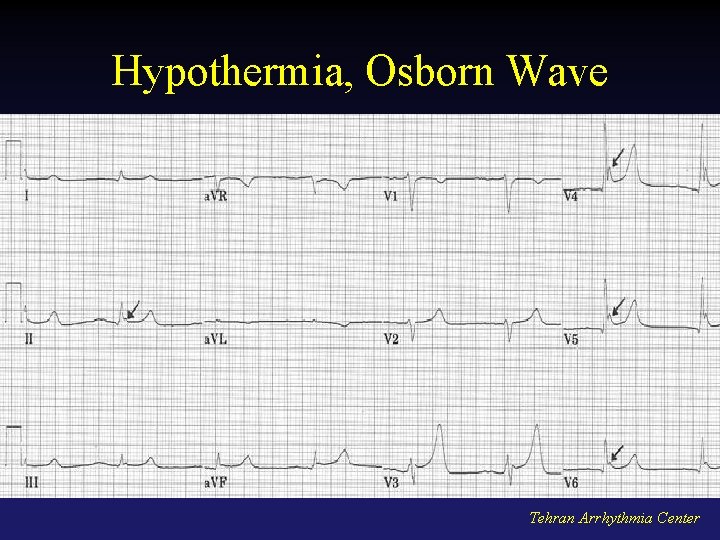
Hypothermia, Osborn Wave Tehran Arrhythmia Center
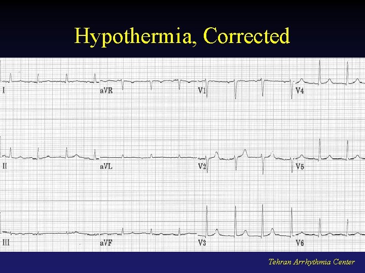
Hypothermia, Corrected Tehran Arrhythmia Center
Tehran Arrhythmia Center
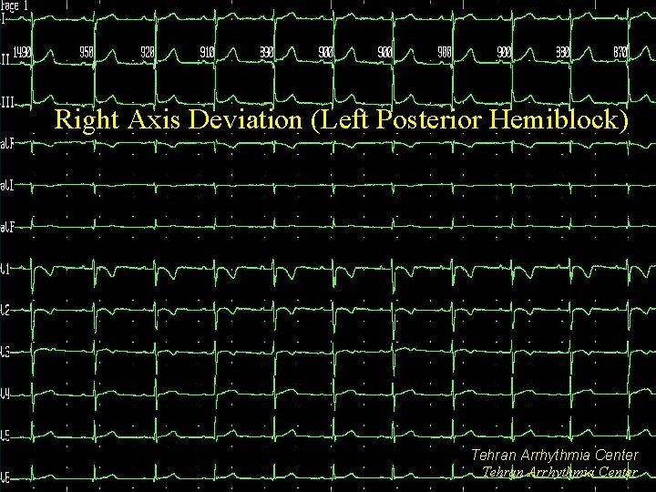
Right Axis Deviation (Left Posterior Hemiblock) Tehran Arrhythmia Center
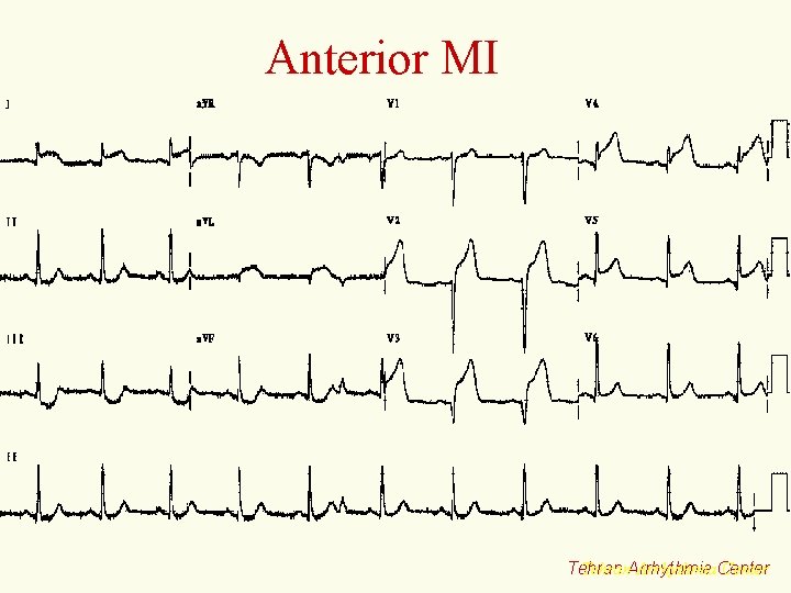
Anterior MI Tehran. Arrhythmia. Center Tehran
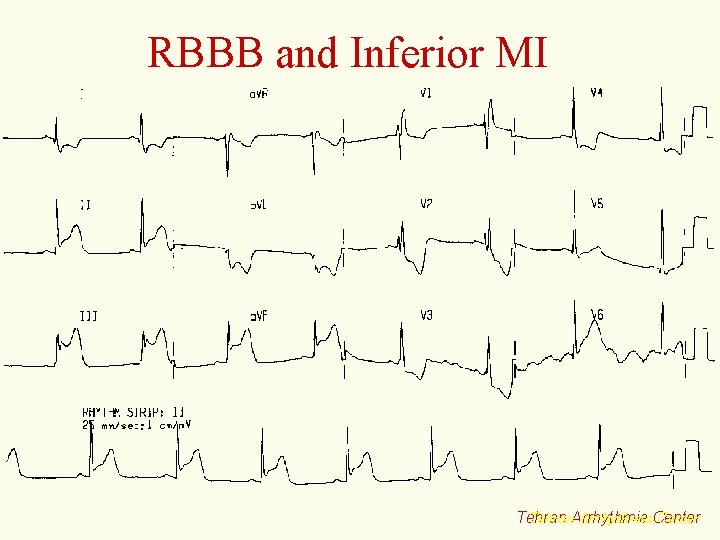
RBBB and Inferior MI Tehran. Arrhythmia. Center Tehran
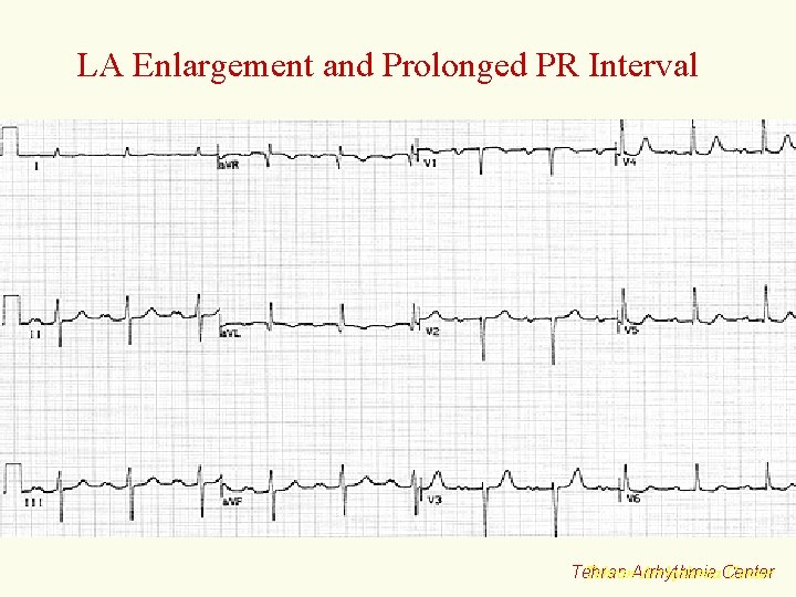
LA Enlargement and Prolonged PR Interval Tehran. Arrhythmia. Center Tehran
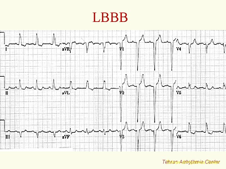
LBBB Tehran. Arrhythmia. Center Tehran
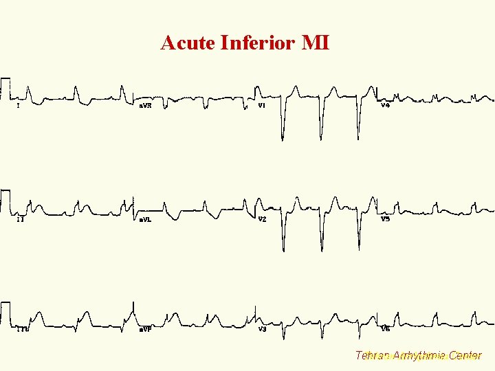
Acute Inferior MI Tehran. Arrhythmia. Center Tehran
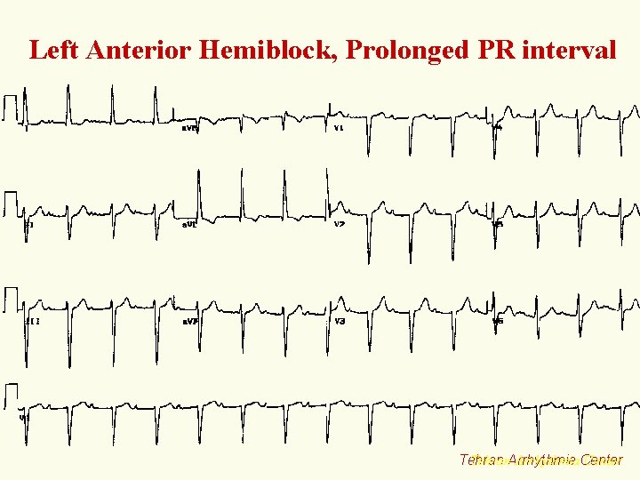
Left Anterior Hemiblock, Prolonged PR interval Tehran. Arrhythmia. Center Tehran
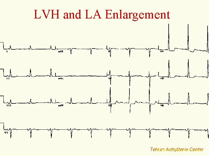
LVH and LA Enlargement Tehran. Arrhythmia. Center Tehran
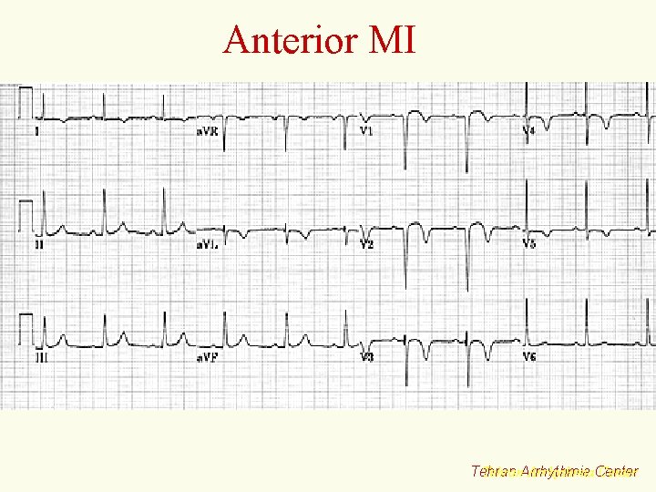
Anterior MI Tehran. Arrhythmia. Center Tehran
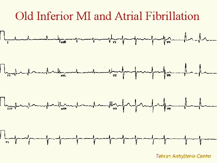
Old Inferior MI and Atrial Fibrillation Tehran. Arrhythmia. Center Tehran
RA Enlargement Tehran. Arrhythmia. Center Tehran
RBBB, LAH, Prolonged PR (Trifascicular Block) Tehran. Arrhythmia. Center Tehran
Tehran Arrhythmia Center WWW. Iran. EP. org info@Iran. EP. org Tehran Arrhythmia Center