Ecology Physiology of Fungi Fungal growth LEC 4


, the former are arranged as a")


 suggested that hyphal growth occurs as the result of a continuously replenished")


")


- Slides: 22
Ecology & Physiology of Fungi Fungal growth LEC= 4
Growth in fungi refers to a positive change in size, volume and number of cell, and that lead to maturation, often over a period of time. • Most fungi are sessile filamentous organisms that grow by extending the tips of hyphae to form an expanding mycelial network. Tip growth, therefore, represents a form of cellular motility and hyphae must be able to coordinate this motility by extending and orientating the trajectory of hyphal extension
Hyphae Composition Fungal species are also further classified based on the hyphal systems they contain. There are four general subtypes: 1 - Monomitic: While virtually all fungal species contain generative hyphae, those with only exhibit this type are referred to as monomitic (e. g. , agaric mushrooms). 2 - Dimitic: A species that contains generative hyphae in addition to one other type of hyphae. The most common combination of dimitic fungi is generative and skeletal. 3 - Trimitic: Species which contain all three types of hyphae (generative, binding, and skeletal). 4 - Sarcodimitic and sarcotrimitic: Sarcodimitic hyphae are fusiform skeletal hyphae bound to generative hyphae. Sarcotrimitic species contain fusiform skeletal hyphae, as well as binding and generative hyphae.
Hyphal growth allows the fungus to interact with its environment through colonization of substrates, secretion of enzymes, nutrient assimilation, and responding to environmental stimuli. Hyphal growth and differentiation must be strictly coordinated with cell wall synthesis, plasma membrane growth, vesicle transport, and cytoplasmic dynamics. The phenomenon of polarized growth via apical extension is not restricted to the fungi
The two fundamental properties of the vegetative fungal hypha , the polarity of both growth and secretion of degradative enzymes , have been known for over a century. Numerous studies have subsequently confirmed that ‘the key to the fungal hypha lies in the apex. Ultrastructural studies have shown that many organelles within the growing hyphal tip are distributed in steep gradients, . The cytoplasm of the extreme apex is occupied almost exclusively by Secretory vesicles and Microvesicles.
In the higher fungi (Asco- and Basidiomycota), the former are arranged as a spherical shell around the latter, and the entire formation is called the Spitzenkörper or ‘apical body The spitzenkörper is an intracellular organelle associated with tip growth. It is composed of an aggregation of membrane-bound vesicles containing cell wall components. The spitzenkörper is part of the endomembrane system of fungi, holding and releasing vesicles it receives from the Golgi apparatus.
The Spitzenkörper may be seen in growing hyphae even with the light microscope. Hyphae of the Oomycota and some lower Eumycota (notably the Zygomycota) do not contain a recognizable Spitzenkörper, and the vesicles are instead distributed more loosely in the apical dome. By modifying certain parameters, it is even possible to generate the somewhat more pointed apex often found in hyphae of Oomycota and Zygomycota
A little behind the apical dome, a region of intense biosynthetic activity and energy generation is indicated by parallel sheets of endoplasmic reticulum (ER) and an abundance of mitochondria. The first nuclei usually appear just behind the biosynthetic zone followed ultimately by a system of ever-enlarging vacuoles. These may fill almost the entire volume of mature hyphal regions, making them appear empty when viewed with the light microscope.
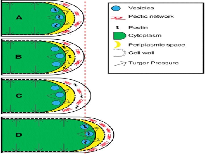
Mechanisms of mycelial growth Hyphae grow at their tips. During tip growth, cell walls are extended by the external assembly and polymerization of cell wall components, and the internal production of new cell membrane • The cell wall at the hyphal tip has viscoelastic properties and yields to the internal turgor pressure within the hypha. • Further behind the tip the wall is rigidified and resistant to the turgor forces resulting from the osmotic flow of water into the hypha. • Turgor pressure generated within the hypha therefore acts as the driving force for hyphal extension. • The vesicles which accumulated in Spitzenkörper region travel to the cell membrane via the cytoskeleton and release their contents outside the cell by the process of exocytosis, where it can then be transported to where it is needed. • Vesicle membranes contribute to growth of the cell membrane while their contents form new cell wall.
• The spitzenkörper moves along the apex of the hyphal strand generates apical growth and branching; the apical growth rate of the hyphal strand parallels and is regulated by the movement of the spitzenkörper. Hyphal extension at the apex requires synthesis and insertion of new wall material and new membranes in a way that does not weaken the tip. This highly organized process is supported by the continuous flow of vesicles generated within the cytoplasm behind the tip, and is coordinated with the growth and replication of all the other cytoplasmic organelles and their migration towards the extending apex.
Synthesis of the cell wall The synthesis of chitin is mediated by specialized organelles termed chitosomes which inactive chitin synthases are delivered to the apical plasma membrane and become activated upon contact with the lipid bilayer. Microvesicles, visible especially in the core region of the Spitzenkörper, are likely to be the ultrastructural manifestation of chitosomes. In contrast, structural proteins and enzymes travel together in the larger secretory vesicles and are discharged into the environment when the vesicles fuse with the plasma membrane.
Wessels (1997) suggested that hyphal growth occurs as the result of a continuously replenished supply of soft wall material at the apex, but there is good evidence that the softness of the apical cell wall is also influenced by the activity of wall-lytic enzymes such as chitinases or glucanases.
Diagrammatic representation of the internal scaffold model of tip growth in fungi proposed . Secretory vesicles and chitosomes are transported along microtubules from their subapical sites of synthesis to the growing apex. The Spitzenkorper forms around a cluster of actin filaments. An actin scaffold inside the extreme apex is linked to rivet-like integrin molecules which are anchored in the rigid subapical cell wall. The apex is further stabilized by spectrin molecules lining the cytoplasmic surface of the plasma membrane.
The properties of the cell wall depend in many ways on the environment in which the hypha grows. 1. when Schizophyllum commune is grown in liquid submerged culture, a significant part of the β-glucan fraction may diffuse into the liquid medium before it is captured by the cell wall, giving rise to mucilage 2. causing problems when growing fungi in liquid culture for experimental purposes, mucilage may cause economic losses when released by Botrytis cinerea in grapes to be used for wine production. 3. secreted polysaccharides, especially of Basidiomycota, may have interesting medicinal properties and are being promoted as antitumour medication both in conventional and in alternative medicine
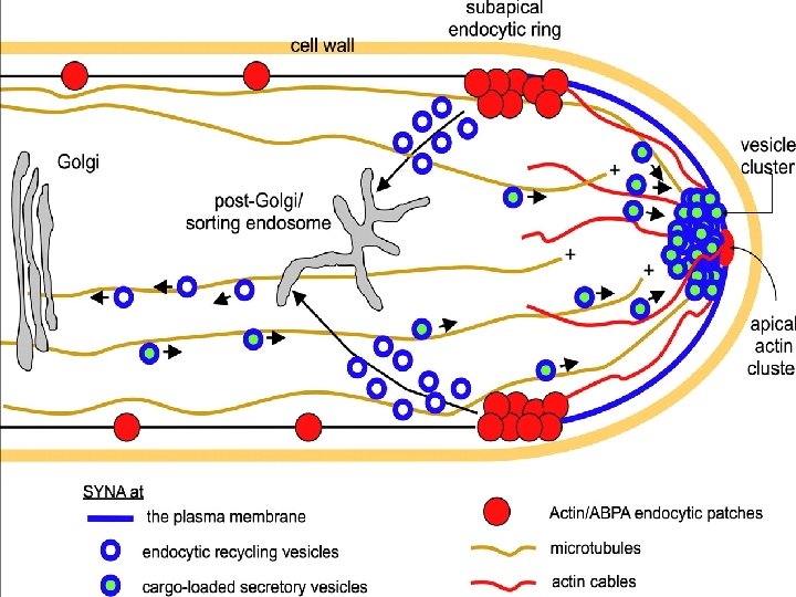
Secretion and membrane traffic One of the most important Ecological roles of fungi, that of Decomposing dead plant matter, requires the secretion of large quantities of hydrolytic and oxidative enzymes into the environment. In liquid culture under optimized experimental conditions, certain fungi are capable of secreting more than 20 g of a single enzyme or enzyme group per litre culture broth within a few days’ growth. The secretory route in fungi begins in the ER. Ribosomes loaded with a suitable messenger RNA dock on to the ER membrane and translate the polypeptide product which enters the ER lumen during its synthesis unless specific internal signal sequences cause it to be retained in the ER membrane.
Vesicles are thought to deliver the main wall-synthetic enzymes (chitin synthase and glucan synthase) to the tip, where they lodge in the plasma membrane as integral membrane proteins. Mannoproteins and other glycoproteins are transported in vesicles from the Endoplasmic reticulum - Golgi secretory system (because the glycosylation of proteins occurs only in the Golgi). Multi vesicular bodies, whose functions are still unclear, may be carried as vesicular cargoes along microtubules.
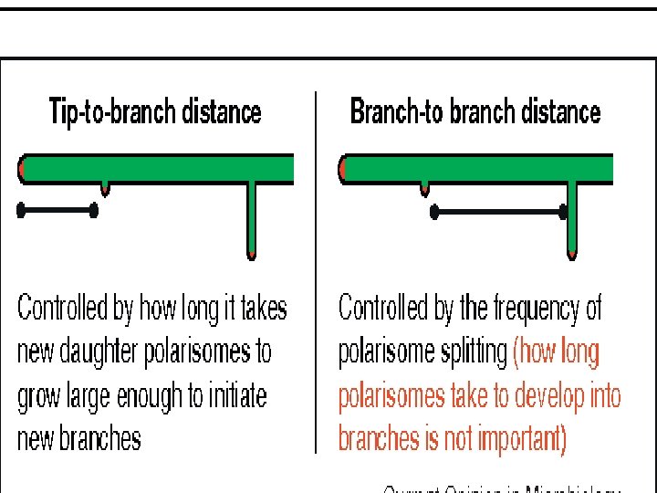
Branching of fungal hyphae 1. Apical branching of fungal hyphae 2. Lateral branching of fungal hyphae
Lateral branching • The predominant branching pattern exhibited by fungal hyphae is lateral branching, whereby new branches emerge from sites distal to the hyphal tip • Occur when a potential site is far enough removed from the tip so as to escape the effects of these factors. • New branches emerge adjacent to septa • appears to be associated with the de novo formation of a Spitzenkorper near the incipient branch site, Apical branching • The emergence of a branch from the hyphal tip is referred to as apical branching • Occurs in response to the abnormal accumulation of exocytic vesicles at the hyphal tip. • They accumulate of vesicles leading to the formation of a new tip • apical branching is triggered by the temporary loss of the Spitzenkorper at the tip