CRANIAL NERVES ANATOMY Keele Neurology Society Aziza Mohamed


- Slides: 22

CRANIAL NERVES ANATOMY Keele Neurology Society Aziza Mohamed Aisha Saleem

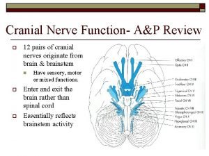
Introduction • Twelve pairs of cranial nerves that originate from the forebrain, brainstem and rostral spinal cord. • Form part of the peripheral nervous system – similar properties to spinal nerves. • Responsible for sensory, motor and/or autonomic function in mainly* functional regions of head and neck. • Integral part of neurological examination. *

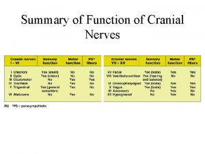
Cranial Nerve Function Overview • Sensory, motor, autonomic or mixed. • Can receive afferents or send impulses locally or to regions such as thoracic and abdominal viscera (e. g. CN X Vagus Nerve). • Sensory - general somatic afferent, general visceral afferent or special afferent. • Motor - general somatic efferent or branchial motor efferents. • General or special visceral efferents are also described as - Parasympathetic efferents.

Internal Cranium Base – Quick Overview

Cranial Nerve Foramina

Cranial Nerve I: Olfactory SENSORY - Special Afferent • FORAMEN - Cribriform Plate of the Ethmoid Bone • Sensory – perception of smell. Transmitted into the frontal lobe from olfactory epithelium.

Cranial Nerve II: Optic SENSORY - Special afferent • FORAMEN - Optic canal • Sensory – perception of vision; detects and transmits light input from optic disc at retina. NB: Contralateral; optic chiasm at the sphenoid wing involves decussation of the nerves into the optic tract.

Cranial Nerve III: Occulomotor MOTOR – occulomotor nerve palsy (down and out – neurogenic ptosis causes drooping of the eyelid due to damage to CN III) PARASYMPATHETIC EFFERENTS • FORAMEN - Superior Orbital Fissure • Motor - Controls four of the six extraoccular muscles ; superior rectus, inferior rectus, medial rectus and inferior oblique muscles. *Also controls levator palpebrae superioris (upper eyelid muscle) • Parasympathetic efferent – innervates sphincter pupillae for pupil constriction and ciliary muscle accommodation of lens at near vision.

Cranial Nerve IV: Trochlear MOTOR • FORAMEN - Superior Orbital Fissure • Motor - controls the movement of the superior oblique muscle of the eye. Aids internal rotation of eye. • *SO 4 – Trochlear (CN IV)

Cranial Nerve V: Trigeminal SENSORY MOTOR - • FORAMEN – Varied: – V 1 [opthalmic]- Superior Orbital Fissure – V 2 [maxillary]- Foramen Rotundum – V 3 [mandibular]- Foramen Ovale • Sensory – touch, temperature perception on different regions of face (testing with soft/crude touch and temperature). • Motor - Muscles of mastication particularly masseter and temporalis

Cranial Nerve VI: Abducens MOTOR • FORAMEN – Superior Orbital Fissure • Motor - Lateral rectus muscle of the eye. Enables abduction of the eye. • *LR 6 – Abducens (CN VI)

Cranial Nerve VII: Facial SENSORY MOTOR PARASYMPATHETIC EFFERENTS • FORAMEN – Internal Acoustic Meatus • Sensory – special sensory is anterior 2/3 taste of tongue and external acoustic meatus (including auricle) • Motor – muscles of facial expressions [5 branches] and neck muscles. • Parasympathetic – submandibular, sublingual and lacrimal salivary glands. Also innervates mucous membranes of nasal cavity. 1. Temporal 2. Zygomatic 3. Buccal 4. Maxillary 5. Cervical

Cranial Nerve VIII: Vestibulocochlear SENSORY - special afferent • FORAMEN – Internal Acoustic Meatus • Sensory – special sensory function: – Vestibular division responsible for balance – Cochlear division responsible for hearing

Cranial Nerve IX: Glossopharyngeal SENSORY MOTOR PARASYMPATHETIC EFFERENTS • FORAMEN – Jugular Foramen • Sensory – input from the carotid body and sinus (detects changes in PCO 2 and pressure). Taste in posterior 1/3 of tongue. • Motor – controls stylopharyngeal muscle for swallowing. • Parasympathetic efferent innervates parotid salivary gland

Cranial Nerve X: Vagus SENSORY MOTOR - dysphagia (often ( seen in patients who’ve suffered from stroke) PARASYMPATHETIC EFFERENTS • FORAMEN – Jugular Foramen • Sensory – varied: – Sensation in larynx, laryngopharynx, including parts of the external acoustic meatus. – Sensory from the aortic body and aortic sinus, thoracic and abdominal viscera. – Taste in the epiglottis and upper pharynx • Motor – innervates only one tongue muscle, varied muscles in pharynx and larynx. – Aid in speech and swallowing • Parasympathetic efferents – innervates smooth muscles and glands in throat region, thoracic viscera and abdominal viscera upto the midgut. (2/3 of Transverse Colon)

Cranial Nerve XI: Accessory MOTOR - • FORAMEN - Jugular Foramen • Motor – innervates sternocleidomastoid muscle and trapezius muscles of neck and shoulder region. Aids rotation and flexion of head and neck; shrugging move scapula and support arm.

Cranial Nerve XII: Hypoglossal MOTOR - “the tongue licks the wound” (damage to the hypoglossal nerve leads to deviation of tongue to ipsilateral side where injury occurred) • FORAMEN - Hypoglossal Canal (lateral to Foramen Magnum) • Motor – control of tongue muscles; including pharynx and larynx (muscles of speech and swallowing).

Origin of Cranial Nerves • • • CN I – Olfactory Bulb (inf. surface of Frontal Lobe) CN – Retina CN III – Midbrain CN IV – Midbrain Majority of cranial nerves CN V – PONS originate from the CN VI – PONS brainstem (CN III – CN XII [excluding CN XI]) CN VII – PONS CN VIII – PONS CN IX – Medulla CN XI – Spinal Cord* CN XII – Medulla

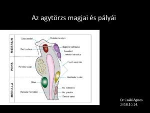
Cranial Nerve Nuclei* Nuclei *Spinal Accessory Nerve XI – Originates from spinal cord directly

Mnemonics • Oh Once One Takes The Anatomy Finals Very Good Vacations Are Heavenly. • Some Say Marry Money, But My Brother Says Big Business Makes Money. • Carl Only Swims South. Silly Roger Only Swims In Infiniti Jacuzzis. Jane Just Hitchhikes. • To Zanzibar By Motor Car* [5 Branches of Facial Nerve]

Useful Websites & Resources • Yale University: http: //www. yale. edu/cnerves/ • fastbleep: http: //www. fastbleep. com/medical-notes/neuroand-psych/2/95/610 • UBC (University of British Colombia): http: //www. neuroanatomy. ca/ • Teach. Me. Anatomy: www. teachmeanatomy. co. uk • Mnemonics: file: ///S: /Downloads/List%20 of%20 mnemonics%20 for%20 the %20 cranial%20 nerves. pdf

References • • • Mike Mahon – Cranial Nerve Neuroanatomy Lecture Neuroanatomy Illustrated Gray’s Anatomy for Students Bear’s Neuroscience Cranial Nerve Neuroanatomy Image 1 http: //d 7 c 2 b 0 wpljtwf. cloudfront. net/var/ezwebin_site/storage/images/media/im ages/e-anatomy/cranial-nerves-anatomy-diagrams/skull-cranial-base-foramencranial-nerves-anatomy-en/2571278 -1 -eng-GB/skull-cranial-base-foramen-cranialnerves-anatomy-en_imagelarge. jpg "Brain human normal inferior view with labels en-2" by Brain_human_normal_inferior_view_with_labels_en. svg: *Brain_human_normal_inferior_view. svg: Patrick J. Lynch, medical illustratorderivative work: Beaoderivative work: Dwstultz (talk) Brain_human_normal_inferior_view_with_labels_en. svg. Licensed under CC BY 2. 5 via Wikimedia Commons http: //commons. wikimedia. org/wiki/File: Brain_human_normal_inferior_view_wit h_labels_en 2. svg#mediaviewer/File: Brain_human_normal_inferior_view_with_labels_en-2. svg
 Sheeps brain
 Aziza boughaf
 Pukpos
 Hypoglossal nerve
 Cranial nerves mnemonic old opie
 Some say money matters cranial nerves
 Brachioradialis reflex
 Cranial nerve 4 function
 On occasion our trusty truck acts funny
 Tractus tectospinalis
 Accessory nerve (xi)
 Cranial nerves labeled with roman numerals
 Testing cranial nerves
 Peripheral nervous system
 Cranial nerves
 On old olympus towering tops
 Light tight dynamite cranial nerve
 Cranial nerves sensory and motor
 Motor cranial nerves
 Hypoglossal nerve test
 First and second cranial nerves
 Cranial nerves
 Middle cranial fossa
