Chapter 15 Signal Transduction and G ProteinCoupled Receptors


- Slides: 50
Chapter 15 – Signal Transduction and G Protein–Coupled Receptors
Chapter 15 – Signal Transduction and G Protein–Coupled Receptors 15. 1 Signal Transduction: From Extracellular Signal to Cellular Response 15. 2 Studying Cell-Surface Receptors and Signal Transduction Proteins 15. 3 G Protein–Coupled Receptors: Structure and Mechanism 15. 4 G Protein–Coupled Receptors That Regulate Ion Channels 15. 5 G Protein–Coupled Receptors That Activate or Inhibit Adenylyl Cyclase 15. 6 G Protein–Coupled Receptors That Trigger Elevations in Cytosolic and Mitochondrial Calcium
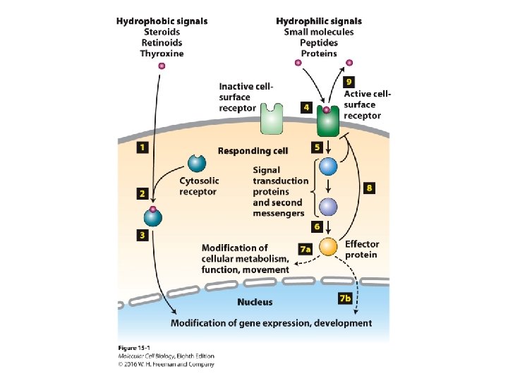
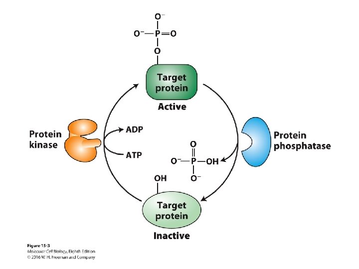
Signal Transduction and G Protein–Coupled Receptors 15. 1 Signal Transduction: From Extracellular Signal to Cellular Response • All cells respond to extracellular signals/stimuli that activate plasma membrane or cytosolic receptors. • Activated receptors function as transcription factors or activate G protein switches that regulate a variety of downstream pathways or induce generation of intracellular second messengers that do so. • Protein phosphorylation by kinases and dephosphorylation by phosphatases regulate protein activity in the cellular pathways and can amplify intracellular signaling.
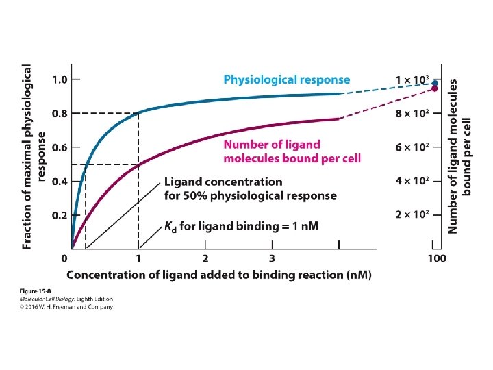
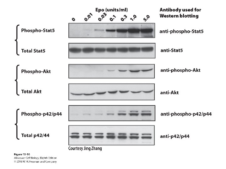
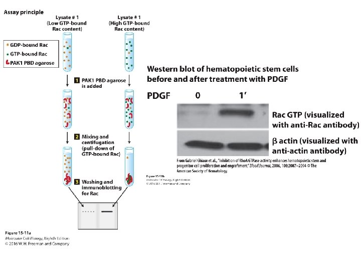
Signal Transduction and G Protein–Coupled Receptors 15. 2 Studying Cell-Surface Receptors and Signal Transduction Proteins • Near-maximal response of a cell to a particular ligand generally occurs at ligand concentrations at which less than 100 percent of its receptors are bound to the ligand. • Signal receptors and pathways are targeted by numerous drugs. • Receptors and signaling pathway intermediates are studied with a variety of experimental approaches including affinity chromatography, Western blotting, immunoprecipitation, and pull-down assays.
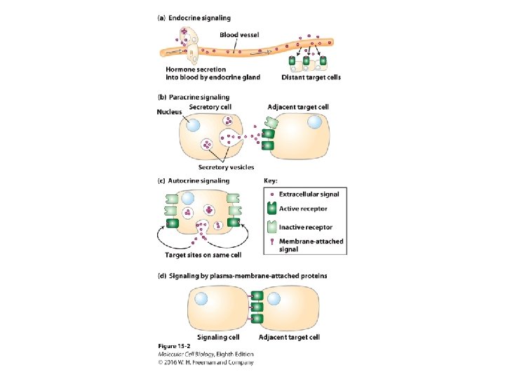
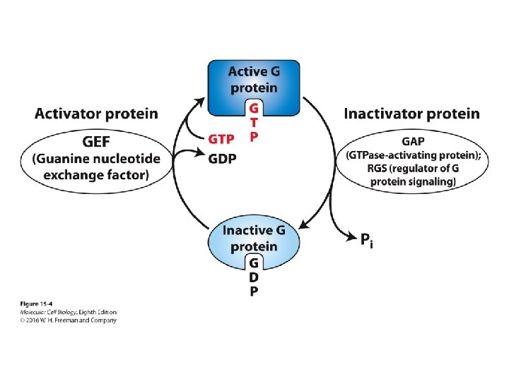
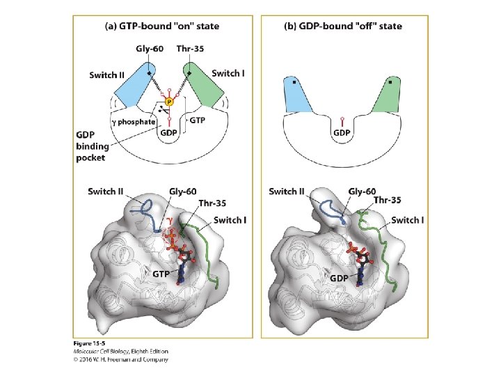
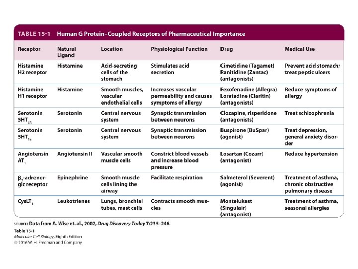
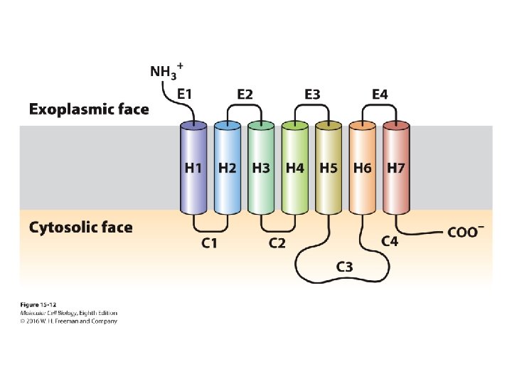
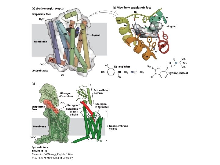
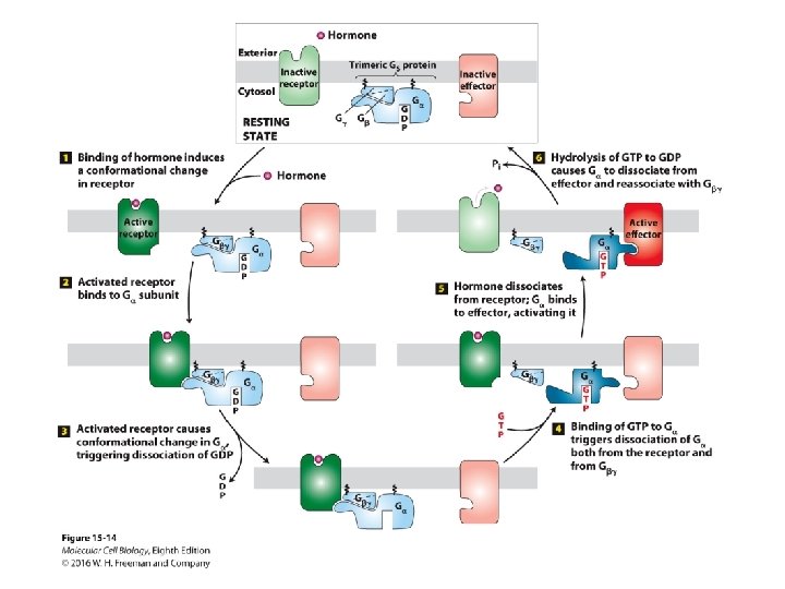
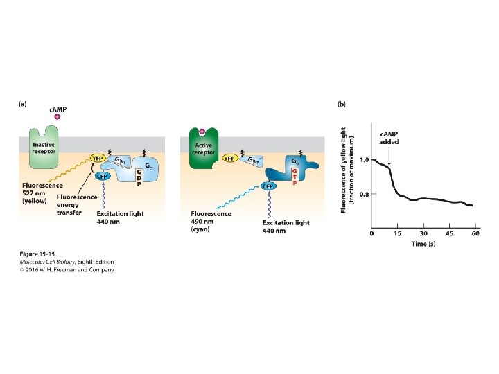
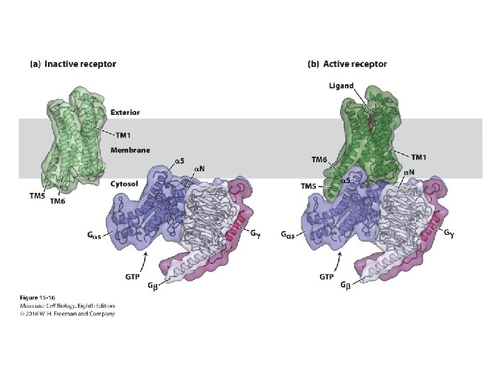
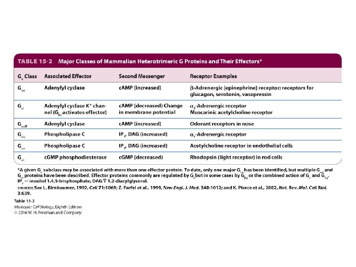
Signal Transduction and G Protein–Coupled Receptors 15. 3 G Protein–Coupled Receptors: Structure and Mechanism • The large diverse family of G protein-coupled receptors (GPCRs) respond to a variety of extracellular signals and activate trimeric G proteins. • G proteins function as On-Off switches for intracellular signaling pathways by activating or inactivating ion channels or effector enzymes that generate second messenger molecules. • GPCR signaling pathways regulate a wide range of cellular activities from metabolism to gene expression.
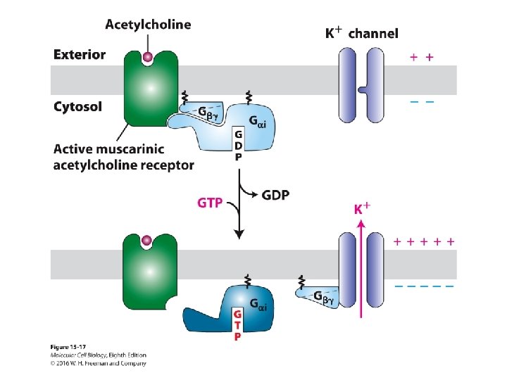
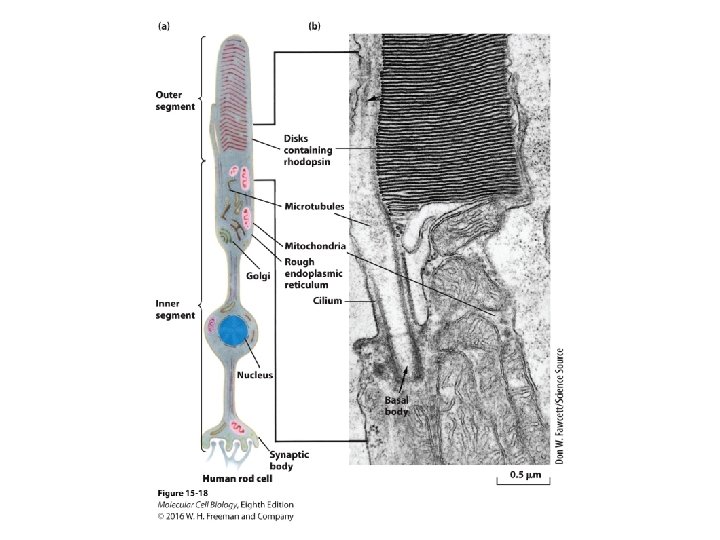
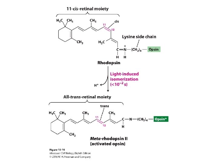
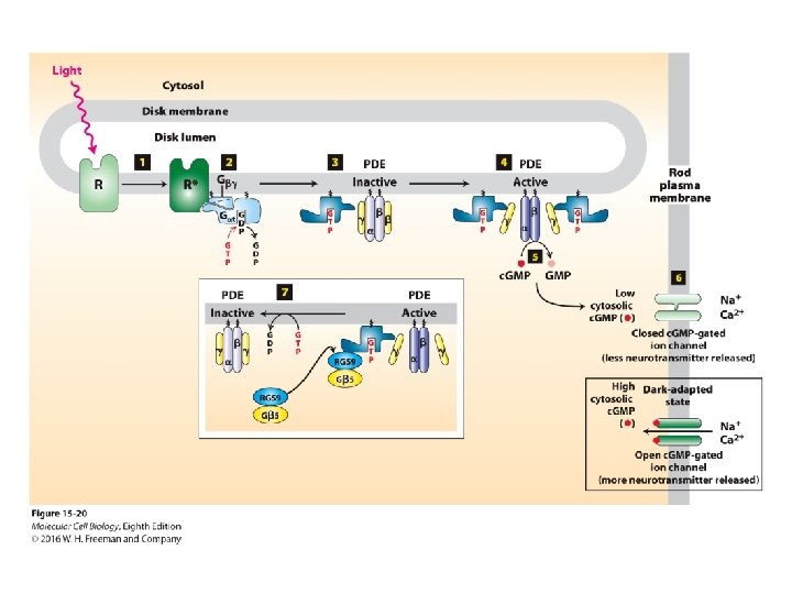
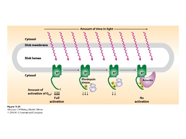
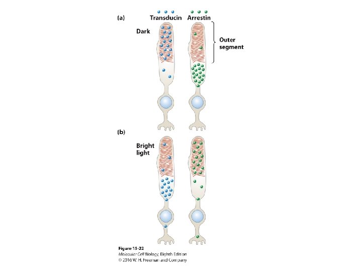
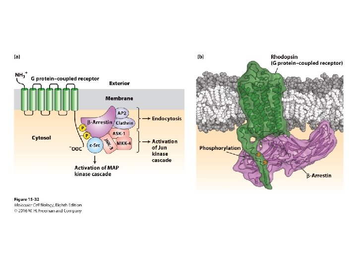
Signal Transduction and G Protein–Coupled Receptors 15. 4 G Protein–Coupled Receptors That Regulate Ion Channels • The cardiac muscarinic acetylcholine GPCR regulates a K+ channel. • Light stimulation of the photosensitive rhodopsin GPCR closes c. GMP-gated Na+/Ca 2+ channels by regulating a c. GMP pathway in retinal cells. • Several mechanisms act to terminate visual signaling. • Adaptation to a wide range of ambient light levels is mediated by movements of the G protein transducin and the inhibitor protein arrestin into and out of the rod-cell outer segment.
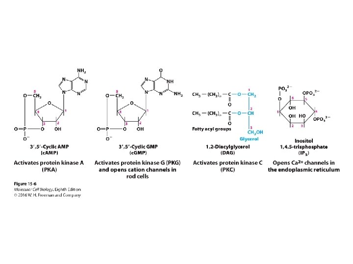
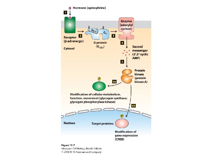
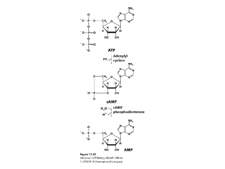
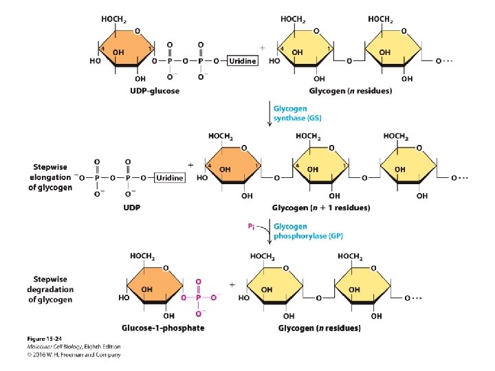
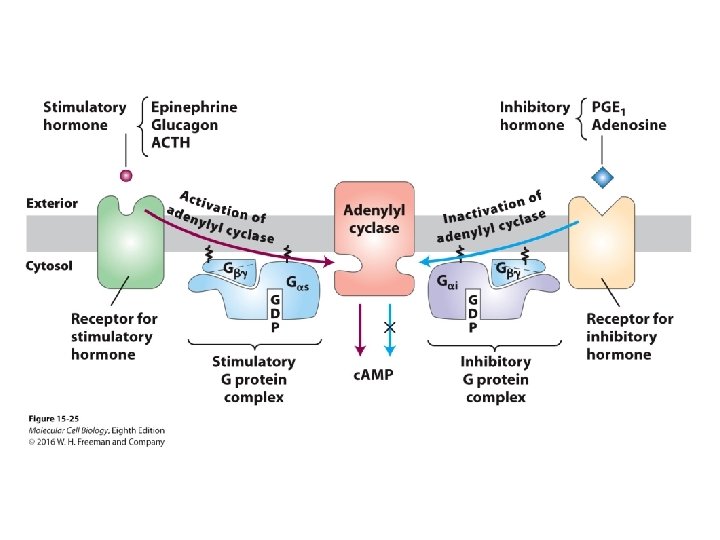
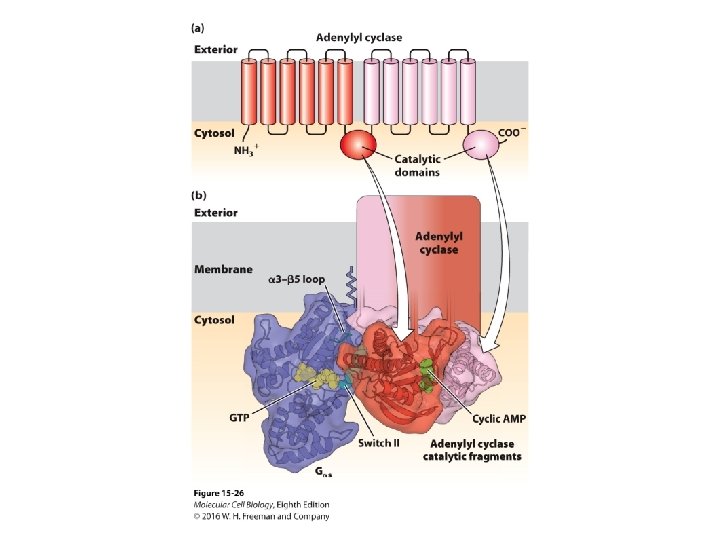
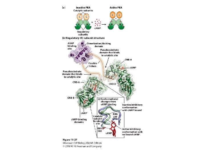
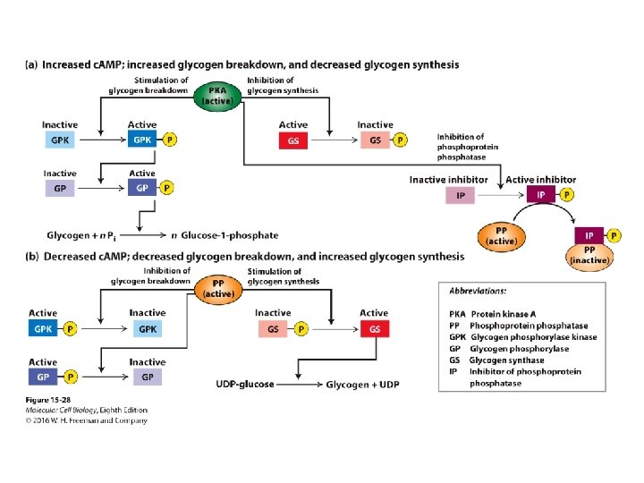
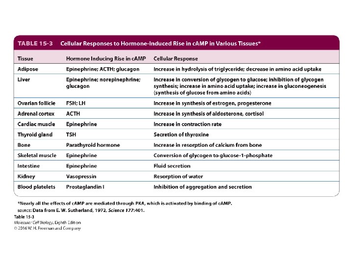
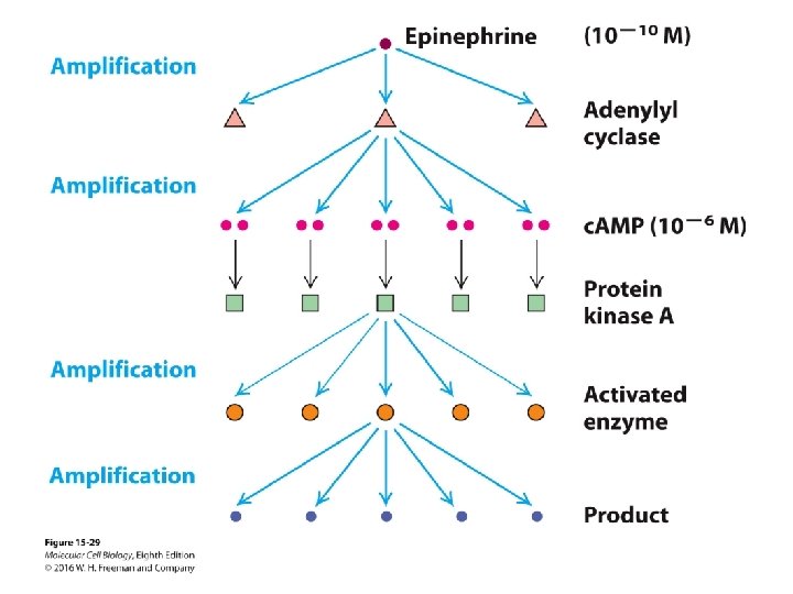
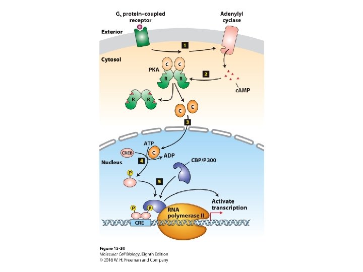
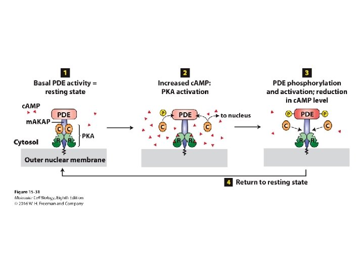
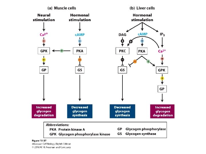
Signal Transduction and G Protein–Coupled Receptors 15. 5 G Protein–Coupled Receptors That Activate or Inhibit Adenylyl Cyclase • GPCRs activate G proteins that activate or inhibit adenylyl cyclase generation of c. AMP from ATP and are regulated by feedback repression. • c. AMP activates protein kinase A (PKA), which phosphorylates-regulates multiple target proteins including enzymes in cells. • Epinephrine activation of its GPCR in liver and muscle cells stimulates glycogen breakdown into glucose by inhibiting glycogen synthesis and stimulating glycogen breakdown via a kinase cascade. • PKA activation can stimulate gene expression.
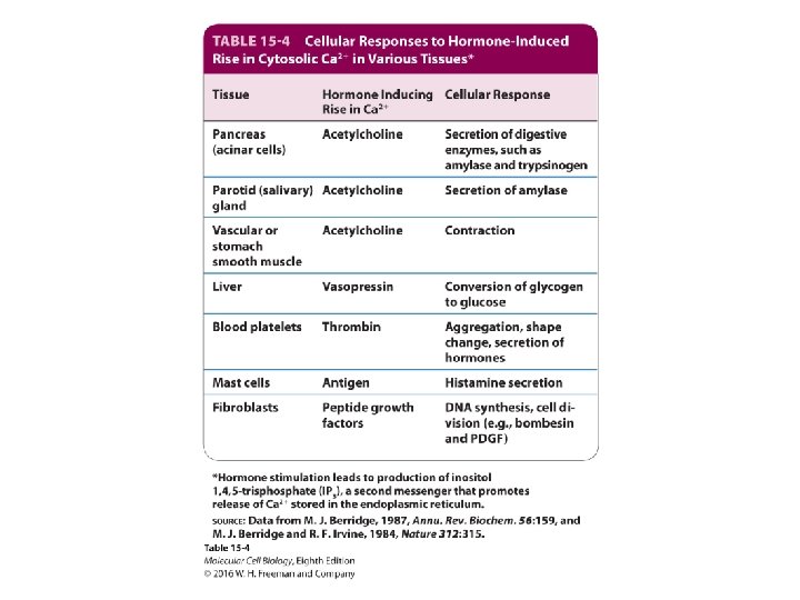
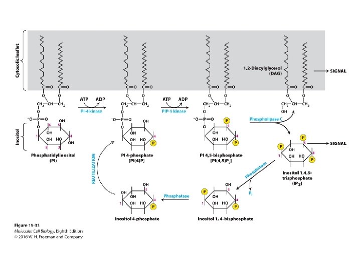
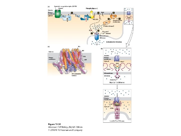
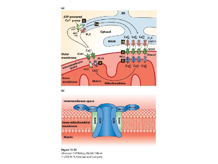
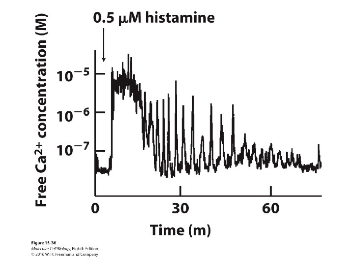
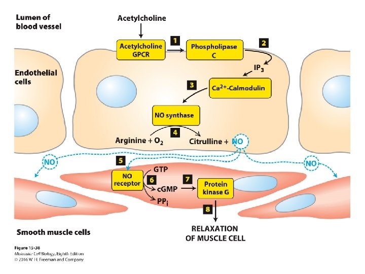
Signal Transduction and G Protein–Coupled Receptors 15. 6 G Protein–Coupled Receptors That Trigger Elevations in Cytosolic and Mitochondrial Calcium • GPCR-G protein activation of phospholipase C generates IP 3 (soluble ) and DAG (membrane bound) second messengers from PIP 2. • IP 3 triggers the opening of IP 3 -gated Ca 2+ channels in the endoplasmic reticulum and elevation of cytosolic free Ca 2+, which activates PKC and calmodulin. • Neural and hormonal stimulation coordinately regulate glycogen breakdown through Ca 2+ and c. AMP. • Acetylcholine activation of its GPCR on endothelial cells induces generation of the NO gaseous signal, which stimulates smooth muscle relaxation and vasodilation.