VISUAL SYSTEM q Perception of shape motion color


 and axons (plexiform layers)")

 connection § the main visual pathway § photoreceptors →")
 § cluster at fovea (macula lutea) § detect color in")

 § ganglion cells that monitor cones § smaller,")

 q LGN is composed of 6 layers q Layers 1")


 runs under the temporal lobe to the occipital")


 q Most LGN axons terminate in V 1 q")


 - area 41")


 and in the position of")


 conscious perception of movement and gravity")


- Slides: 52
VISUAL SYSTEM q Perception of Ø shape Ø motion Ø color q Two pathways Ø retina – cortex • visual perception retina – brainstem, diencephalon Ø • • eye movements circadian photoentrainment accommodation pupillary reflexes
Light passes through the cornea, aqueous humor, lens, and vitreous body to form an image on the retina.
Macula lutea + fovea centralis = areas of the highest visual acuity Fundus oculi
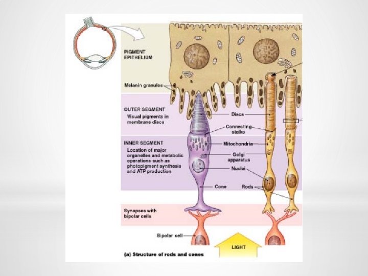
RETINA 10 layers: mainly separated by cell bodies (nuclear layers) and axons (plexiform layers)
q 5 main cell types: § photoreceptors § bipolar cells § horizontal cells § amacrine cells § ganglion cells q Photoreceptors: § rods and cones § involved in transduction converting the light signal into a nerve impulse
q neurons with serial (vertical) connection § the main visual pathway § photoreceptors → bipolar cells → ganglion cells q neurons with parallel (horizontal) connection § modulation of the visual information by retina § horizontal cells § amacrine cells
q Cones (7 million) § cluster at fovea (macula lutea) § detect color in bright light = photopic vision q Rods (100 million) § outside the fovea § sensitive to shape and movement = scotopic vision
CONES q 3 different types with three different photopigments: blue, green and red q Each type is maximally sensitive to the wavelength that corresponds to the specific color range (spectral sensitivity)
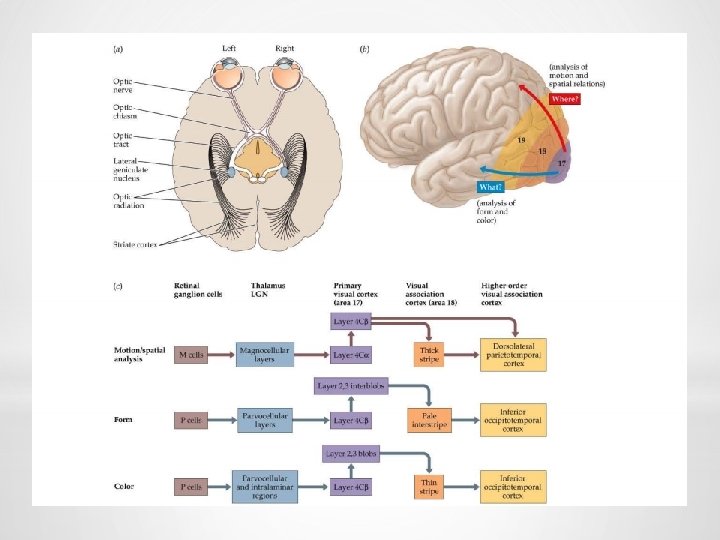
GANGLION CELLS q P cells (80%) § ganglion cells that monitor cones § smaller, more numerous § axons end on parvocellular laminae of LGN § provide information about fine detail and color q M cells (10%) § ganglion cells that monitor rods § relatively large § axons end on magnocellular laminae of LGN § provide information about a general form of an object, motion, and shadows in dim light q non-P non-M cells (10%) § projection to subcortical nuclei, koniocellular cells of LGN
PRIMARY VISUAL PATHWAY q The primary visual pathway connects the retina with lateral geniculate nucleus and primary visual cortex (retinogeniculostriate pathway) q It is responsible for detection of shape, movement and color Primary visual cortex 1 st neuron (photoreceptors) 2 nd neuron (bipolar cells) Optic tract CN II 3 rd neuron (ganglion cells) Optic radiation Optic chiasm LGN
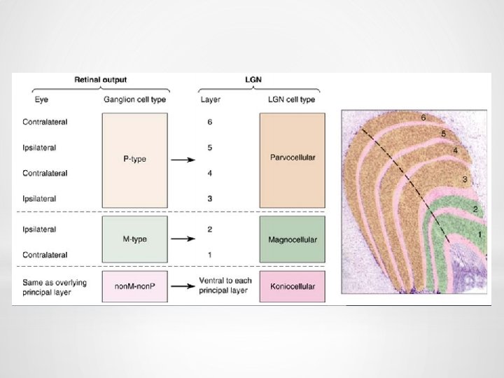
LATERAL GENICULATE NUCLEUS (LGN) q LGN is composed of 6 layers q Layers 1 and 2 contain larger neurons q Layers 3 - 6 contain smaller neurons
q Ipsilateral input enters layers 2, 3 and 5 q Contralateral input enters layers 1, 4 and 6
q LGN contains the topographic representation of what the retina “ sees”. This retinotopic map is sent to the cortex. q LGN modulates and regulates the flow of visual information to the primary visual cortex q cortex can control efficiency of thalamic input
GENICULOSTRIATE PATHWAY optic radiation (geniculocalcarine fibres) runs under the temporal lobe to the occipital lobe
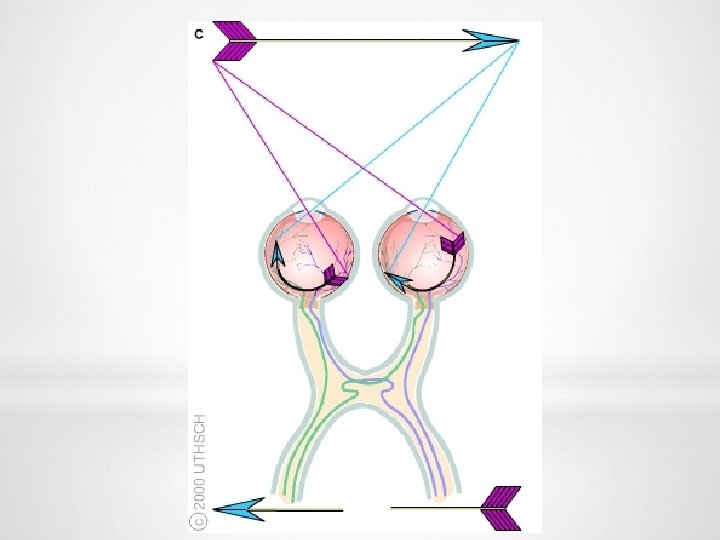
RETINOTOPIC REPRESENTATION q Nasal and temporal visual fields q Reversed to opposite halves of retinal representative fields (hemiretinas) q Inverted and reversed q Nasal visual fields project to temporal hemiretinas and their axons do not cross at the optic chiasm q Temporal visual fields project to nasal hemiretinas and their axons cross at the optic chiasm
RETINOTOPY q Most of the visual field is shared by the two eyes (binocular field) q Representation of different parts of the visual field is disproportionate in size
VISUAL CORTEX
PRIMARY VISUAL CORTEX (V 1) q Most LGN axons terminate in V 1 q All V 1 neurons respond to visual stimuli exclusively q Ablating V 1 results in blindness in the contralesional hemifield (homonymous hemianopsia) q Electrical stimulation of V 1 elicits visual sensations
VISUAL ASSOCIATION CORTEX Dorsal Stream Ø spatial orientation Ø binocular fusion/depth perception Ø the location, the movement and the movement direction and velocity of objects in space Ventral Stream Ø recognize objects and colors Ø read text Ø learn and remember visual objects (e. g. , words and their meanings)
VISUAL PATHWAYS TO SUBCORTICAL STRUCTURES q to the suprachiasmatic nucleus of hypothalamus q to the pretectum of the midbrain q to the superior colliculus
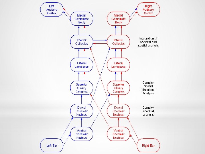
1 st order neuron § bipolar neuron of the spiral ganglion § dendrites make synapses with hair cells § axons form the cochlear part of CN VIII
2 nd order neuron § ventral cochlear nucleus → trapezoid body → lateral lemniscus § dorsal cochlear nucleus → lateral lemniscus 3 rd order neuron § nucleus of inferior colliculus → brachium c. i. 4 th order neuron § medial geniculate nucleus → radiatio acustica (internal capsule)
PRIMARY AUDITORY CORTEX gyrus temporalis superior (gyri temporales transversi of Heschl) - area 41 + 42
Two functionally significant features: q tonotopical organization q bilateral projection
DESCENDING PATHWAYS q feedback system processing ascending information q enhance signals q supress noise q mainly functions of the superior olivary complex q focus on a particular speaker and inhibit other voices
q changes in the motion of the head (kinetic) and in the position of the head with respect to gravity (static) q 3 afferent sources: the eyes, general proprioceptive receptors throughout the body, and the vestibular receptors in the inner ear q to maintain equilibrium, to direct the gaze of the eyes, and to preserve a constant plane of vision
VESTIBULAR APPARATUS q Labyrinth of static apparatus § macula utriculi – orientation in horizontal position § macula sacculi – orientation in vertical position q Labyrinth of kinetic apparatus § cristae ampullares of semicircular ducts
q Hair cells in the maculae of the saccule and the utricle respond to linear acceleration (gravity). q Hair cells in the cristae ampullares in the semicircular ducts respond to angular acceleration (rotation of the head).
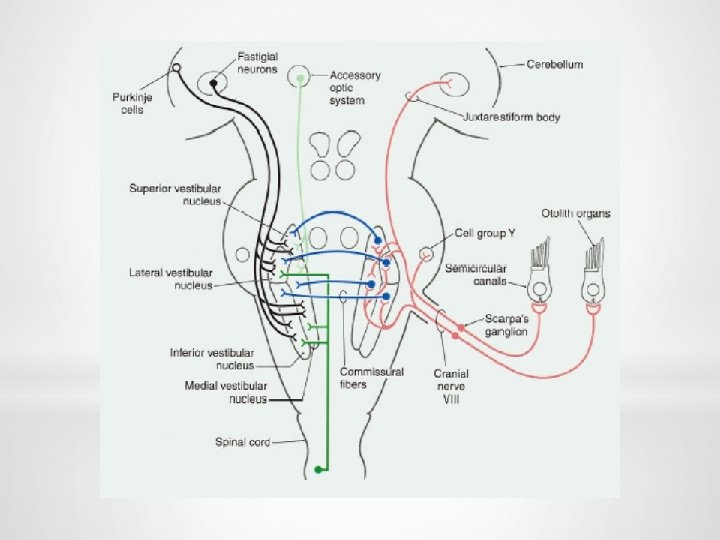
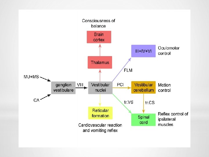
VESTIBULAR PATHWAY q 1 st order neuron – vestibular ganglion (utriculoampullar nerve, saccular nerve, posterior ampullar nerve) q 2 nd order neuron – vestibular nuclei (superior, inferior, medial, lateral)
Connections with the cerebellum q vestibular portion of the CN VIII – inferior cerebellar peduncles – ipsilateral vestibulocerebellum q vestibular nuclei – inferior cerebellar peduncles – vestibulocerebellum maintenance of balance
Connections with the spinal cord to motoneurons that innervate axial and proximal limb muscles q lateral vestibulospinal tract § from lateral vestibular nucleus § uncrossed § terminating at all levels of the spinal cord § excitatory influences for extensors q medial vestibulospinal tract § from medial vestibular nucleus § uncrossed § descends in the MLF § terminates mainly at cervical levels § coordination of head position and eye movements
Connections with the brain stem q ascending portion of MLF Ø CN III, IV, VI Ø Darkschewitsch and Cajal nuclei Ø coordination of eye movements in response to head movements
Connection with the thalamus (cortex) conscious perception of movement and gravity
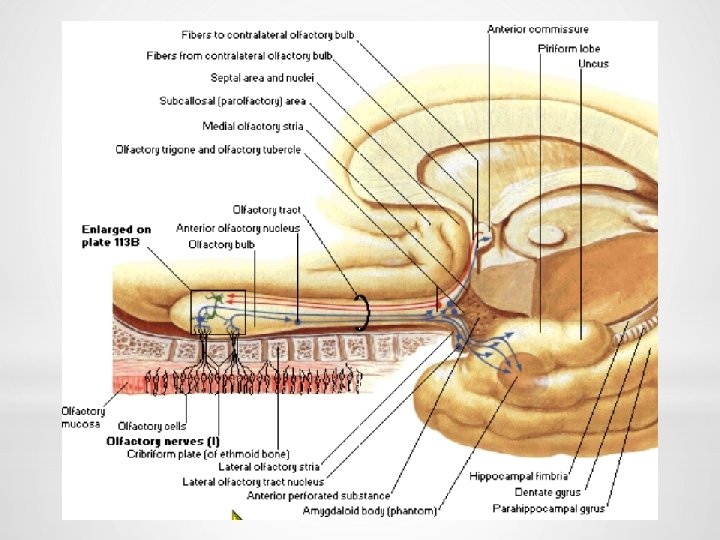
Olfactory region
1 st order neuron – bipolar olfactory neurons 2 nd order neuron – mitral cells – olfactory tract
3 rd order neuron – olfactory tubercle 4 th order neuron – dorsomedial nucleus of thalamus Orbitofrontal cortex (perception of olfactory information)
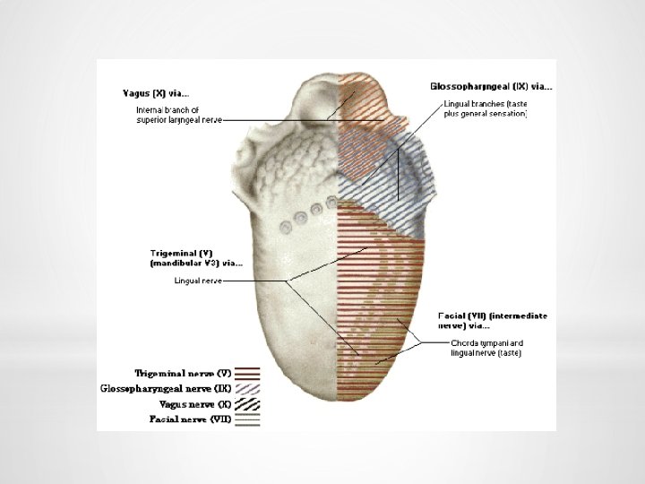
Taste buds q receptor cells (replaced about every 9 -10 days by differentiating basal cells) q supportive columnar cells q basal cells
1 st order neuron – § CN VII –geniculate ganglion Ø via lingual nerve and chorda tympani Ø via greater petrosal nerve § CN IX – inferior ganglion of CN IX § CN X – inferior ganglion of CN X
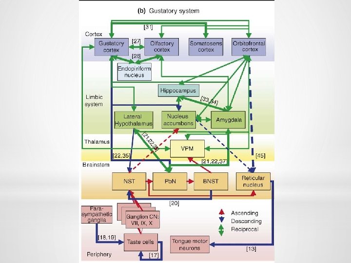
2 nd order neuron - rostral part of the solitary nucleus 3 rd order neuron – ventral posteromedial nucleus of thalamus
Primary gustatory cortex q a. 43 in the postcentral gyrus q insula
Illustrations were copied from: Neuroscience Online, the Open-Access Neuroscience Electronic Textbook.