REGULATION of RESPIRATION Dr M B Bhat Control

REGULATION of RESPIRATION Dr. M. B. Bhat.
![Control of breathing[regulation] n n n Can be classified into – Central control of](http://slidetodoc.com/presentation_image/cfad12891d6db0e4376d66316ff8c903/image-2.jpg "Control of breathing[regulation] n n n Can be classified into – Central control of")
Control of breathing[regulation] n n n Can be classified into – Central control of breathing – by the neurons (centers) present in the brain – again divided into Voluntary central control & Involuntary or autonomic central control Peripheral control of breathing – Control of breathing by nervous (reflexes) & chemical mechanisms

Central control of breathing n n Voluntary control system – Center –Neocortex through motor cortex – Efferent through Corticospinal tract – to respiratory motor neurons of the spinal cord Involuntary control system – Center – Respiratory centers located in Pons & Medulla Efferent to spinal respiratory motor neurons lie between lateral & ventral corticospinal tracts

n n n 1. 2. By both the central control mechanisms -When motor neurons supplying inspiratory muscles are active, the motor neurons supplying expiratory muscles are inhibited & Vice versa This is done by – Descending pathways which excite agonist muscles inhibit antagonist muscles Reciprocal innervation property of the spinal reflexes

Respiratory centers n n n Two types Medullary respiratory centers –contain the main components of the ‘respiratory control generator’ of autonomic respiration Pontine respiratory centers – control the autonomic respiration by influencing the medullary centers activity.
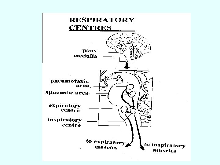

Medullary Respiratory centers n n n Two groups of respiratory neurons Dorsal respiratory group ofneurons or (DRG neurons) or (Inspiratory) neurons Ventral respiratory groupof neurons (VRG neurons) or E (expiratory) neurons
 or Inspiratory neurons Located in & near Nucleus")
n n Dorsal group neurons (DRG) or Inspiratory neurons Located in & near Nucleus Tractus Solitarius (NTS) DRG –produce inspiratory ‘Ramp signal’ – Efferent -- from these neurons –project monosynaptically to phrenic motor neurons & motor neurons supply external inter-costal muscles -- Also to VRG & Apneustic center of pons & stimulate them Afferent –receive from peripheral chemoreceptors
 The impulses")
Ramp signal n n n Produced by DRG (Dorsal respiratory group neurons) The impulses (depolarization) generate here weakly first & increases steadily in a ramp fashion for about 2 seconds. Abruptly ceases for 3 seconds & then the cycle repeats again & again Significance –there is a steady increase in lung volume (rather than inspiratory gasps). Initiated by “pre-BÖtzinger complex” (recent concept) [Old concept DRG has spontaneous discharge) Controlled by the factors that regulate respiratory rate by controlling the limiting point at which ramp signal suddenly ceases.
 n n n It has got ‘pace maker cells’ Discharges rhythmically")
Pre-BÖtzinger complex (pre-BÖTC) n n n It has got ‘pace maker cells’ Discharges rhythmically & responsible for initiation of rhythmic respiration Located between nucleus ambiguus & lateral reticular formation on either side of the medulla DRG & VRG respiratory neurons project to pre. BÖTC It sends rhythmic impulses to phrenic motor neurons
 or Expiratory neurons n n n n Located about 5")
Ventral group neurons (VRG) or Expiratory neurons n n n n Located about 5 mm anterior & lateral to DRG It is a long column of neurons extends through the nucleus ambiguus & retro ambiguus in the ventro-lateral medulla The long column of neurons has got rostral & caudal ‘E’ neurons, middle ‘I’ neurons They are stimulated by impulses from DRG They in turn inhibit DRG In normal quiet respiration, their peripheral output are almost inactive When sufficiently stimulated by higher respiratory drive (ventilation greater than normal), they stimulate expiratory muscles.
")
Pontine respiratory centers n n Control the medullary respiratory centers activity Two types (Upper) Pneumotaxic center (Lower) Apneustic center

Apneustic center n n n Present in lower pons – at the level of striae acusticae Send stimulatory impulses to – DRG thereby prevent “switch off” the ramp signal –cause prolonged depth of respiration (Apneusis) & also Pneumotaxic center Receive impulses from – DRG –which is stimulatory & Pneumotaxic center –which is inhibitory Also from periphery (lungs) through vagus –which also is inhibitory
")
Pneumotaxic center n n n n Located in the upper pontine region of (medial) nucleus parabrachialis & Kolliker-Fuse nuclei of the dorso-lateral pons These neurons are stimulated by Apneustic center In turn, they inhibit Apneustic center (when sufficiently stimulated). Significance – control of normal rhythmic respiration by sending impulses to apneustic center -With weak signal increase respiratory rate With strong signal decrease respiratory rate Damage of this area –respiration becomes slower & deeper (tidal volume is increased)
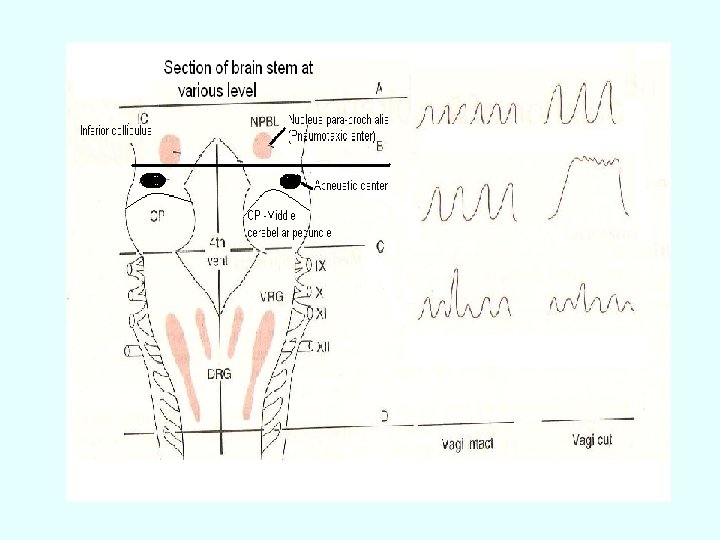

Section at various level of brain stem n n A — Above pons (both respiratory centers are intact)– normal respiration B – Mid pons (Pneumotaxic center of pons is removed)– Normal respiration –with intact vagus; Apneustic type of breathing without vagus C – Between pons & medulla (only medullary center is present)– Air hunger breathing (shallow rapid breathing) D – Below medulla (medullary center also removed) – Respiration ceases
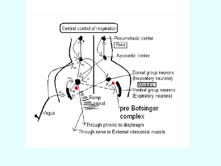

Concept of production of Rhythmic respiratory cycle n n n Recent view: The rhythmic respiration is initiated by Pre. BÖtzinger complex, which has got pace maker cells which discharge rhythmically & send impulses to DRG. The DRG neurons of medulla by firing ramp signal spontaneously (old concept) send these impulses to phrenic nerve to external inter-costal muscle bring about inspiration by producing slow & steady increase in lung volumes. At the same time DRG stimulates both apneustic neurons & VRG neurons This DRG ramp signals are strengthened by apneustic center activity. Apneustic center also stimulate pnemotaxic center

n n n Once pneumotaxic center is sufficiently stimulated; it in turn inhibits apneustic center. [In higher lung volumes (of > 1 liter), the apneustic center can be inhibited through vagus (Herring-Brewer reflex)] Once apneustic center is inhibited, its drive on inspiratory center is stopped. Now with expiratory center (VRG) activity; the DRG neurons are inhibited which brought about abrupt stop of ramp signals. Expiration follows passively Next cycle repeats by producing ramp signal by DRG (initiated by Pre-BÖTC) In this, the respiratory rhythm is modified by both nervous (reflex) & chemical regulations [peripheral regulation of respiration].

Nervous regulation of respiration or Reflex regulation of respiration or Non-chemical regulation of respiration

Types of nervous regulation of respiration 1. 2. 3. 4. 5. From respiratory tract through vagal afferents from receptors in the airways & lungs Afferent from baroreceptors (arterial, atrial, ventricular & pulmonary) Afferents from higher centers (cerebral, hypothalamus & limbic system) Afferents from periphery through proprioceptors & visceral receptors Effect of sleep on respiration

Role of vagal afferents from receptors in airways & lungs n 1. 2. Afferent fibers passing through the vagus nerve are – Aortic nerve from aortic arch of baroreceptor & aortic body of chemoreceptor Myelenated A δ fibers from airways – from a. Slow adapting stretch receptor; b. Rapid adapting stretch receptor; C. Irritant receptors 3. Non-myelenated C-fibers from lungs – from. ‘J’ receptor of alveoli & bronchi

Reflexes brought through vagal afferents 1. 2. 3. 4. Hering-Breuer reflexes Head’s paradoxical reflex Reflexes through irritant receptors ‘J’ receptor reflex

Hering-Breuer reflexes n n n n I. Hering-Breuer inflation reflex – Inflation of lung (with higher volume >1 liter) causes reflex arrest of inspiration & expiration starts. Mechanism – Stimulus – stretching of alveoli due to lung inflation Stretch receptor (slow adapting receptor) –present in the alveoli Afferent – through myelenated A δ fibers Center –Apneustic center & Inspiratory center (DRG) – inhibition on both Effect – Inspiration stops & expiration starts passively Significance – previously, it was considered, the normal rhythmic respiration is brought about by this reflex. Now it is proved, that this reflex acts only at higher volumes; hence helps to limit respiration in higher volumes (as in the case of exercise etc).

n n n n II. Hering-Breuer deflation reflex – If the lungs are deflated forcefully, inspiration starts Stimulus – forceful deflation of lungs Receptor – Rapid adapting receptor (lung irritant receptor) Afferent -- through myelenated A δ fibers Center –Inspiratory center (DRG) Effect -- stimulation
, when the lungs are inflated, instead of")
Head’s paradoxical reflex (After warming the vagi), when the lungs are inflated, instead of inhibition, inspiration is stimulated. n Indicating -- Presence of ‘positive feed back’ by separate group of nerve fibers
 n n 1. 2. 3. n Located")
Reflexes through irritant receptors (rapid adapting receptors) n n 1. 2. 3. n Located in air ways Stimulus – Inhalation of chemical & mechanical irritant gases & aerosols (ammonia & ether), cigarette smoke & carbon dust Contraction of air way smooth muscle (by histamine or aerosols) Collapse of alveoli (which pulls air ways)– due to pneumothorax, pulmonary congestion (The following reflexes are elucidated)

n n n n 1. Cough reflex –protective reflex; cough is a sudden forcible expiratory act. Stimulation of irritant receptors present in epiglottis, larynx, trachea by chemical & mechanical irritants. Afferent – Vagal A δ fibers Effect --Cough begins with a deep inspiration followed by forced expiration against closed glottis (intra pleural pressure goes up to >100 mm Hg) With sudden opening of glottis producing explosive outflow of air at velocities up to 1000 km/hr (3 to 4 times of PEFR) 2. Sneezing reflex –similar expiratory effort with continuously open glottis Significance of these two reflexes – help expel irritants & keep air ways clear

Juxta pulmonary-capillary receptors or Juxta capillary receptors or ‘J’ receptors n n n Present in lung parenchyma close to capillary endothelium (Dawes, Mott & Widdicome in 1951) Innervated by unmyelenated ‘C’ fibers (Paintal in 1958) Stimulated by hyperinflation of the lungs & also by pulmonary edema Effect – Apnoea followed by rapid shallow breathing & also bradycarida & hypotension & also inhibit spinal stretch reflex (limit exercise) {also has chemo receptor effect – injection of capsaicin, nicotine, 5 -hydorxythryptamine, phenyl biguanide stimulate these receptors and elucidate the same effect}

Afferent from baroreceptor n n Located in carotid sinuses, aortic arch, (aslo in atria & ventricles) Afferent –Sino-aortic nerves (buffer nerves) Center –Respiratory center, Vasomotor center & Cardio-inhibitory center of medulla Effect – Inhibition of respiration, vasodilation & decrease of Heart rate

n n n Yawning –is a peculiar ‘infectious’ respiratory act –of deep inspiration followed by prolonged expiration. Physiological basis not known. However, under ventilated alveoli tend to collapse, by yawing (deep inspiration), the alveoli is stretched & open thereby prevent development of atelectasis. Yawning also increase venous return May be primitive ‘nonverbal signal’ for communication in animals Sighing – also have similar function

n n n Afferent from propioceptors of muscles, tendons & joints (goes through dorsal column tract) – excite respiratory center Afferent from visceral receptors – Swallowing, vomiting & gagging –reflexly inhibit respiration Voluntary & involuntary abdominal muscles cause inhibition of respiration Hiccup –is spasmodic contraction of diaphragm that produces an inspiration during which the glottis suddenly closes. (Closure of glottis responsible for characteristic sensation & sound)

Role of sleep in respiration n n Respiration is less rigorously controlled during sleep; because, During sleep – CO 2 sensitive to respiratory drive is decreased, stimuli from various proprioceptors are reduced Sleep-apnea syndrome – occur at any age Symptoms – morning headache, fatigue, in extreme case lead to respiratory failure with normal lungs). Patients are polycythemic, hypoxemic, hypercapnic Cause – failure during sleep of the genioglossus muscles to contract during inspiration, resulting the tongue falls back & obstruct air way. Apneic spells are common in premature infants Sudden infant death syndrome (SIDS) –form of sleep apnea & apparently healthy infants are found dead in sleep.

Role of Higher centers in respiration n n Stimulation of motor cortex –acceleration of respiration Stimulation of medial surface of anterior cingulate gyrus & ventral surface of orbito-insulo -temporal polar cortex –inhibition of respiration Limbic system (emotions) & Hypothalamus (pain) –affect respiration Voluntary control – from neocortex – to motor cortex to respiratory muscles via pyramidal tract

Ondine’s curse n n n Loss of automatic control without loss of voluntary control Disease due to bulbar poliomyelitis or disease process that compress medulla, inadvertent surgical cut during antero-lateral cordotomy for releving pain Ondine –in German legend, was a unfaithful mortal lover of a water nymph. The king of water nymph by curse took away all his autonomic function.

End
- Slides: 36