Ankle Joint Type of joint Articular parts Lower


- Slides: 18


Ankle Joint

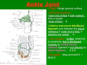
Type of joint Articular parts Lower end of Tibia Medial surface of Lateral malleolus trochlea of the talus Typical hinge synovial joint Lateral surface of Medial malleolus

Superior trochlear surface for lower end of tibia Articular surface of Talus Comma shaped articular surface for medial malleolus Triangular articular surface for lateral malleolus

Lower end of Tibia Anterior view The talus Lateral malleolus Medial malleolus

Ligaments of Ankle Joint

Prevents over Eversion of the foot and maintain the medial longitudinal arch. Neck of talus Tuberosity of Navicular bone Medial (deltoid) ligament Medial malleolus Very strong ligament Body of talus Spring ligament plantar calcaneo-navicular ligament Sustentaculum tali of calcaneus

2 - Posterior talofibular Lateral malleolus 1 - Anterior talofibular Lateral ligament 3 - Calcaneofibular ligament Resists inversion of the foot and may be torn (inversion injury) during an ankle sprain

Relations of ankle joint

Anterior relations Dorsiflexion Tibialis anterior Extensor Hallucis longus Anterior tibial vessel , nerve. Extensor digitorum longus Peroneus tertius Extensor retinacula Tom Has Very Nice Dark Pen

Posteromedial relations Plantar flexion Flexor hallucis longus Tibialis posterior Flexor digitorum longus Posterior tibial vessels Posterior Tibial nerve Flexor retinaculum Tendocalcaenus Tom Designs Very Nice House

Posterior relations tendon of Peroneus longus within its synovial sheath Lateral relations superior peroneal retinaculum inferior peroneal retinaculum Tendocalcaenus tendon of Peroneus brevis within its synovial sheath

• • • v Tendocalcaneus (Tendo-Achilis) It receives insertion of Gastrocnemius and soleus muscles. It ends in the middle of the posterior surface of the calcaneus. Tendocalcaneus is the strongest and thickest tendon of the body. The soleus muscle has a very strong but slow action (like the 1 st gear of car). When movement is under way, the quicker acting gastrocnemius increases the speed (like the top gear of the car) e. g. in running. The 2 heads of gastrocnemius and soleus are called triceps surae muscle. Soleus muscle contains a rich venous plexus which drains superficial veins and pumps it to the deep veins against gravity (peripheral heart). So, it liable to deep venous thrombosis Embolism especially with old age (fracture neck of femur), bed rest for a long, injury of the vein after bone fracture Rupture of tendocalcaneus leading to walking disability and running is impossible. Rupture of the tendon of plantaris leading to a sudden and severe pain.

Joints of the foot

phalanx 3 cuneiform bones Navicular Talus Dorsal aspect bones of foot Metatarsal bones cuboid Calcaneus

Talocalcaneo-navicular joint Ball & socket synovial Head of talus (ball) Articular surface; a- Ball is formed by the head of the talus. b- Socket is formed by navicular bone, upper surface of the spring ligament, sustentaculum tali and superior surface of the calcaneus. Navicular bone Spring ligament Sustentaculum tali Superior surface of the calcaneus

Inversion and eversion of the foot 1 - Inversion; medial rotation of the foot and the sole looks inwards. It is done by a) Tibialis anterior. b) Tibialis posterior. c) Flexor hallicus longus. d Extensor hallicus longus. 2 - Everson: Lateral rotation of the foot and the sole looks outwards (laterally); It is done by a) Peroneus longus. b) Peroneus brevis. c) Peroneus tertius. ** Significance: They occur at talocalcaneonavicular joint, mainly during walking on the rough ground. N. B: - The Lateral malleolus is Lower than the medial malleolus; - This bony projection resists the eversion - So Inversion is wider than eversion.

 Esr raised causes
 Chapter 19 worksheet the ankle and lower leg
 Chapter 15 worksheet the ankle and lower leg
 Ankle joint anatomy
 Ankle joint
 Membrane tectoria
 Test de abdominales en 1 minuto tabla
 Dr dinu gabriel
 Test de movilidad articular
 Anomalia monstruosidade
 Promontory of sacrum
 Diagrama de cuerpo libre biomecanica
 Radio ulnar joint
 Dr nabil khouri
 Differential diagnosis of articular syndrome
 Thoracic cage anterior view
 Costal facet
 Teres minor muscle
 Shoulder anatomy
