Achalasia John Rosen MD Miguel Saps MD Ann

- Slides: 18
Achalasia John Rosen, MD Miguel Saps, MD Ann & Robert H. Lurie Children’s Hospital of Chicago 2013 Resident Education Series Reviewed by Diana Riera, MD of the Professional Education Committee
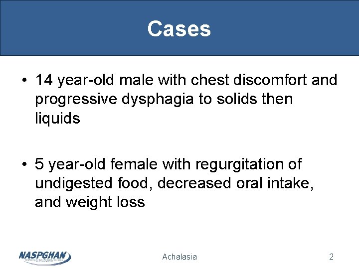
Cases • 14 year-old male with chest discomfort and progressive dysphagia to solids then liquids • 5 year-old female with regurgitation of undigested food, decreased oral intake, and weight loss Achalasia 2
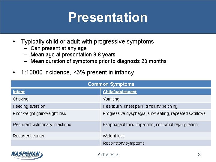
Presentation • Typically child or adult with progressive symptoms – Can present at any age – Mean age at presentation 8. 8 years – Mean duration of symptoms prior to diagnosis 23 months • 1: 10000 incidence, <5% present in infancy Common Symptoms Infant Child/adolescent Choking Vomiting Feeding aversion Heartburn, chest pain, difficulty belching Poor weight gain/weight loss Progressive dysphagia, slow eating, repeated swallows Recurrent pulmonary infections Esophageal food impaction, nocturnal regurgitation Recurrent cough Weight loss Respiratory symptoms Achalasia 3
Presentation • Differential Diagnosis – Esophageal obstruction (tumors in adults) – Surgical complications – Chagas disease – Eosinophilic esophagitis – Anorexia nervosa – Other motility disorders – Scleroderma – Rumination – Peptic stricture Achalasia 4
Presentation • Associated disorders – Trisomy 21 – Allgrove/Triple A Syndrome • (achalasia, alacrima, ACTH deficiency) – Rozychi Syndrome • (achalasia, deafness, short stature, vitiligo, and muscle wasting) – – – Sarcoidosis Hirschsprung disease Pyloric stenosis Hodgkin disease Pierre-Robin sequence Achalasia 5
Classification • Idiopathic esophageal motility disorder – Lack of esophageal peristalsis – Partial or incomplete LES relaxation – May have increased LES pressure • Absent or abnormal inhibitory innervation in distal esophageal myenteric plexus – Mixed inflammatory process precedes neural degeneration • Etiologic theories include: – Infection: similar clinical and manometric findings in Chagas disease – Autoimmune: nonspecific esophageal antimyenteric antibodies – Genetic: clustered in families and syndromic associations Achalasia 6
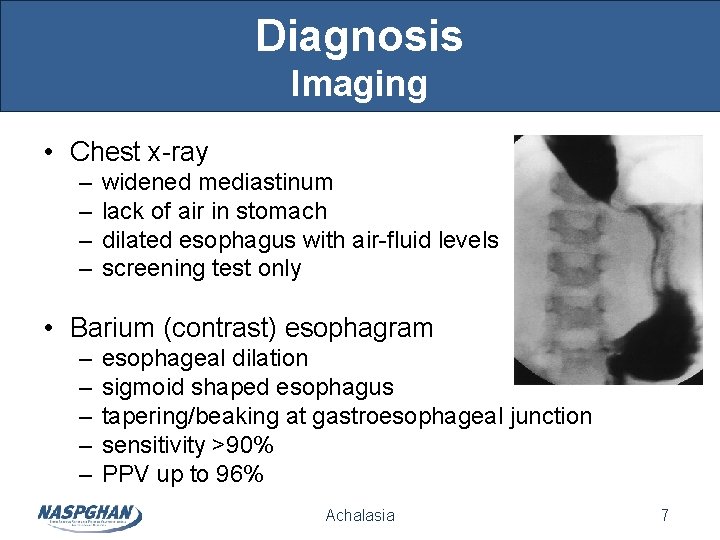
Diagnosis Imaging • Chest x-ray – – widened mediastinum lack of air in stomach dilated esophagus with air-fluid levels screening test only • Barium (contrast) esophagram – – – esophageal dilation sigmoid shaped esophagus tapering/beaking at gastroesophageal junction sensitivity >90% PPV up to 96% Achalasia 7
Diagnosis Imaging • Timed barium esophagram – height of column at 1 and 5 min is delayed • Both BE and timed BE correlate poorly with severity of symptoms • Esophageal transit scintigraphy – T 99 m sulfur colloid nuclide – PPV is 95% Achalasia 8
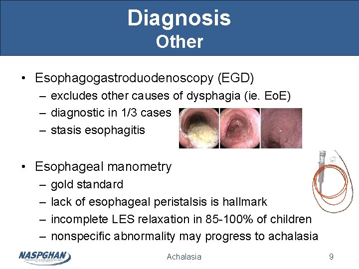
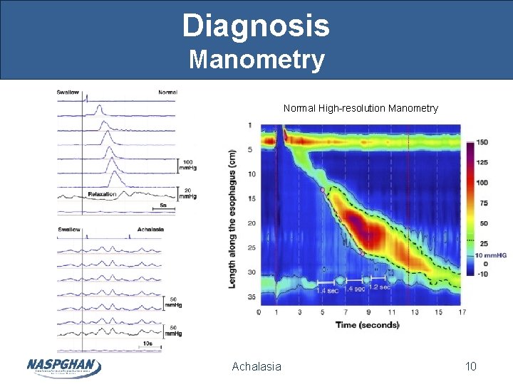
Diagnosis Other • Esophagogastroduodenoscopy (EGD) – excludes other causes of dysphagia (ie. Eo. E) – diagnostic in 1/3 cases – stasis esophagitis • Esophageal manometry – – gold standard lack of esophageal peristalsis is hallmark incomplete LES relaxation in 85 -100% of children nonspecific abnormality may progress to achalasia Achalasia 9
Diagnosis Manometry Normal High-resolution Manometry Achalasia 10
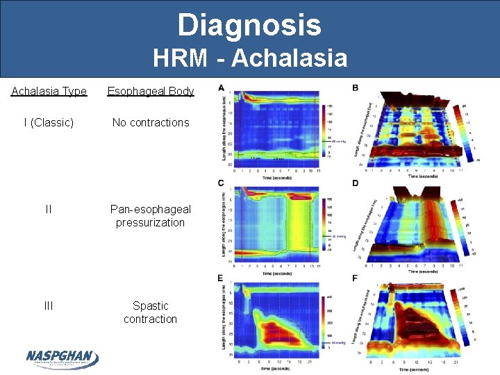
Diagnosis HRM - Achalasia Type Esophageal Body I (Classic) No contractions II Pan-esophageal pressurization III Spastic contraction Achalasia 11
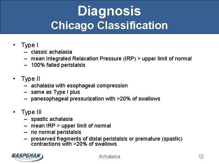
Diagnosis Chicago Classification • Type I – classic achalasia – mean Integrated Relaxation Pressure (IRP) > upper limit of normal – 100% failed peristalsis • Type II – achalasia with esophageal compression – same as Type I plus – panesophageal pressurization with >20% of swallows • Type III – – spastic achalasia mean IRP > upper limit of normal no normal peristalsis preserved fragments of distal peristalsis or premature (spastic) contractions with >20% of swallows Achalasia 12
Treatment • No cure • Pneumatic dilation • Esophageal myotomy – per-oral esophageal myotomy (POEM) • Less effective – – calcium channel blockers nitrates sildenafil botulinum toxin Achalasia 13
Treatment Pneumatic Dilation • Controlled tear of lower esophageal sphincter (LES) • Postdilation pressure of LES predicts success – some advocate multiple dilations in (1 -3 week) time span • Possible postdilation GER or esophagitis • Perforation risk 0. 5 -6% – – severe chest pain, unexplained tachycardia, fever XR: subcutaneous/mediastinal emphysema, left pleural effusion diagnosis with water soluble contrast esophagram treatment with TPN, PPI, IV antibiotics • surgery may be needed if mediastinal contamination or sepsis Achalasia 14
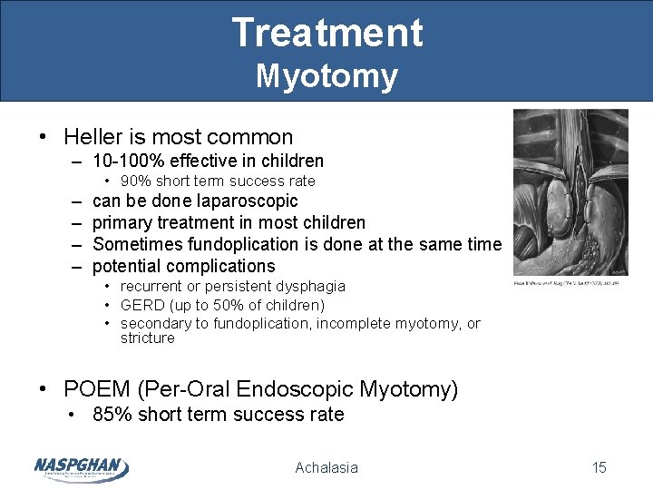
Treatment Myotomy • Heller is most common – 10 -100% effective in children • 90% short term success rate – – can be done laparoscopic primary treatment in most children Sometimes fundoplication is done at the same time potential complications • recurrent or persistent dysphagia • GERD (up to 50% of children) • secondary to fundoplication, incomplete myotomy, or stricture • POEM (Per-Oral Endoscopic Myotomy) • 85% short term success rate Achalasia 15
Postoperative Prognosis • Continued risk of aspiration, dysphagia, GERD • No change in esophageal motor function • Esophageal malignancy – occurs even after years symptom free – cause unknown – squamous cell carcinoma frequency of 0. 02%-5% – occurs at mean age 48 years Achalasia 16
Summary • Achalasia is an idiopathic esophageal motility disorder with lack of both peristalsis and LES relaxation • Diagnosis is by esophageal manometry • Myotomy is the primary treatment in children • Treatment may relieve symptoms, but does not address underlying disorder • Long-term risk of esophageal adenocarcinoma is unchanged regardless of treatment Achalasia 17
References Boudewijn F. Kessing, , Albert J. Bredenoord, André J. P. M. Smout. Erroneous Diagnosis of Gastroesophageal Reflux Disease in Achalasia. Clin Gastroenterol and Hepatol. 2011 Dec; 9(12): 1020 -24. Bredenoord AJ, Fox M, Kahrilas PJ, Pandolfino JE, Schwizer W, Smout AJ & The International High Resolution Working Group. Chicago classification criteria of esophageal motility disorders defined in high resolution esophageal pressure topography. Neurogastroenterol Motil. 2012; 24: 57 -65. Chiu PW, Wu JC, Teoh AY, Chan Y, et al. Peroral endoscopic myotomy for treatment of achalasia: from bench to bedside (with video). Gastrointest Endosc. Oct 2012. PAP. Gold B, Frem J. Other Neuromuscular Disorders; Achalasia. In: Kleinmen R, Goulet O, Mieli-Vergani G et al. eds. Walker’s Pediatric Gastrointestinal Disease. 5 th ed. Sheldon, CT: People’s Medical Publishing House; 2008: 77 -81. Nurko S. , et al: Other motor disorders. In: Walker W. , Durie P. , Hamilton J. , ed. Pediatric Gastrointestinal Disease, Pathophysiology, Diagnosis and Management, Vol. 1, 3 rd ed. . St. Louis, MO: Mosby; 2000: 317 -350. Pandolfino JE, Kwiatek MA, Nealis T, Bulsiewicz W, Post J, Kahrilas PJ. Achalasia: A new clinically relevant classification by high-resolution manometry. Gastroenterol. 2008; 135(1): 1526 -33. Patti MG, Albanese CT, Holcomb GW 3 rd, Molena D, Fisichella PM, Perretta S, Way LW. Laparoscopic Heller myotomy and Dor fundoplication for esophageal achalasia in children. J Pediatr Surg. 2001 Aug; 36(8): 1248 -51. Sood M, Rudolph C. Achalasia and other motor disorders. In: Wyllie R, Hyams J. eds. Pediatric Gastrointestinal and Liver Disease. 4 th ed. Online; Elsevier; 2012. Walzer N, Hirano I. Achalasia. Gastroenterol Clin N Amer. 2008; 37(4): 807 -25. Achalasia 18