Vision Our most dominant sense Our Essential Questions


 transform into")


 – approx.")


 – approx.")


 Theory • Cones are “tuned” to be sensitive to red, green")


- Slides: 58
Vision Our most dominant sense
Our Essential Questions • What are the major parts of the eye? • How does the eye translate light into neural impulses?
Vision Purpose of the visual system – transform light energy into an electro-chemical neural response – represent characteristics of objects in our environment such as size, color, shape, and location
Light: The Visual Stimulus
Light: The Visual Stimulus • Wavelength of a light is the distance of one complete cycle of the wave. • Visible light : 400 nm - 700 nm. • Wavelength of light is related to its perceived color
The Structure of the Visual System So how does this stimulus (light) transform into messages in our brain?
Cornea • The clear bulge on the front of the eyeball • Begins to focus the light by bending it toward a central focal point • Protects the eye
Parts of the Eye – Cornea
Iris • Colored portion of the eye – Does color affect vision? • A ring of muscle tissue that regulates the size of the pupil – Allows more or less light to enter the eye
Parts of the Eye - Iris
Pupil • Opening in the center of the eye • Controls the amount of light entering the eye – bright conditions - iris expands, pupil gets smaller – dark conditions - iris contracts, pupil gets larger
Parts of the Eye - Pupil
Lens • A transparent structure behind the pupil • Focuses the image on the back of the eye – Muscles change thickness of the lens change how light is bent focuses the image • Glasses or contacts correct problems
Parts of the Eye - Lens
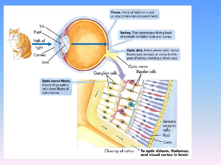
Retina • At the back of the eyeball • Light-sensitive surface with cells that convert light energy to neural impulses – This is where the magic happens!
Parts of the Eye - Retina
Fovea • The central focal point of the retina • The spot where vision is best (most detailed)
Parts of the Eye - Fovea
Receptor Cells • In sight they change light into neural impulses the brain can understand • Visual system has two types of receptor cells – rods and cones
Distribution of Rods and Cones • Cones—concentrated in center of eye (fovea) – approx. 6 million • Rods—concentrated in periphery – approx. 120 million • Blind spot—region with no rods or cones
Differences Between Rods and Cones • Cones – allow us to see in bright light – allow us to see fine spatial detail – allow us to see different colors • Rods – allow us to see in dim light – can not see fine spatial detail – can not see different colors
Receptive Fields and Rod vs. Cone Visual Acuity • Cones—in the fovea, one cone often synapse onto only a single ganglion cell • Rods—the axons of many rods synapse onto one ganglion cell • This allows rods to be more sensitive in dim light, but it also reduces visual acuity
Let’s Review • Cone Characteristics • Rod Characteristics
Rods • Located in the retina • Can only detect black and white • Respond to less light than do cones
• Located in the retina • Can detect sharp images and color • Need more light than the rods • Many cones are clustered in the fovea Cones
Let’s do an experiment now • What do you see in your peripheral vision (that’s the stuff on the side)?
Get into groups of 3
• Pick an A, B, and C
• • The Experiment A will look straight ahead B will look A in the eyes – to make sure that A doesn’t cheat! C will move various colored pieces of paper in A’s peripheral vision A will guess the color – Note: if the person is consistently guessing correctly then they are cheating!
Write up the results… 1. Results – correct guess versus bad 2. Your conclusion - What do your results tell you about our vision? - How do the different kinds of receptor cells affect our vision?
Distribution of Rods and Cones • Cones—concentrated in center of eye (fovea) – approx. 6 million • Rods—concentrated in periphery – approx. 120 million • Blind spot—region with no rods or cones
Let’s Compare… Cones – allow us to see in bright light – allow us to see fine spatial detail – allow us to see different colors Rods – allow us to see in dim light – can not see fine spatial detail – can not see different colors
Visual Processing in the Retina
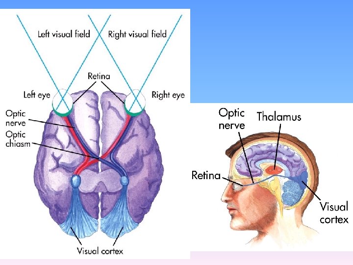
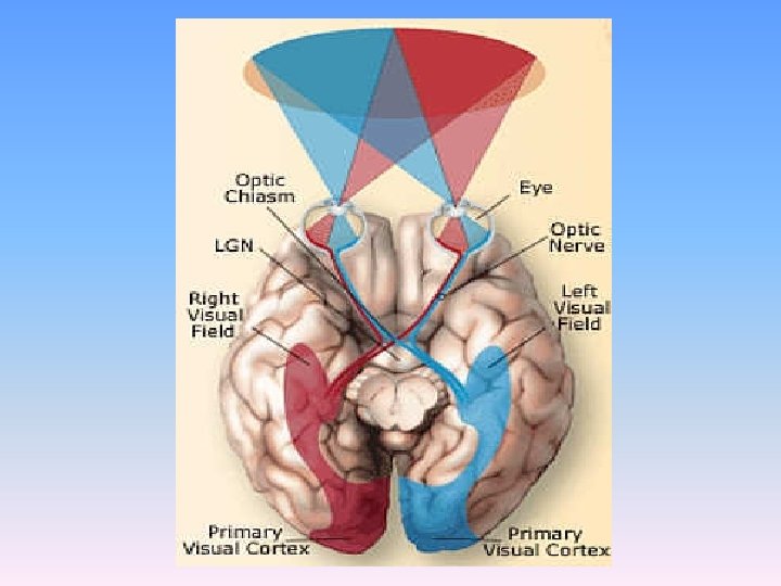
Optic Nerve • The nerve that carries visual information from eye occipital lobes
Parts of the Eye – Optic Nerve
Blind Spot • The point which the optic nerve travels Blindat Spot through the retina to exit the eye • There are no rods and cones at this point
Parts of the Eye – Blind Spot
What do you see in your blind spot?
The Visual System: Color Vision How do we see color?
Color Vision • Differences in wavelength of light = color • Rods are color blind, but cones can see different colors – We have only one type of rod but three types of cones
Color Vision • Two theories of color vision: –Trichromatic Theory –Opponent-Process Theory
Trichromatic (3 -Color) Theory • Cones are “tuned” to be sensitive to red, green and blue light • All the colors we see are a combination of these 3 colors • Similar to the design of a color TV
Opponent-Process Theory • Sensory receptors in the retina come in pairs: – Red/Green – Yellow/Blue – Black/White • Only one side is “on” at a time
Opponent Process Theory ON” red green blue yellow black white “OFF” green red yellow blue white black
Opponent-Process Theory • If one sensor is stimulated, the other is inhibited • If one sensor is over-stimulated, and fatigues, the paired sensor will be activated, causing an afterimage
Afterimage Effect
Can you see what is in the middle?
Red-Green Color Blindness
Color Deficient Vision • People who lack one of the three types of cones • Usually the red or green receptors are missing • Usually referred to as color blindness • Inherited and found more in males
Overview of Visual System • The eye is like a camera; instead of using film to catch the light, we have rods and cones. • Cones allow us to see fine spatial detail and color but cannot function well in dim light.
Overview of Visual System • Rods enable us to see in dim light but at the loss of color and fine spatial detail. • Our color vision is based on the presence of 3 types of cones, each maximally sensitive to a different range of wavelengths.