Tishk International University Dentistry Faculty Oral Maxillofacial Surgery


")


- Slides: 32
Tishk International University Dentistry Faculty Oral & Maxillofacial Surgery Department Oral Pathology- Dent 496 TOPIC: Practical: Part 1 4 th Grade- Spring Semester 2019 -2020 Instructor: Dr. Wafaa Khalil Ph. D. , OMFS
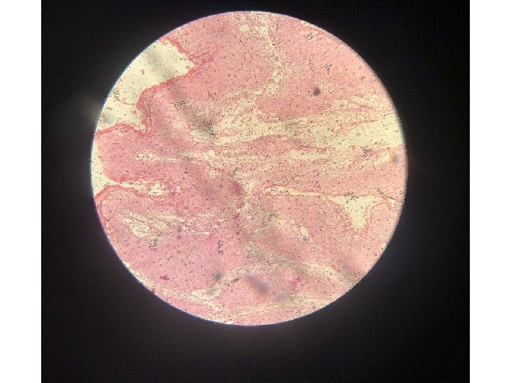
Slide no. 1 Epithelial dysplasia
Features of epithelial dysplasia 1. Nuclear and cellular pleomorphism, cells and nuclei of different shape and size. 2. Increase in nuclear/cytoplasmic ratio. 3. Nuclear hyperchromatism. 4. Prominent nucleoli. 5. Increase and abnormal mitosis. 6. Disturbed polarity of the basal cell layer or loss of cellular orientation. 7. Basal cell hyperplasia. 8. drop-shape rete pegs. 9. Irregular epithelial stratification. 10. Abnormal keratinization within the stratum spinosum. 11. Loss or reduction of intercellular adhesion.
Individual cell alteration found in epithelial dysplasia
Degree of dysplasia is subjectively assessed using terms: 1. Mild dysplasia 2. Moderate dysplasia. 3. Severe dysplasia.
Mild epithelial dysplasia There are hyperchromatic & slightly pleomorphic nuclei are noted in basal and parabasal cell layer of stratified seq. epithelia.
Moderate epithelial dysplasia Dysplastic changes extended to mid point of epithelium & characterized by nuclear hyperchromatism, pleomorphism & cellular crowding.
Severe epithelial dysplasia Cellular crowding & disordered arrangement noted throughout most of epi. Thickness, although slight maturation & flattening of the cells appear to be present at epi. Surface.
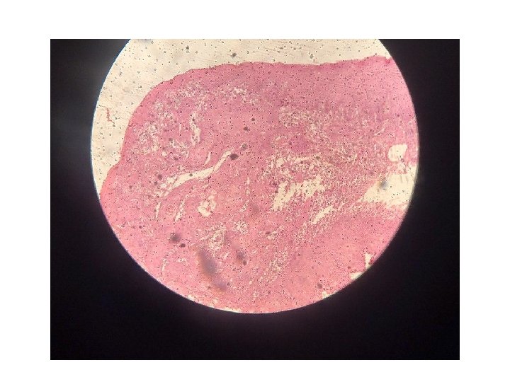
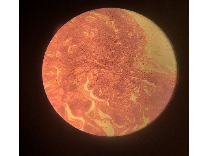
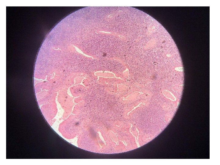
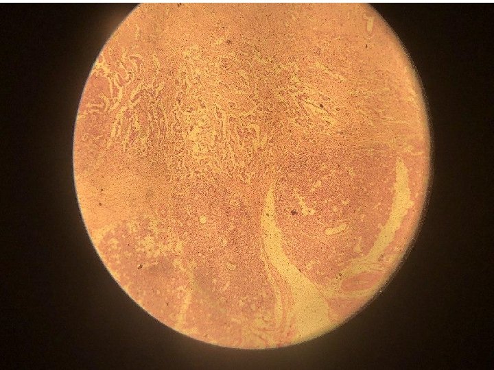
Slide no. 2 Warthin’s tumor (Adenolymphoma)
HISTOPATHOLOGICAL FEATURES OF WARTHIN’S TUMOR Tumor composed of mixture of ductal epithelium and lymphoid stroma. The epithelium forming uniform rows of cells surrounding cystic space and are arranged in two layers the inner luminal layer consist of tall columnar cells with centrally placed nuclei, Warthin’s tumour. Tall columnar cells surround lymphoid tissue and line a convoluted cystic space.
HISTOPATHOLOGICAL FEATURES OF WARTHIN’S TUMOR Beneath this a second layer of cuboidal or polyhydral cells. The lining epithelium demonstrate multiple papillary infolding that protrude into the cystic space. The epithelium is supported by a lymphoid stroma that frequently shows germinal center formation. Warthin’s tumour. At higher power, the tall columnar epithelial cells are readily identified and beneath them the lymphoid tissue.
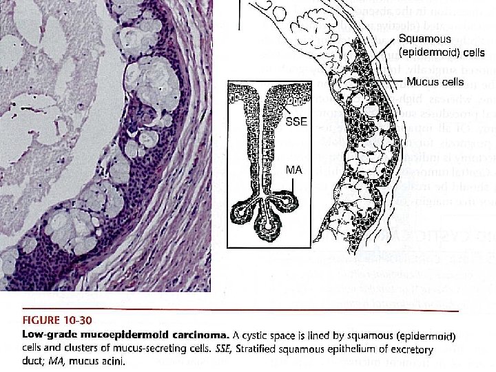
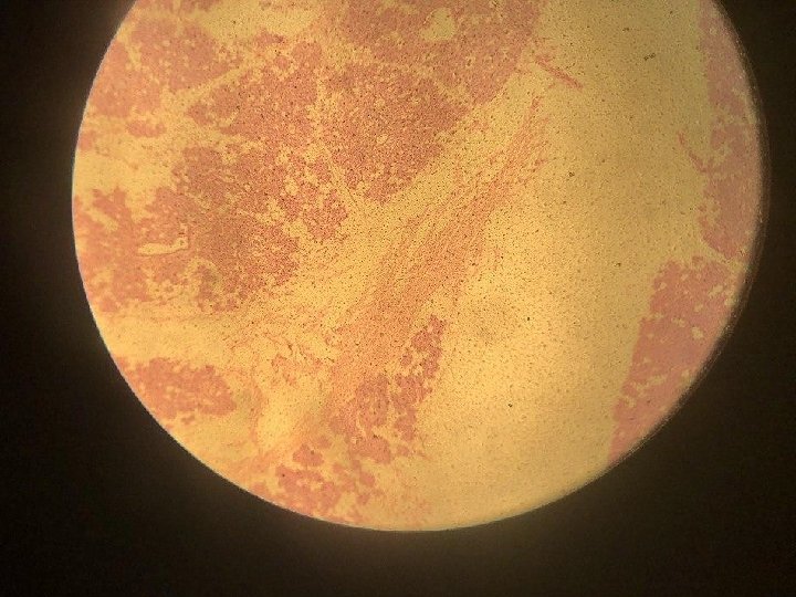
Slide no. 3 Mucoepidermoid carcinoma
Histopathological features of Mucoepidermoid carcinoma The tumor composed of a mixture of mucus producing cells and squamous cells, mucus secreting cells vary in shape but contain abundant foamy cytoplasm that stain positively with mucus stain, the epidermoid cells are squamoid feature, often demonstrating polygonal shape. In addition third type of cells is typically present, the intermediat cells these cells are basaloid in appearance
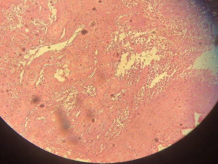
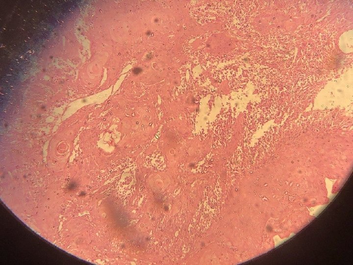
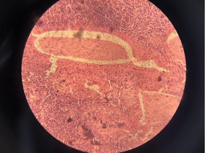
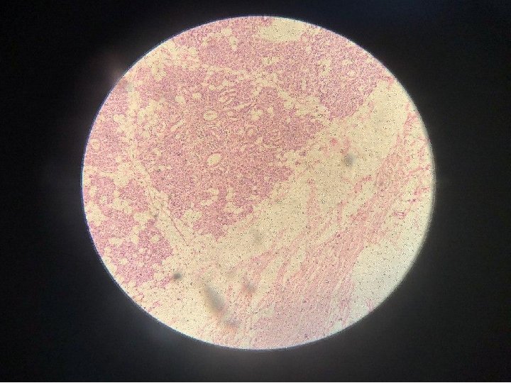
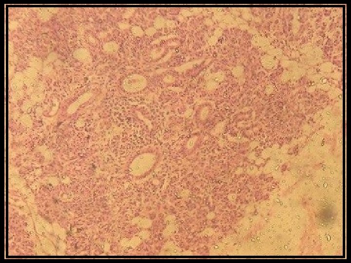
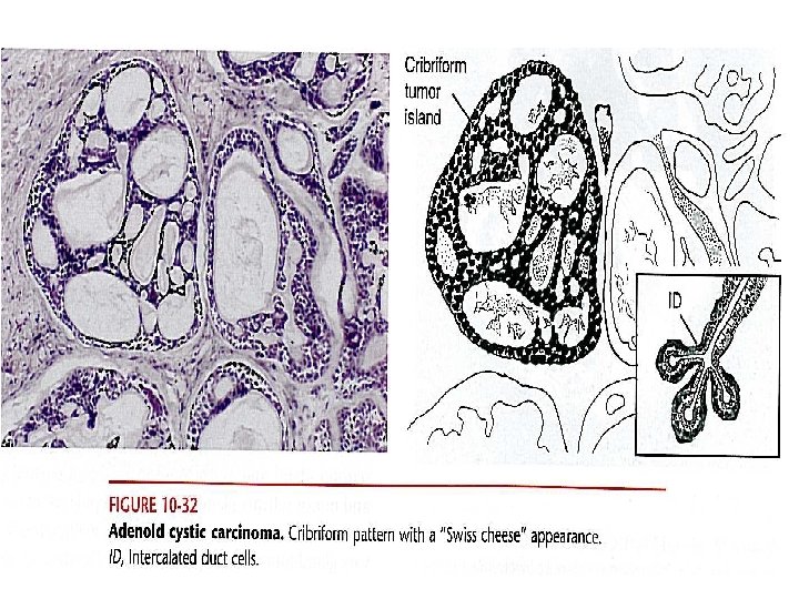
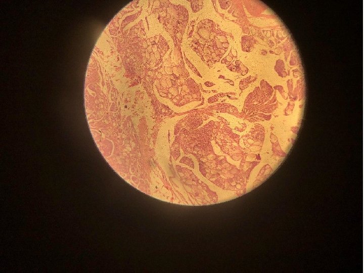
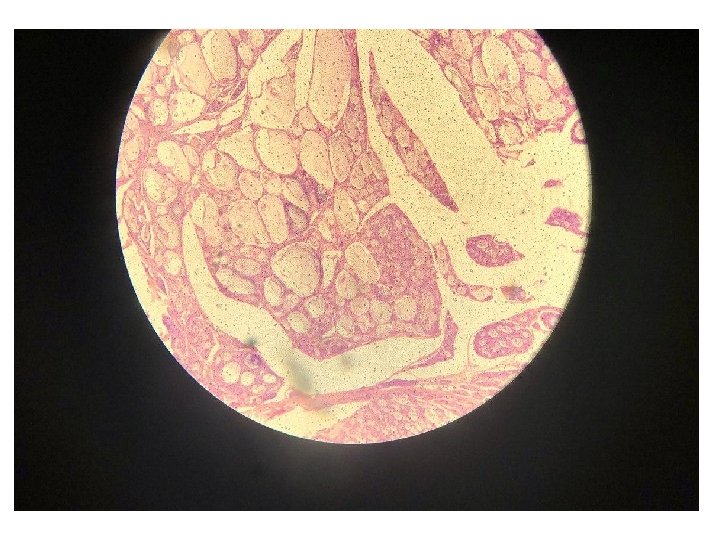
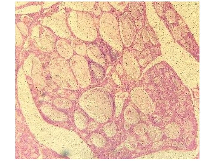
Slide no. 4 Adenoid cystic carcinoma
Histopathological features of Adenoid cystic carcinoma Composed of mixture of myoepithelial cells and ductal cells. That have varied arrangement. Three major patterns are recognized: 1. Cribriform 2. Tubular 3. solid Adenoid cystic carcinoma. The small darkly staining cells of the adenoid cystic carcinoma form cribriform islands with large holes which have been likened, rather inappropriately, to