Spinal cord reflexes Dr Altdorfer tract Kahler Ascending


 and Descending (left) Tracts of the Spinal Cord")


 -heat, pain")
 Stretch reflex (or monosynaptic, proprioceptive, myotatic) 2) Withdrawal reflex (or polisynaptic,")
 Stretch reflex (or monosynaptic, proprioceptive, myotatic) 2) Withdrawal reflex (or polisynaptic,")


 Stretch reflex (or monosynaptic, proprioceptive, myotatic) 2) Withdrawal reflex (or polisynaptic,")
- Slides: 18
Spinal cord - reflexes Dr. Altdorfer tract
Kahler
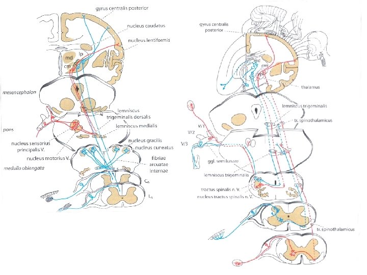
Ascending (right) and Descending (left) Tracts of the Spinal Cord
The most mobile parts of the spinal cord are the most vulnerable for injury and degeneration. In the cervical region the cervical enlargement almost completely fill the vertebral canal: the smallest displacement of the vertebrae will result in damages of the spinal cord. . The lumbar region is loaded with almost the complete weight of the trunk – leading to frequent degenerative disorders (disc herniation, spondilosis). Cervical myopathy is compression of the spinal cord The cord get squeezed And this causes numbness and /or weakness in the arms and legs
Disc herniation Radiculopathy: the location of the symptoms helps determine diagnosis Symptoms of spinal nerve injury. Pain or sensory loss of the given dermatome, weaker or missing reflexes and paresis of the muscles innervated by the spinal nerve affected. The most commonly injured segments are: L 4, L 5, S 1
Dermatomes
Clinically important monosynaptic reflexes Reflex Effector-Muscle Segment Bicepsreflex M. biceps brachii C 5 -C 6 Tricepsreflex M. triceps brachii C 6 -C 7 Patellareflex M. quadriceps femoris L 2 -L 4 Achillesreflex M. triceps surae S 1 Masseterreflex M. masseter Brainstem (pons-mesencephalon)
In case of complete transsection of the spinal cord, there is a complete sensory loss under the damage, on both side, and complete paresis including all the muscles! If the site of the damage is above C 4 segment the patient could die (phrenic n!).
Brown-Séquard Syndrome In case of hemisection of the spinal cord (Brown-Séquard syndrome) -heat, pain and basic touch information is lost under the damage on the opposite side -epicritical sensitivity is lost under the damage on the same side.
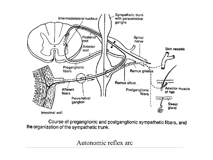
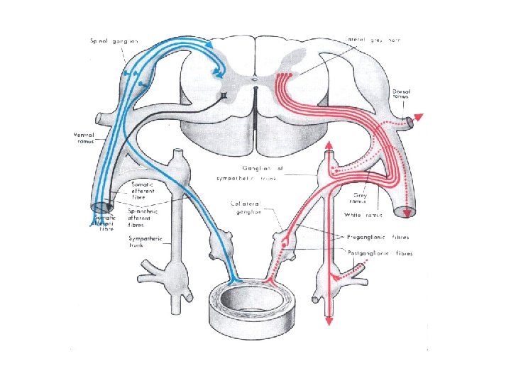
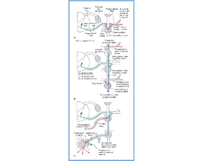
Spinal reflexes 1) Stretch reflex (or monosynaptic, proprioceptive, myotatic) 2) Withdrawal reflex (or polisynaptic, nociceptive, flexor…) 3) Autonomous reflex (or vegetative) • Receptor • Sensory neuron • (Interneuron) • Effector neuron • Effector organ
Spinal reflexes 1) Stretch reflex (or monosynaptic, proprioceptive, myotatic) 2) Withdrawal reflex (or polisynaptic, nociceptive) 3) Autonomous reflex (or vegetative) • Receptor • Sensory neuron • (Interneuron) • Effector neuron • Effector organ
Nociceptive reflex arc • Synonyms: flexor reflex, withdrawal reflex, polysynaptic spinal reflex • Ancient reflex that protects the body from potentially damaging, noxious stimuli.
Nociceptive reflex arc • Nociceptors of the skin and mucous membranes → • A delta and/or C fibres → • DRG (ganglion) → • spinal cord: dorsal horn → interneuron • numerous collaterals: excitacion of motoneurons of flexors and inhibition of extensor on the side of stimulus and stimulation of extensors together with inhibition of flexors on the contralateral side (flexor reflex with crossed extensor response).
Spinal reflexes 1) Stretch reflex (or monosynaptic, proprioceptive, myotatic) 2) Withdrawal reflex (or polisynaptic, nociceptive) 3) Autonomous reflex (or vegetative) • Receptor • Sensory neuron • (Interneuron) • Effector neuron • Effector organ