
Exercise 21 22 Spinal Cord Spinal nerves Reflexes



- Slides: 21
Exercise 21 & 22 Spinal Cord, Spinal nerves & Reflexes
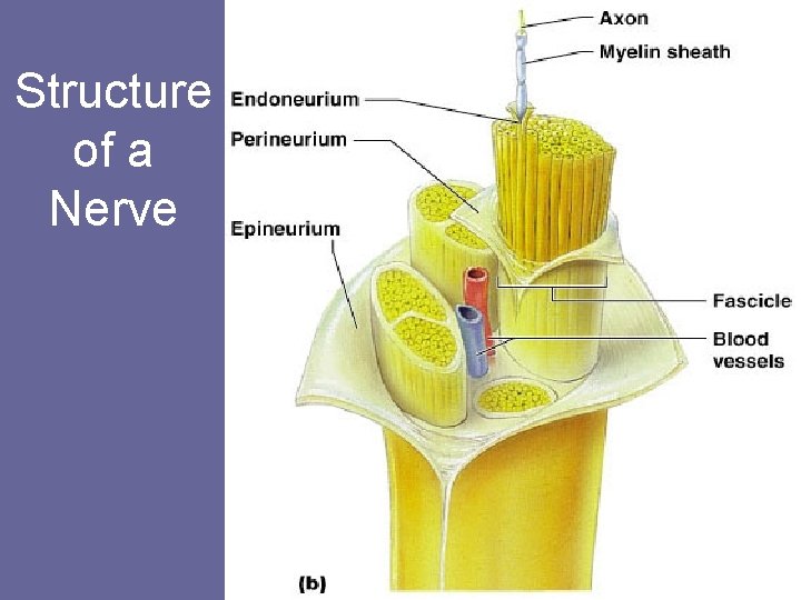
Structure of a Nerve Figure 13. 3 b
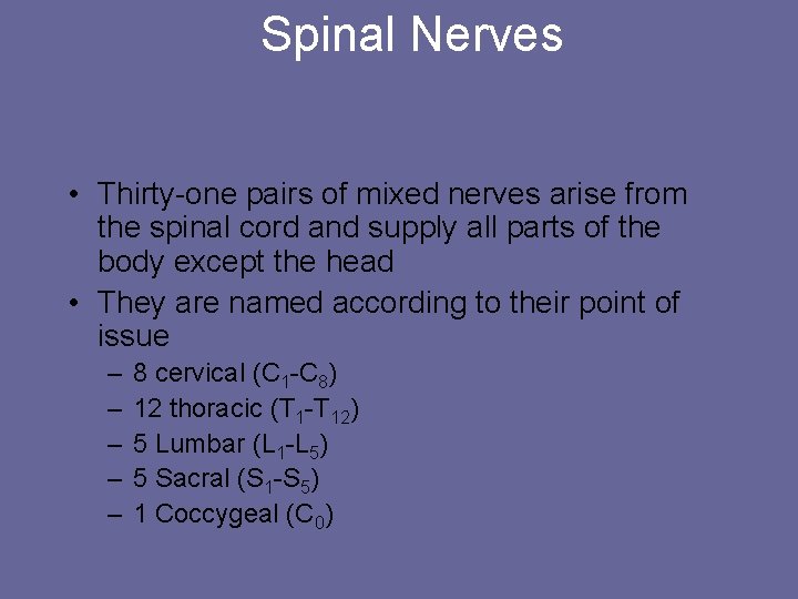
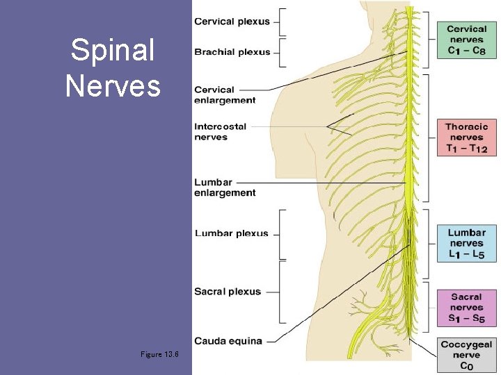
Spinal Nerves • Thirty-one pairs of mixed nerves arise from the spinal cord and supply all parts of the body except the head • They are named according to their point of issue – – – 8 cervical (C 1 -C 8) 12 thoracic (T 1 -T 12) 5 Lumbar (L 1 -L 5) 5 Sacral (S 1 -S 5) 1 Coccygeal (C 0)
Spinal Nerves Figure 13. 6
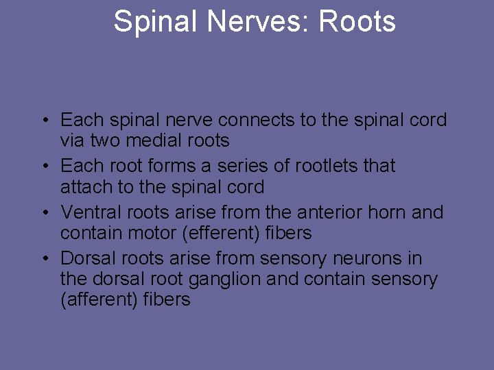
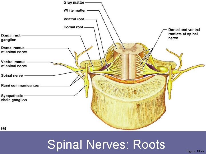
Spinal Nerves: Roots • Each spinal nerve connects to the spinal cord via two medial roots • Each root forms a series of rootlets that attach to the spinal cord • Ventral roots arise from the anterior horn and contain motor (efferent) fibers • Dorsal roots arise from sensory neurons in the dorsal root ganglion and contain sensory (afferent) fibers
Spinal Nerves: Roots Figure 13. 7 a
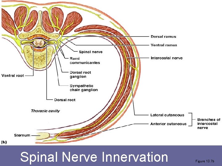
Spinal Nerve Innervation Figure 13. 7 b
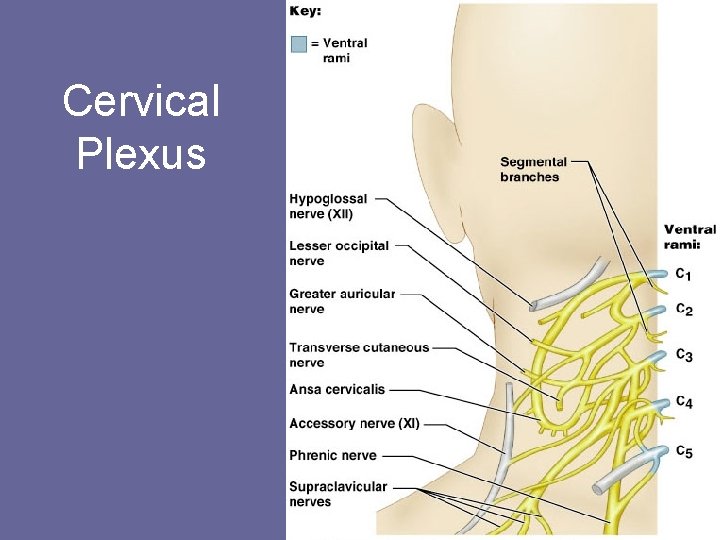
Cervical Plexus Figure 13. 8
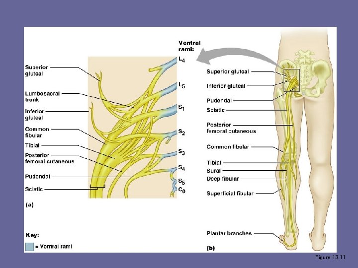
Sacral Plexus Figure 13. 11
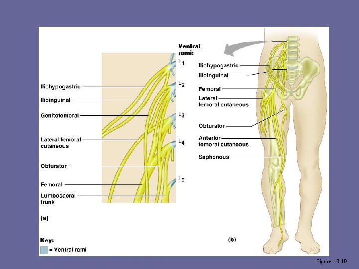
Lumbar Plexus Figure 13. 10
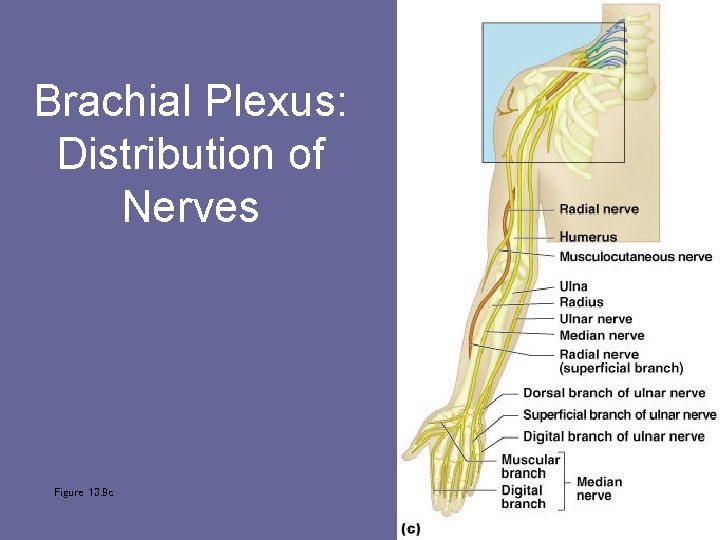
Brachial Plexus: Distribution of Nerves Figure 13. 9 c
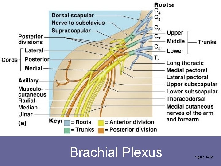
Brachial Plexus Figure 13. 9 a
Reflexes • A reflex is a rapid, predictable motor response to a stimulus • Reflexes may: – Be inborn (intrinsic) or learned (acquired) – Involve only peripheral nerves and the spinal cord – Involve higher brain centers as well
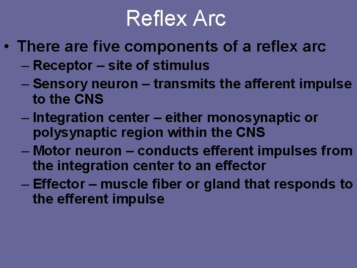
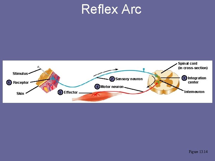
Reflex Arc • There are five components of a reflex arc – Receptor – site of stimulus – Sensory neuron – transmits the afferent impulse to the CNS – Integration center – either monosynaptic or polysynaptic region within the CNS – Motor neuron – conducts efferent impulses from the integration center to an effector – Effector – muscle fiber or gland that responds to the efferent impulse
Reflex Arc Spinal cord (in cross-section) Stimulus 1 2 Sensory neuron Receptor 3 Integration center 4 Motor neuron Skin 5 Effector Interneuron Figure 13. 14
Crossed Extensor Reflex Figure 13. 19
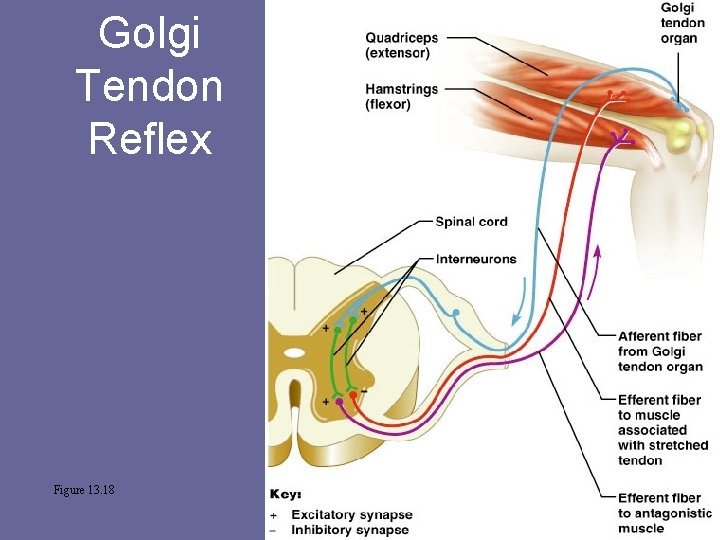
Golgi Tendon Reflex Figure 13. 18
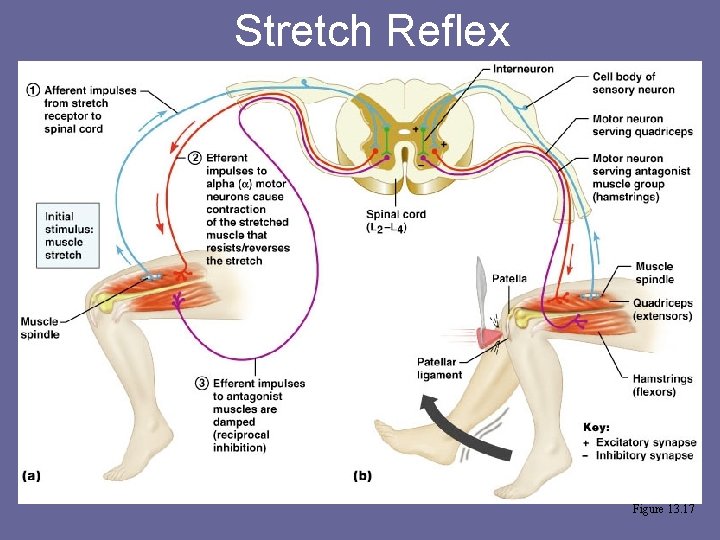
Stretch Reflex Figure 13. 17
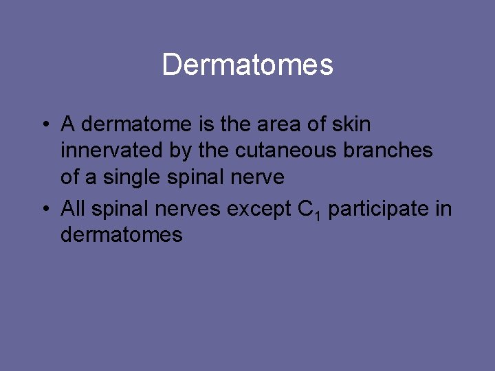
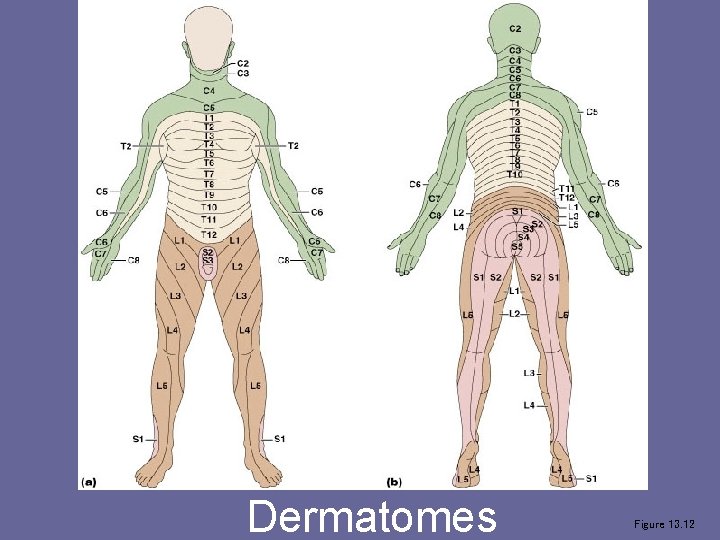
Dermatomes • A dermatome is the area of skin innervated by the cutaneous branches of a single spinal nerve • All spinal nerves except C 1 participate in dermatomes
Dermatomes Figure 13. 12