Radiological Category Gastrointestinal Principal Modality 1 Fluoroscopy Principal


- Slides: 17

Radiological Category: Gastrointestinal Principal Modality (1): Fluoroscopy Principal Modality (2): CT Case Report #0420 Submitted by: Nicholas Beckmann, M. D. Faculty reviewer: Agnes Guthrie, M. D. Date accepted: 14 April 2007

Case History 34 year-old Hispanic female with no significant prior medical history presenting with gradual onset shortness of breath and dysphagia.

Radiological Presentations CT Chest, Mediastinal Window

Radiological Presentations CT Chest, Mediastinal Window

Radiological Presentations CT Chest, Mediastinal Window

Radiological Presentations CT Chest, Mediastinal Window

Radiological Presentations Chest CT, Lung Window

Radiological Presentations CT Chest, Lung Window

Radiological Presentations CT Chest, Lung Window

Radiological Presentations Barium Swallow

Radiological Presentations Barium Swallow

Test Your Diagnosis Which one of the following is your choice for the appropriate diagnosis? After your selection, go to next page. Bronchoesophageal Fistula secondary to: • Carcinoma • Granulomatous Infection • Crohn’s Disease • Congenital Malformation • Ulceration from Barrett’s Esophagus

Findings and Differentials Findings: CT: Consolidation of the posterior aspects of the right upper and lower lobes with multiple areas of cavitation within the consolidated lung. Incidental note is made of a right lower lobe calcified granuloma. A tree-in-bud appearance to the lungs can be seen bilaterally. Multiple enlarged pre- and subcarinal lymphnodes are present as well. The esophagus has an irregular appearance inferiorly with a possible tract seen between the esophagus and the right lower lung lobe. No esophageal wall thickening is present. Barium Swallow: A fistula is present between the distal esophagus and a segmental bronchus of the right lower lung lobe. No narrowing of esophageal lumen is seen. Differentials: Bronchoesophageal Fistula secondary to: • Granulomatous Infection • Carcinoma, Bronchogenic or Esophageal • Chrohn’s Disease • Congenital • Ulceration from Barrett’s Esophagus

Discussion Granulomatous Infection: Granulomatous infection leads to bronchoesophageal fistula formation from chronic inflammation secondary to mediastinal lymphadenitis. Tuberculosis is the most common granulomatous infection to cause a bronchoesophageal fistula with other fungal infections such as candidiasis and histoplasmosis being common causes as well. A tree-in-bud pattern of the lungs is classic for Tuberculosis and may also be seen in many fungal infections. Lung cavitation can also be commonly caused by pulmonary Tuberculosis, although, in this case the lung cavities are caused by an anaerobic pneumonia arising due to the fistula. Carcinoma, Bronchogenic or Esophageal: Only 1% of bronchogenic carcinomas will develop a bronchoesophageal fistula. These fistulas usually form from a necrotic bronchogenic mass. Esophageal carcinoma is the most common cause of acquired bronchoesophageal fistulas, however esophageal wall thickening should be seen on CT with narrowing of esophageal lumen and shouldering of esophageal mucosa identified on fluoroscopy. Crohn’s Disease: A bronchoesophageal fistula can form secondary to chronic inflammation of Chrohn’s Disease, although, it would be very unusual for a person to present with a fistula without a history of long standing Inflammatory Bowel Disease.

Discussion Congenital: While congenital development of a trachoesophageal fistula is relatively common, a congenital bronchoesophageal fistula is rare. It is unusual for a fistula to go undiagnosed until adulthood, and classifying a bronchoesophageal fistula as congenital is a diagnosis of exclusion. Ulceration from Barrett’s Esophagus: This is a very rare causes of bronchoesophageal fistula. Evidence of an esophageal mucosal defect representing ulceration at the site of fistula formation with irregular mucosa in the distal esophagus should be seen on barium swallow.

Diagnosis Bronchoesophageal fistula secondary to mediastinal lymphadenitis from presumed fungal infection.

References Eisenhuber E. The Tree-in-Bud Sign. Radiology. 2002; 222: 771 -772 Hsin-Hui C. et al. Coexistence of a Tuberculosis Bronchoesophageal Fistula and Intracranial Tuberculosis in an Immunocompetent Patient. Southern Medical Journal. 2007; 100: 225 -226 Lim Y C. et al. A 52 year-old woman with recurrent hemoptysis. Chest. 2001; 119: 955 -957 Nigro J. et al. Perforating Barrett’s Ulcer Resulting in a Life-Threatening Esophageal Fistula. Ann of Thoracic Surgery. 2002; 73: 302 -304 Pickhardt P. Bhalla S. Balfe D. Aquired Gastroinestinal Fistulas: Classification, Etiologies & Imaging Evaluation. Radiology. 2002; 223: 9 -23 Rowe W. Inflammatory Bowel Disease. e. Medicine. com. Dec. 5, 2006 Shin J. et al. Esophagorespiratory Fistula: Long-term Results of Palliative Treatment with Covered Expandable Metallic Stents in 61 Patients. Radiology. 2004; 232: 252 -259
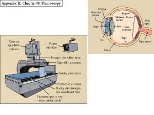
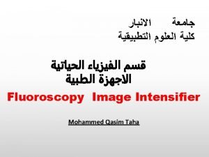
 Digital fluoroscopy vs conventional fluoroscopy
 Erate category 1
 Radiological dispersal device
 Tennessee division of radiological health
 Center for devices and radiological health
 National radiological emergency preparedness conference
 Spot film device
 Carm fluoroscopy
 Optical coupling in fluoroscopy
 Aspurbi
 Mesenterio
 Gastrointestinal sistem hormonları
 Intestinal villus
 Diagram of alimentary canal
 Composition of stomach
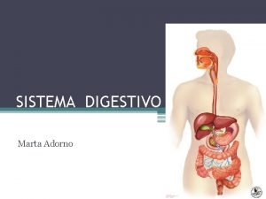
 Gastrointestinal tract
 Gastrointestinal hormones
 Anatomia do rim


