
Carm Fluoroscopy Developing a Method for Orthotic Analysis


- Slides: 17
C-arm Fluoroscopy: Developing a Method for Orthotic Analysis Lisa Friedland Department of Medical Biophysics University of Western Ontario London , Ontario
Acknowledgements �Thomas Jenkyn, Ph. D. , P. Eng �Megan Balsdon, MSc. Candidate �Kristen Bushey, MSc. Candidate
Question How does a podiatrist/orthotist know that a custom made orthotic is functioning effectively during dynamic gait? Create a method to analyze the orthotic in real time motion.
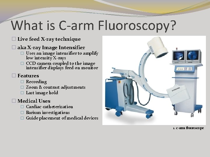
What is C-arm Fluoroscopy? � Live feed X-ray technique � aka X-ray Image Intensifier � Uses an image intensifier to amplify low intensity X-rays � CCD camera coupled to the image intensifier displays feed on monitor � Features � Recording � Zoom & contrast adjustments � Last image hold � Medical Uses � Cardiac catheterization � Barium investigations � Guide placement of medical devices 1. c-arm fluoroscope
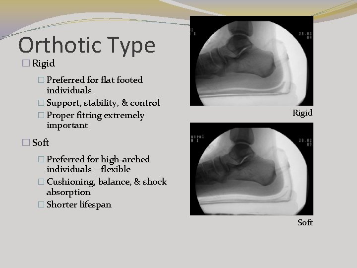
Orthotic Type � Rigid � Preferred for flat footed individuals � Support, stability, & control � Proper fitting extremely important Rigid � Soft � Preferred for high-arched individuals—flexible � Cushioning, balance, & shock absorption � Shorter lifespan Soft
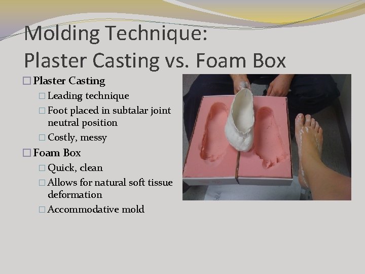
Molding Technique: Plaster Casting vs. Foam Box �Plaster Casting � Leading technique � Foot placed in subtalar joint neutral position � Costly, messy �Foam Box � Quick, clean � Allows for natural soft tissue deformation � Accommodative mold
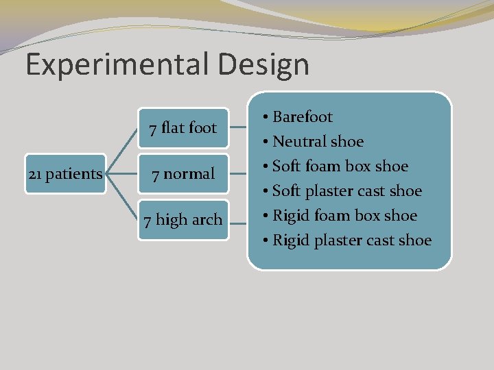
Experimental Design 7 flat foot 21 patients 7 normal 7 high arch • Barefoot • Neutral shoe • Soft foam box shoe • Soft plaster cast shoe • Rigid foam box shoe • Rigid plaster cast shoe
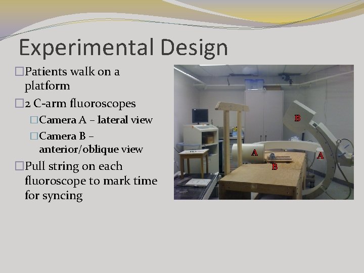
Experimental Design �Patients walk on a platform � 2 C-arm fluoroscopes B �Camera A – lateral view �Camera B – anterior/oblique view �Pull string on each fluoroscope to mark time for syncing A A B
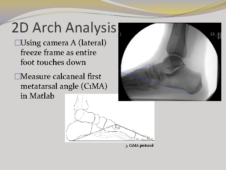
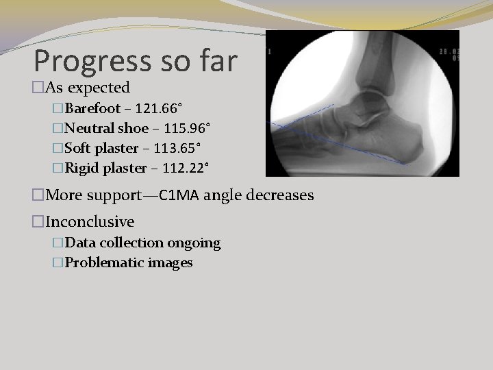
2 D Arch Analysis �Using camera A (lateral) freeze frame as entire foot touches down �Measure calcaneal first metatarsal angle (C 1 MA) in Matlab 3. C 1 MA protocol
Barefoot Dynamic
Rigid Foam Box Dynamic
Progress so far �As expected �Barefoot – 121. 66° �Neutral shoe – 115. 96° �Soft plaster – 113. 65° �Rigid plaster – 112. 22° �More support—C 1 MA angle decreases �Inconclusive �Data collection ongoing �Problematic images
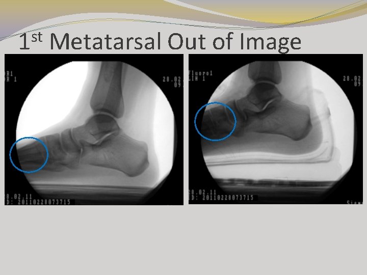
st 1 Metatarsal Out of Image
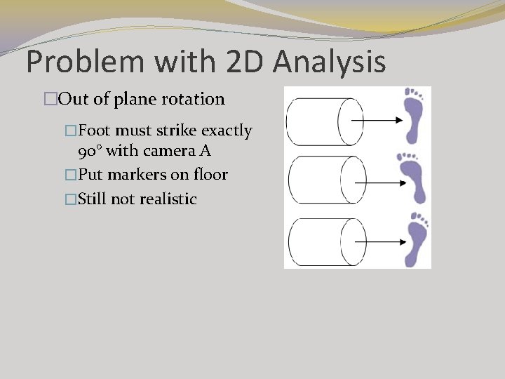
Problem with 2 D Analysis �Out of plane rotation �Foot must strike exactly 90° with camera A �Put markers on floor �Still not realistic
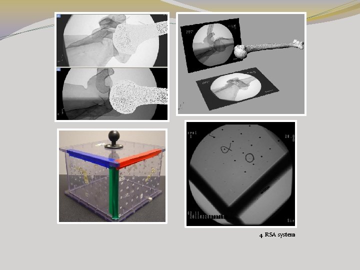
Future 3 D Analysis �Radiostereometric Analysis (RSA) �Calibration box �Image from cameras A & B—calibrate using Matlab �Digitize the image frames in Adobe Photoshop �Import 3 D CT image into Rhinoceros modelling software �Bone match CT image to Fluoroscope image
4. RSA system
Images 1. c-arm fluoroscope: http: //www. industrymedical. com/mymedical/ajmedical/prodetail 2284/AJ 4501_High_Frequency_Mobile_CArm_Xray_Imaging_System. html 2. normal/high/flat: http: //www. footlogics. ie/pronation-flatfeet-fallen-arches. html & http: //www. youcanbefit. com/shoes. html 3. C 1 MA protocol: Murley, G. , Menz, H. , & Landorff, K. (2009). A protocol for classifying normal-and flatarched foot posture for research studies using clinical and radiographic measurements. Journal of Foot and Ankel Research, 2, 22, 1 -13. 4. Allen, A, M. (2009). Development and validation of a markerless RSA system. Thesis. Biomedical Engineering, University of Western Ontario: London.