q Fertility peaks when a woman is in


")


- Slides: 44
q. Fertility peaks when a woman is in her late teens and early twenties qbegins to decline at age thirty qdropping more rapidly after age 35 years q. Fertility plummets after age 40 and pregnancy After age 45 is rare botros et. al infertility and assisted reproduction - 2008 infertility rate : overall : 2. 4% after age 34 : 11% after age 40 : 33% by the age 45 : 87% Speroff et. al Clinical Gynecologic Endocrinology & Infertility, 7 th Edition -2005
↓fertility rates : 25 -29 age : 4% to 8% ↓ 30 -34 age : 15% to 19% ↓ 35 -39 age : 26% to %46%↓ 40 -45 age : 95%↓ Factors Involved in Female Fertility Loss • Egg Quantity Egg quantity refers to the number of eggs that you have in your ovaries Germ cells in the female are not replenished during life the number of oocytes and follicles is determined in utero and declines following an exponential curve from the second trimester to menopause Speroff et. al Clinical Gynecologic Endocrinology & Infertility, 7 th Edition -2005
During fetal life , germ cells rapidly proliferate by mitosis to yield approximately 6 to 7 million oogonia by 16 -20 weeks of pregnancy Transformed to oocytes after entering the first meiotic division, the number of germ cells falls to between 1 and 2 million at birth and to about 300, 000 to 500, 000 by the onset of puberty Over the next 35 -40 years of reproductive life, only about 400 to 500 oocytes will ovulate Until age 37 -38 : approximately 25, 000 oocytes remain At the time of menopause , fewer than 1, 000 follicles remain Speroff et. al Clinical Gynecologic Endocrinology & Infertility, 7 th Edition -2005
2. Egg Quality refers to how ready and able your eggs are to become fertilized. Ø Every woman carries a certain number of eggs in her ovaries ready to be released for fertilization Ø These eggs need to have the right shape, health, and chromosomes in order to be able to develop into an embryo and, eventually, a baby. Ø Unfortunately, egg quality also changes over time Ø As you age, your eggs become weaker, and less able to form a healthy embryo Ø Your eggs also begin to decrease in number, leaving fewer and fewer quality eggs available for fertilization. A woman of 40 typically has lower egg quality than a woman of 20
Complications of Poor Egg Quality Poor egg quality can lead to a vareity of complications, including: 1. IVF or IUI failure 2. repeated miscarriages 3. chromosomal abnormalities Female Age and Egg Quality q Age is one issue, but the real fertility issue is egg quality and quantity and not the number in a woman's age. q Egg quantity and quality in an individual woman can be average for her age, better than average, or worse than average. We know that egg quantity and quality tends to decline significantly in the mid to late 30 s and fall faster in the late thirties and early 40 s.
Testing Fertility Loss as You Age The following ovarian reserve screening tests are used by fertility specialists to predict the "remaining egg supply" and the ability (reserve) of the ovaries to respond to stimulation with drugs. These tests are helpful. However, they predict the quantity of eggs remaining - rather than the quality of those eggs. Do ovarian reserve tests check egg quantity, quality, or both? Ovarian reserve testing can tell us quite a lot about the remaining quantity of eggs a woman has, but it tells us little about the quality of those eggs. 1. Day 3 FSH testing 2. AMH levels 3. Antral follicle counts
Age is the best "test" that we have at this time for egg quality If your FSH levels were run using a different assay, you can not compare your results to those shown below with confidence. For example, with some assays an FSH of 12 is normal. Day 3 FSH level FSH interpretation for DPC Immulite assay Less than 9 Normal FSH level. Expect a good response to ovarian stimulation 9 - 11 Fair. Response is between normal and somewhat reduced (response varies widely). Overall, a slightly reduced live birth rate 11 - 15 Reduced ovarian reserve. Expect a reduced response to stimulation and some reduction in embryo quality with IVF. Reduced live birth rates on the average 15 - 20 Expect a more marked reduction in response to stimulation and usually a further reduction in embryo quality. Low live birth rates. Antral follicle count is an important variable Over 20 This is pretty much a "no go" level in our center. Very poor (or no) response to stimulation. "No go" levels should be individualized for the particular lab assay and IVF center.
What is AMH? ØAMH production is highest in the preantral and small antral stages (less than 4 mm diameter) of follicle development. ØProduction decreases and then stops as the follicles grow larger. ØThere is almost no AMH made in human follicles over 8 mm in size. Because of this, the levels are quite constant and the AMH test can be done on any day of a woman's cycle. How can AMH hormone levels be a fertility test? ØSince AMH is produced only in small ovarian follicles, blood levels of this substance have been used to attempt to measure the size of the pool of growing follicles in women. ØResearch shows that the size of the pool of growing follicles is heavily influenced by the size of the pool of remaining primordial follicles (microscopic follicles in "deep sleep"). ØTherefore, AMH blood levels are thought to reflect the size of the remaining egg supply - or ovarian reserve
ØWith increasing female age, the size of their pool of remaining microscopic follicles decreases. Likewise, their blood AMH levels and the number of ovarian antral follicles visible on ultrasound also decreases. ØWomen with many small follicles, such as those with polycystic ovaries have high AMH hormone values and women that have few remaining follicles and those that are close to menopause have low anti-mullerian hormone levels AMH levels and pregnancy chances with in vitro fertilization AMH levels probably do not reflect egg quality, but having more eggs at egg retrieval gives us more to work with - so we are more likely to have at least one high quality embryo available for transfer back to the female partner's uterus.
Ovarian reserve testing methods Anti mullerian hormone is one potential test of ovarian reserve.
What Are Antral Follicles? Ø Antral follicles are small follicles (about 2 -8 mm in diameter) Ø Vaginal ultrasound is the best way to accurately assess and count these small structures. Ø In my opinion, the antral follicle counts (in conjunction with female age) are by far the best tool that we currently have for estimating ovarian reserve and/or chances for pregnancy with in vitro fertilization 1. When there an average (or high) number of antral follicles, we tend to get a "good" response with many mature follicles. Pregnancy rates are higher than average. 2. When there are few antral follicles, we tend to get a poor response with few mature follicles. 3. When the number of antral follicles is intermediate, the response is not as predictable.
Antrals and IVF Success - Female age under 35 IVF live birth rates are reduced with low antral follicle counts The average antral follicle count in the under 35 age group was 21
From our own observations and experience, here are some general guidelines:
Antrals and IVF Success - Female age 35 -37 The average antral follicle count in this group was 16
Antrals and IVF Success - Female age 38 -40 The average antral follicle count at age 38 to 40 was 13
Antrals and IVF Success - Female age 41 -42 the average antral count in this group was only 11 Advanced Fertility Center of Chicago
Menstural characteristic q. Menstrual characteristics in older women correlate with the number of follicles remaining q. The ovaries of regularly menstruating older women contain 10 -fold more follicles than those of perimenopausal women having irregular and infrequent menses qthe interval from loss of menstrual regularity to menopause is approximately 5 years q As age increases and FSH levels rise, the follicular phas becomes shorter, LH levels and luteal phase duration remain unchanged q. Cycles remain regular, but overall cycle length and cycle variability decrease q. Over the subsequent 8 - 10 years preceding the menopause , average cycle length and variability steadily increase as ovulations become less regular and frequent
q Circulating follicular phase inhibin-B levels decrease as or even before FSH concentrations begin to increase q serum follicle-stimulating hormone (FSH) levels begin to increase; luteinizing hormone (LH) concentrations remain unchanged q rise in circulating FSH concentrations : 1. age-related changes in the pattern of pulsatile gonadotropinreleasing hormone (Gn. RH) secretion 2. progressive follicular depletion and lower levels of feedback inhibition on pituitary FSH secretion by ovarian hormones q Later, luteal phase serum inhibin-A levels decline as well Speroff et. al Clinical Gynecologic Endocrinology & Infertility, 7 th Edition -2005
q. The initial rise in FSH levels precedes any measurable decrease in estradiol levels, by several years q. Follicular phase estradiol levels in older cycling women are generally similar to those in younger women and often even higher q. However, careful studies have shown that the earlier acute rise in estradiol levels results not from accelerated follicle growth but from advanced follicular development at the beginning of the cycle and earlier selection of the dominant follicle q. Luteal phase progesterone levels in older and younger cycling women are also similar Speroff et. al Clinical Gynecologic Endocrinology & Infertility, 7 th Edition -2005
Female Age and Miscarriage and Fertility q. Numerous studies have documented the increased risk for miscarriage and increase in infertility as women age Maternal Age and Pregnancy Loss Rate
Miscarriages botros et. al infertility and assisted reproduction - 2008
The main reason for the increased risk for miscarriage in "older" women is due to the increase in chromosomal abnormalities (abnormal karyotype) in their eggs Chromosomal Problems in Aging Eggs Many studies have documented the increased rate of chromosomal abnormalities in women of advanced reproductive age. ● The meiotic spindle is a critical component of eggs that is involved in organizing the chromosome pairs so that proper division of pairs can occur as the egg is developing ● When the chromosomes line up in a straight line on the spindle, the division process should proceed normally ● However, with a disordered arrangement on an abnormal spindle, the division process may be uneven with an unbalanced chromosomal situation resulting
Older eggs are significantly more likely to have abnormal spindles and an abnormal spindle predisposes to development of chromosomally abnormal eggs. �here are 2 general types of chromosomal abnormalities: T ● Numerical abnormalities where there is an abnormal number of chromosomes q. This situation is called aneuploidy q. Such as a missing (monosomy) or an extra chromosome (trisomy) q. An example of monosomy is Turner Syndrome and a well known example of trisomy is Down Syndrome ● Structural abnormalities where there is a problem with the structure of a chromosome q. Examples include translocations, duplications and deletions of part of a chromosome
q 17% of the eggs studied from women 20 -25 years old were found to have an abnormal spindle appearance and at least one chromosome displaced from proper alignment. q 79% of the eggs studied from women 40 -45 years old were found to have an abnormal spindle appearance and at least one chromosome displaced from proper alignment. Maternal age <35 40 43 Aneuploidy 10 30 50 >45 100
Recipient Age
Summary There is a significant reduction in implantation rates in egg donor recipients 45 -50 years of age There is a significant increase in miscarriage rates in egg donor recipients 45 -50 years of age Discussion Uterine receptivity might decline with advanced age due to unknown biochemical and/or molecular aberrations of the endometrium This decline could also be the result of a higher incidence of other pathological conditions in the uterus such as myomas, synechiae or polyps Hypertension and other systemic disorders are more common in older women and could be contributing to the reduced potential for implantation and successful pregnancy outcome
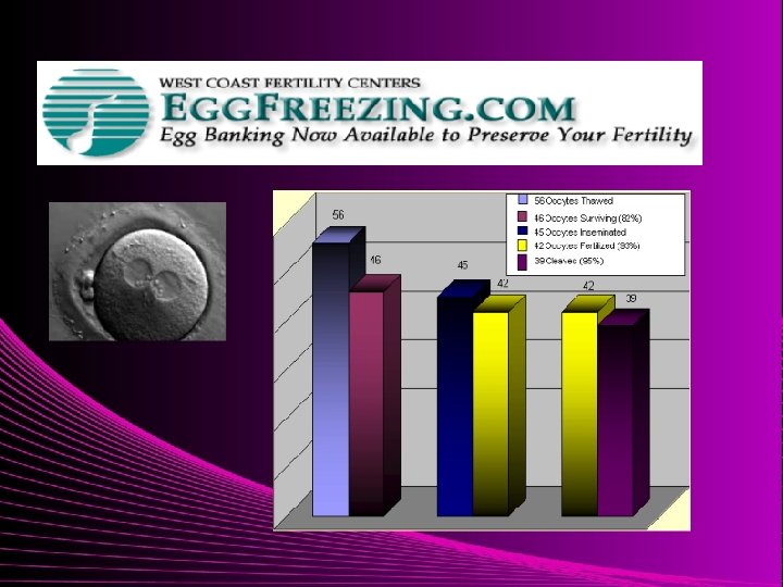
Aging and female fertility 1. Fertility Drugs For women 40 and older, success rates with this form of infertility treatment are very low, and IVF should be considered relatively soon 2. ART egg freezing for preservation of fertility q This study involved 40 cycles in women (average 35. 5) Ø The ongoing pregnancy rate (beyond 12 weeks of pregnancy) with vitrified eggs was 30% per cycle. ■ Study by L Rienzi, et al, Human Reproduction; January 2010
● A 2009 study of 23 IVF cycles using frozen eggs (average 31. 5) q. There were 14 pregnancies, 1 miscarriage and 13 ongoing pregnancies (57% per transfer) ■ Study by J Grifo and N Noyes, Fertility and Sterility; May 2009
Clinical Applications of Egg Freezing Oocyte cryopreservation could be a clinical tool for: q. Women at risk of losing ovarian function q. Women desiring fertility preservation (e. g. delayed maternity) q. Eliminating ethical concerns of embryo cryopreservation
Future considerations q. Oocyte cryopreservation q“Pausing the biological clock” q. Cytoplasmic transfer q. Donation of enucleated oocytes q. Reproduction without gametes q. Use of nuclear material from somatic cells q. Donated or synthetic cytoplasm q. Reconstituted oocytes
ovum donation in older women pregnancy outcome after ovum donation in older women : Pregnancy in advanced maternal age women after ovum donation carrying twins is associated with significant maternal and fetal complications, with increased risks of prematurity and lower birthweight. Possibly, the aged uterus is less suitable for carrying a multifetal pregnancy than a younger uterus. Therefore, the alternative of transferring a single, good-quality embryo should be the preferred option. Human Reproduction 2009 24(10): 2500 -2503
Who are candidates for egg donation ? q. Premature ovarian failure q. Ovarian insufficiency (e. g. FSH>15 ) q. Physiologic menopause : Maternal age over 43 q. History of poor egg/embryo quality or multiple IVF failures Human Reproduction 2009 24(10): 2500 -2503
How old is too old ? q. Danger to mother q. Decreased life expectancy of parents q. Is 55 a “physiological limit” Human Reproduction 2009 24(10): 2500 -2503
Pregnancy in the Sixth Decade of Life: Obstetric Complications Pre-eclampsia : 35% Background Incidence : 3 -10% Gestational Diabetes : 20% Background Incidence : 5% Human Reproduction 2009 24(10): 2500 -2503
Pregnancy in the 6 th decade of life : Conclusion There does not appear to be any definitive medical reason for excluding these women from attempting pregnancy on the basis of age alone Human Reproduction 2009 24(10): 2500 -2503
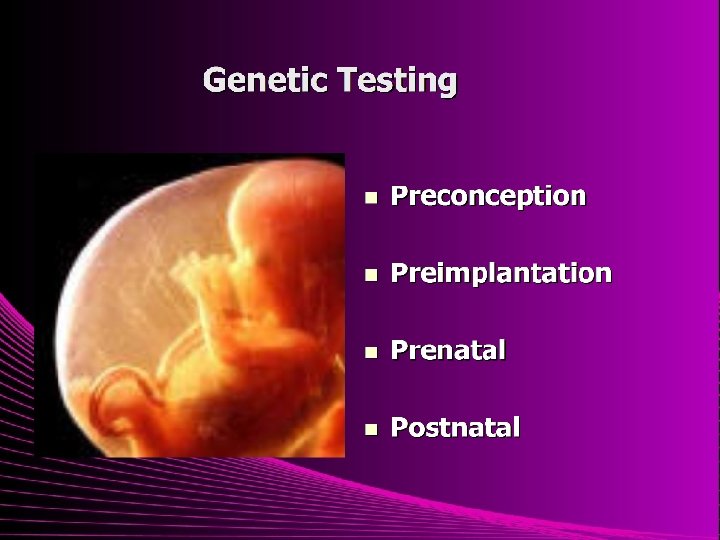
PGD
In the general population, the risk for a live birth with a chromosomal abnormality is about: The published studies on IVF with PGD success rates thus far have generally shown: ● No improvement in clinical pregnancy rates with PGD - it appears at this time that PGD actually results in lower pregnancy rates ● No improvement in live birth success rates with PGD - it appears at this time that PGD actually results in lower live birth rates ● Questionable improvement in implantation rates with PGD - and the possibility of actually having lower implantation rates. Not uncommonly, fewer embryos are transferred after PGD "Implantation rate" is the percentage of transferred embryos that implant in the uterus and are seen with early pregnancy ultrasound ● Some studies are showing a somewhat lower rate of miscarriage after PGD, other studies are not showing any difference (see details of studies discussed above on this page)
Are PGD test results reliable? PGD test results are not always correct ● Sometimes the embryo has abnormal chromosomes, but PGD testing shows a normal result ● Sometimes the embryo has normal chromosomes, but PGD testing shows an abnormal result ● Therefore, some chromosomally normal embryos will be discarded, and some chromosomally abnormal embryos will be transferred after PGD
PGD – Clinical Indications q. Single gene defects q. Balanced translocations q. Advanced maternal age (aneuploidy) q. Repetitive IVF failure q. Recurrent pregnancy loss q. Embryo selection
IVF with preimplantation genetic diagnosis for reasons of advanced maternal age or in couples with unexplained recurrent pregnancy loss can increase implantation rates and decrease miscarriage risk but the modest improvements in live birth rates achieved thus far cannot justify the associated costs in couples without other specific indications for IVF. Speroff et. al Clinical Gynecologic Endocrinology & Infertility, 7 th Edition -2005