Practical class Immunopathology Dr Irena Nedelea Case study




- Slides: 30
Practical class Immunopathology Dr. Irena Nedelea
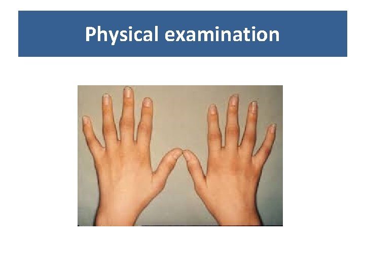
Case study • 26 year-old female patient presents with: - pain and stiffness of the small joints of the hands (metacarpophalangeal, MCP), onset 2 months - feeling unwell and tired for the last 6 months • Family history: non-contributory • Personal history: non-contributory • Occupational history: dentist
Physical examination
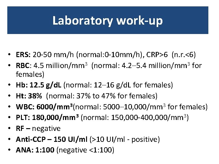
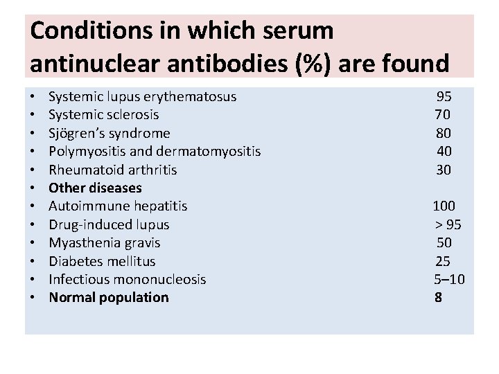
Laboratory work-up • ERS: 20 -50 mm/h (normal: 0 -10 mm/h), CRP>6 (n. r. <6) • RBC: 4. 5 million/mm 3 (normal: 4. 2– 5. 4 million/mm 3 for females) • Hb: 12. 5 g/d. L (normal: 12– 16 g/d. L for females) • Ht: 38% (normal: 37% to 47% for females) • WBC: 6000/mm 3(normal: 5000– 10, 000/mm 3 for females) • PLT: 180, 000/mm 3 (normal: 150, 000 -400, 000/mm 3) • RF – negative • Anti-CCP – 150 UI/ml (>10 UI/ml - positive) • ANA: 1: 100 (negative <1: 100)
Diagnosis?
Rheumatoid arthritis • Is an autoimmune disease causing a chronic symmetrical polyarthritis with systemic involvement. • Its course is extremely variable and it is associated with nonarticular features.
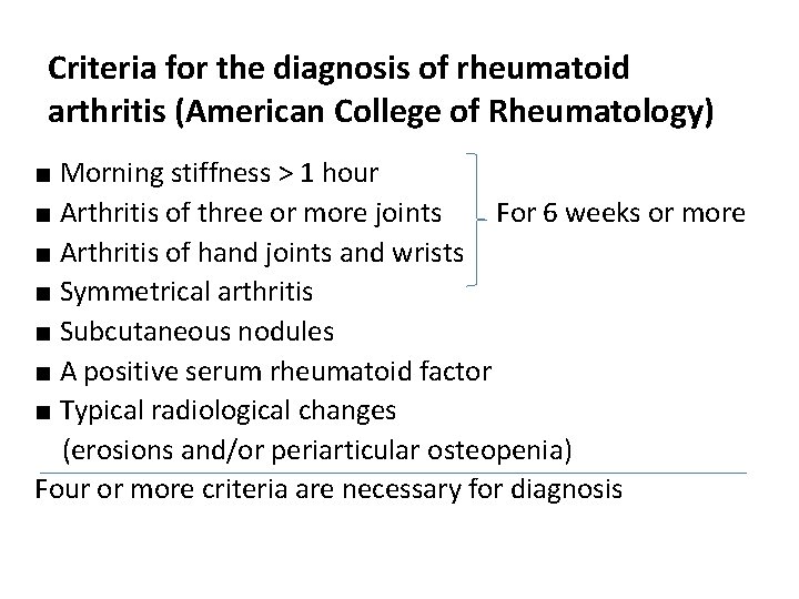
Criteria for the diagnosis of rheumatoid arthritis (American College of Rheumatology) ■ Morning stiffness > 1 hour ■ Arthritis of three or more joints For 6 weeks or more ■ Arthritis of hand joints and wrists ■ Symmetrical arthritis ■ Subcutaneous nodules ■ A positive serum rheumatoid factor ■ Typical radiological changes (erosions and/or periarticular osteopenia) Four or more criteria are necessary for diagnosis
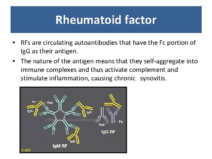
Rheumatoid factor • RFs are circulating autoantibodies that have the Fc portion of Ig. G as their antigen. • The nature of the antigen means that they self-aggregate into immune complexes and thus activate complement and stimulate inflammation, causing chronic synovitis.
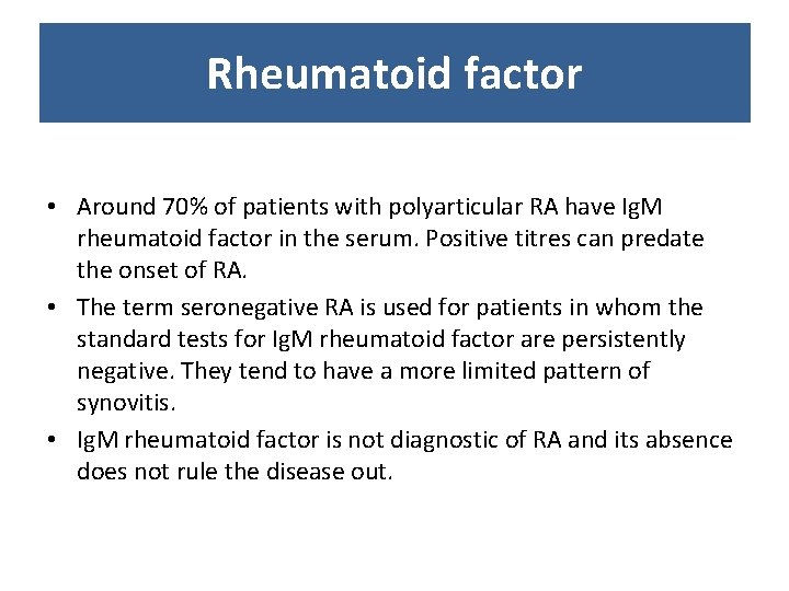
Rheumatoid factor • Around 70% of patients with polyarticular RA have Ig. M rheumatoid factor in the serum. Positive titres can predate the onset of RA. • The term seronegative RA is used for patients in whom the standard tests for Ig. M rheumatoid factor are persistently negative. They tend to have a more limited pattern of synovitis. • Ig. M rheumatoid factor is not diagnostic of RA and its absence does not rule the disease out.
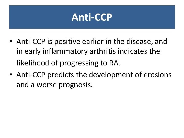
Anti-CCP • Anti-CCP is positive earlier in the disease, and in early inflammatory arthritis indicates the likelihood of progressing to RA. • Anti-CCP predicts the development of erosions and a worse prognosis.
Conditions in which serum antinuclear antibodies (%) are found • • • Systemic lupus erythematosus 95 Systemic sclerosis 70 Sjögren’s syndrome 80 Polymyositis and dermatomyositis 40 Rheumatoid arthritis 30 Other diseases Autoimmune hepatitis 100 Drug-induced lupus > 95 Myasthenia gravis 50 Diabetes mellitus 25 Infectious mononucleosis 5– 10 Normal population 8
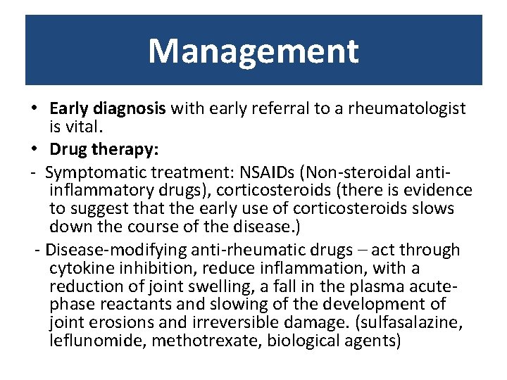
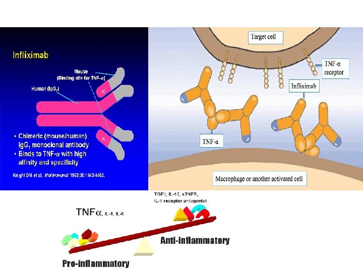
Management • Early diagnosis with early referral to a rheumatologist is vital. • Drug therapy: - Symptomatic treatment: NSAIDs (Non-steroidal antiinflammatory drugs), corticosteroids (there is evidence to suggest that the early use of corticosteroids slows down the course of the disease. ) - Disease-modifying anti-rheumatic drugs – act through cytokine inhibition, reduce inflammation, with a reduction of joint swelling, a fall in the plasma acutephase reactants and slowing of the development of joint erosions and irreversible damage. (sulfasalazine, leflunomide, methotrexate, biological agents)
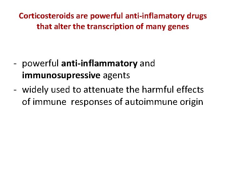
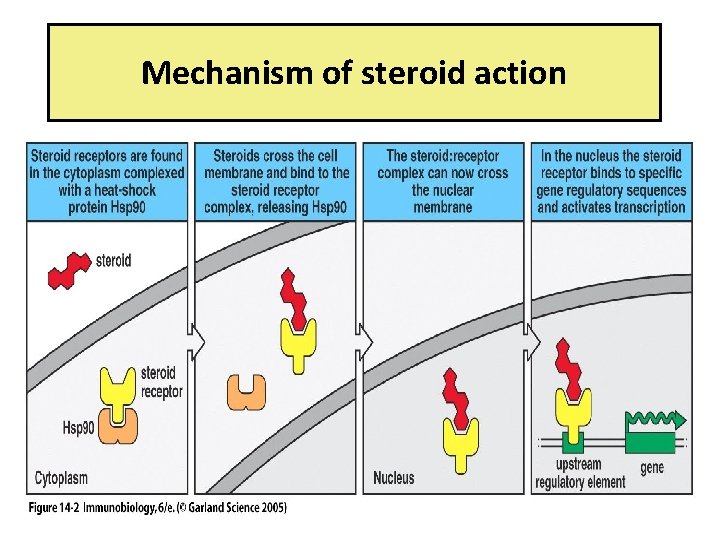
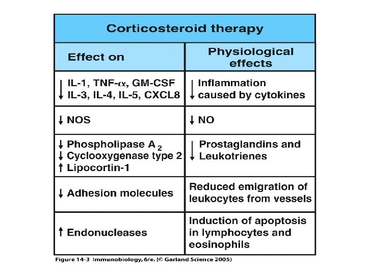
Corticosteroids are powerful anti-inflamatory drugs that alter the transcription of many genes - powerful anti-inflammatory and immunosupressive agents - widely used to attenuate the harmful effects of immune responses of autoimmune origin
Figure 14 -2 Mechanism of steroid action
Figure 14 -3
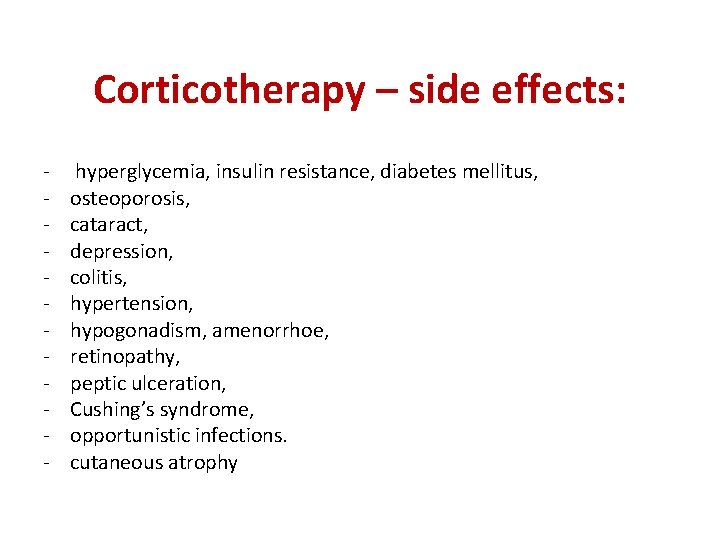
Corticotherapy – side effects: - hyperglycemia, insulin resistance, diabetes mellitus, osteoporosis, cataract, depression, colitis, hypertension, hypogonadism, amenorrhoe, retinopathy, peptic ulceration, Cushing’s syndrome, opportunistic infections. cutaneous atrophy
…back to our case • Symptomatic treatment – NSAIDs • DMARDs - Methotrexate and Sulfasalazine did not improve the symptoms
Case study
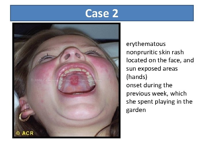
Case 2 erythematous nonpruritic skin rash located on the face, and sun exposed areas (hands) onset during the previous week, which she spent playing in the garden
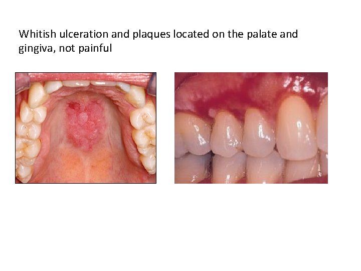
Whitish ulceration and plaques located on the palate and gingiva, not painful
What is the most likely diagnosis?
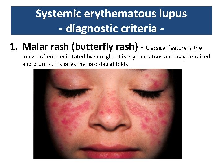
Systemic erythematous lupus - diagnostic criteria 1. Malar rash (butterfly rash) - Classical feature is the malar: often precipitated by sunlight. It is erythematous and may be raised and pruritic. It spares the naso-labial folds
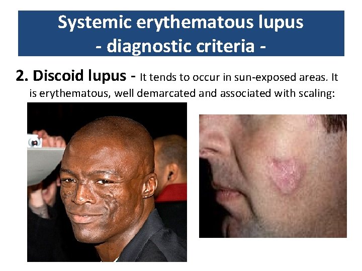
Systemic erythematous lupus - diagnostic criteria 2. Discoid lupus - It tends to occur in sun-exposed areas. It is erythematous, well demarcated and associated with scaling:
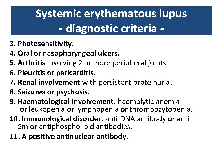
Systemic erythematous lupus - diagnostic criteria 3. Photosensitivity. 4. Oral or nasopharyngeal ulcers. 5. Arthritis involving 2 or more peripheral joints. 6. Pleuritis or pericarditis. 7. Renal involvement with persistent proteinuria. 8. Seizures or psychosis. 9. Haematological involvement: haemolytic anemia or leukopenia or lymphopenia or thrombocytopenia. 10. Immunological disorder: anti-DNA antibody or anti. Sm or antiphospholipid antibodies. 11. A positive antinuclear antibody.
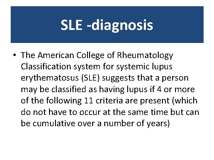
SLE -diagnosis • The American College of Rheumatology Classification system for systemic lupus erythematosus (SLE) suggests that a person may be classified as having lupus if 4 or more of the following 11 criteria are present (which do not have to occur at the same time but can be cumulative over a number of years)
…. Back to our patient • What laboratory work-up would you consider necessary to establish the diagnosis?
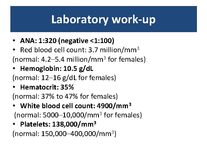
Laboratory work-up • ANA: 1: 320 (negative <1: 100) • Red blood cell count: 3. 7 million/mm 3 (normal: 4. 2– 5. 4 million/mm 3 for females) • Hemoglobin: 10. 5 g/d. L (normal: 12– 16 g/d. L for females) • Hematocrit: 35% (normal: 37% to 47% for females) • White blood cell count: 4900/mm 3 (normal: 5000– 10, 000/mm 3 for females) • Platelets: 138, 000/mm 3 (normal: 150, 000– 400, 000/mm 3)
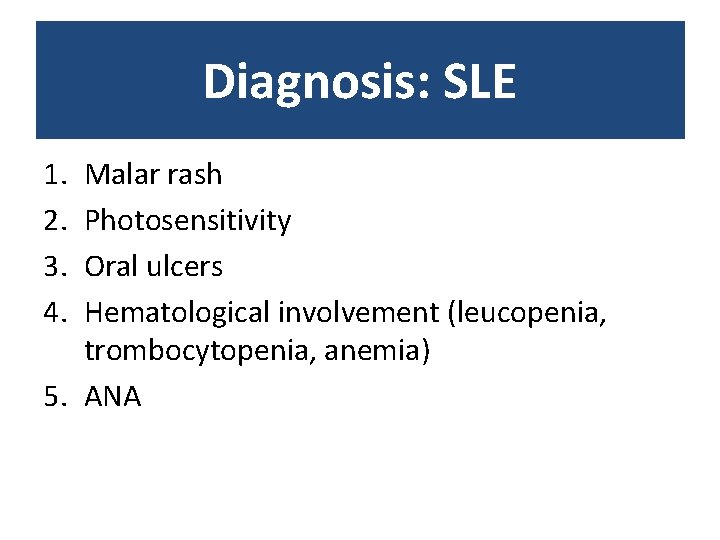
Diagnosis: SLE 1. 2. 3. 4. Malar rash Photosensitivity Oral ulcers Hematological involvement (leucopenia, trombocytopenia, anemia) 5. ANA
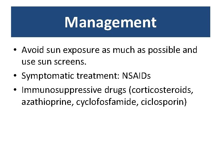
Management • Avoid sun exposure as much as possible and use sun screens. • Symptomatic treatment: NSAIDs • Immunosuppressive drugs (corticosteroids, azathioprine, cyclofosfamide, ciclosporin)