Physiology of Consciousness Dr Taha Sadig Ahmed Physiology


- Slides: 19
Physiology of Consciousness Dr Taha Sadig Ahmed Physiology Department , College of Medicine , King Saud University , Riyadh 1
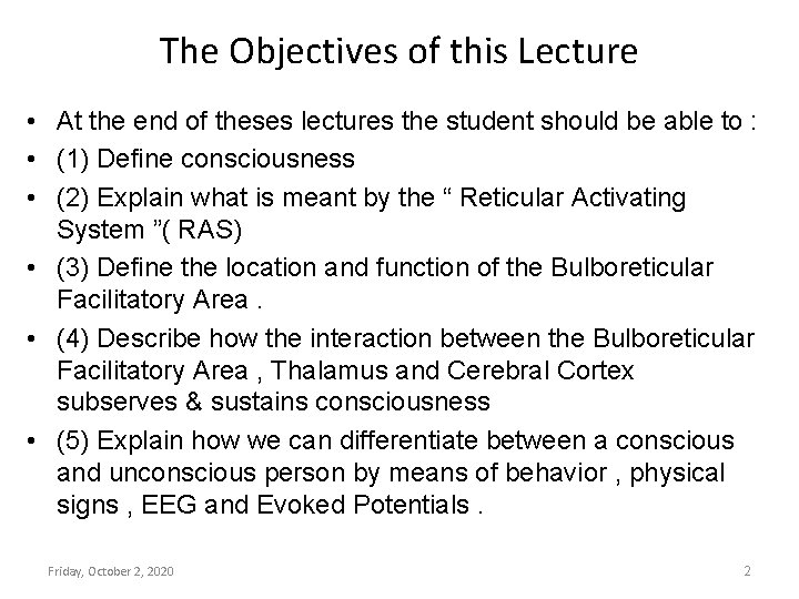
The Objectives of this Lecture • At the end of theses lectures the student should be able to : • (1) Define consciousness • (2) Explain what is meant by the “ Reticular Activating System ”( RAS) • (3) Define the location and function of the Bulboreticular Facilitatory Area. • (4) Describe how the interaction between the Bulboreticular Facilitatory Area , Thalamus and Cerebral Cortex subserves & sustains consciousness • (5) Explain how we can differentiate between a conscious and unconscious person by means of behavior , physical signs , EEG and Evoked Potentials. Friday, October 2, 2020 2
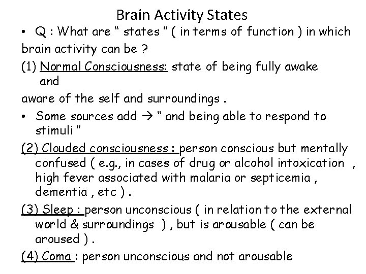
Brain Activity States • Q : What are “ states ” ( in terms of function ) in which brain activity can be ? (1) Normal Consciousness: state of being fully awake and aware of the self and surroundings. • Some sources add “ and being able to respond to stimuli ’’ (2) Clouded consciousness : person conscious but mentally confused ( e. g. , in cases of drug or alcohol intoxication , high fever associated with malaria or septicemia , dementia , etc ). (3) Sleep : person unconscious ( in relation to the external world & surroundings ) , but is arousable ( can be aroused ). (4) Coma : person unconscious 3 and not arousable
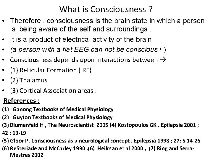
What is Consciousness ? • Therefore , consciousness is the brain state in which a person is being aware of the self and surroundings. • It is a product of electrical activity of the brain • (a person with a flat EEG can not be conscious ! ) • Consciousness depends upon interactions between • (1) Reticular Formation ( RF). • (2) Thalamus • (3) Cortical Association areas. References : (1) Ganong Textbooks of Medical Physiology (2) Guyton Textbooks of Medical Physiology (3) Blumenfeld H , The Neuroscientist 2005 (4) Kostopoulos GK. Epilepsia 2001 ; 42 : 13 -19 (5) Gloor P. Consciousness as a neurological concept. Epilepsia 1998 ; 27: S 14 -26 (6) Re. Steriade and Mc. Carley 1990 , (6) Heilman et al 2000 , (7) Ring and Serra. Mestres 2002
Reticular Activating System ( RAS ) (Bulboreticular Facilitatory Area + Thalamus) 5
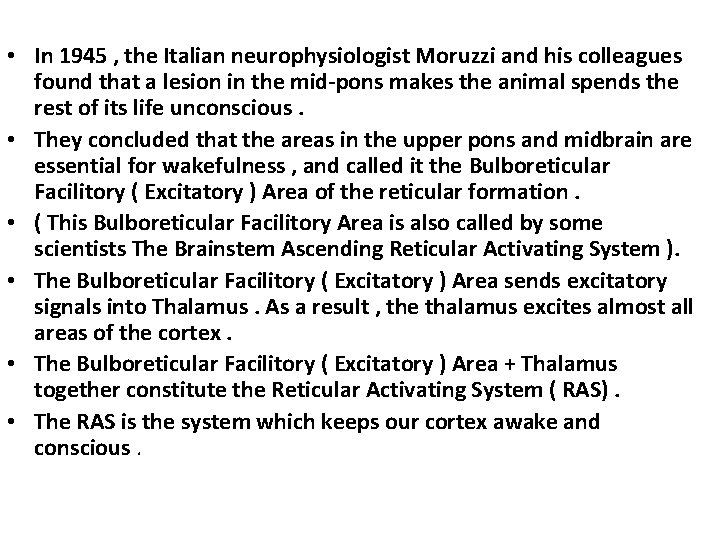
• In 1945 , the Italian neurophysiologist Moruzzi and his colleagues found that a lesion in the mid-pons makes the animal spends the rest of its life unconscious. • They concluded that the areas in the upper pons and midbrain are essential for wakefulness , and called it the Bulboreticular Facilitory ( Excitatory ) Area of the reticular formation. • ( This Bulboreticular Facilitory Area is also called by some scientists The Brainstem Ascending Reticular Activating System ). • The Bulboreticular Facilitory ( Excitatory ) Area sends excitatory signals into Thalamus. As a result , the thalamus excites almost all areas of the cortex. • The Bulboreticular Facilitory ( Excitatory ) Area + Thalamus together constitute the Reticular Activating System ( RAS). • The RAS is the system which keeps our cortex awake and conscious.
RAS
• The awakening action of the RAS is mediated by fibers secreting Acetylcholine ( Ach) and Norepinephe (NE) • The RAS provides the main drive that maintains effective cortical excitability level , & interruption of this ascending pathway ( e. g. , by a tumor ) causes the subject to go into unremitting coma lasting for the remainder of life. • The level of consciousness is largely influenced by : (1) peripheral sensory inputs , and by (2) Thalamocortical sectors.
• Peripheral Sensory Inputs Increase the Level of Excitation of the Bulboreticular Faciltatory Area : • The level of activity of the Bulboreticular Facilitatory area and , consequently , the level of alertness and arousal is largely determined by the number and type of sensory ( afferent ) signals that enter the CNS from the periphery. • Pain signals , in particular , increase activity in this excitatory area and therefore strongly excite the brain to attention. • Role of Thalamo-Cortical Sectors : • Not only do excitatory signals pass to the cerebral cortex from the RAS , but feedback signals also return from the cortex back to the Thalamus ( which is part of the RAS ).
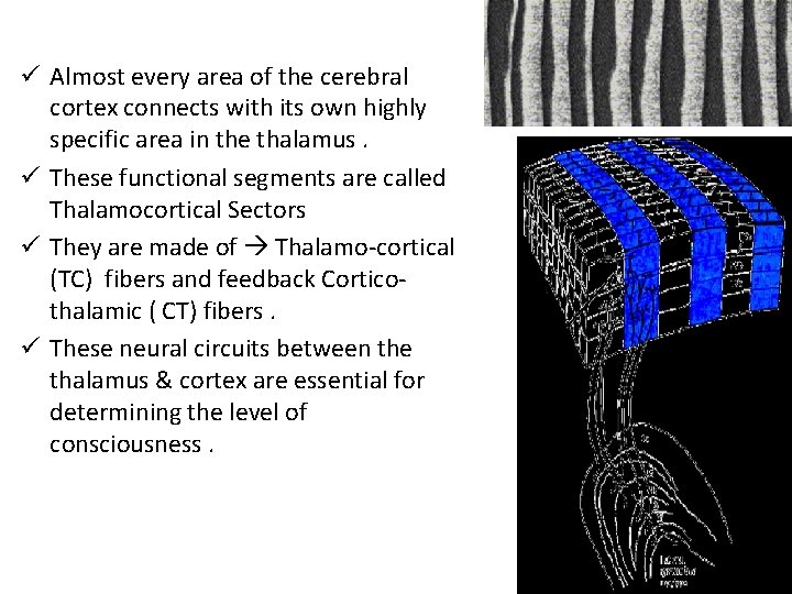
ü Almost every area of the cerebral cortex connects with its own highly specific area in the thalamus. ü These functional segments are called Thalamocortical Sectors ü They are made of Thalamo-cortical (TC) fibers and feedback Corticothalamic ( CT) fibers. ü These neural circuits between the thalamus & cortex are essential for determining the level of consciousness.
Indices ( indicators) of Level of Consciousness • • Appearance & Behavior : posture ( sitting , standing ? ) , open eyes ? . Facial expression ? , responds to stimuli ( including the examiner’s questions about name , orientation in time & place ? & other general Qs like who is the president ? ) Physical signs : pulse , BP, respiration , pupils , reflexes , particularly brainstem reflexes , etc ) EEG Each of these states ( wakefulness , sleep , coma and death ) has specific EEG patterns. Evoked potentials ( in cases of Brain Death ). 11
EEG ( Electroencephalogram( Routine EEG ( Electroencephalogram ) is recording of cortical activity from scalp surface 12
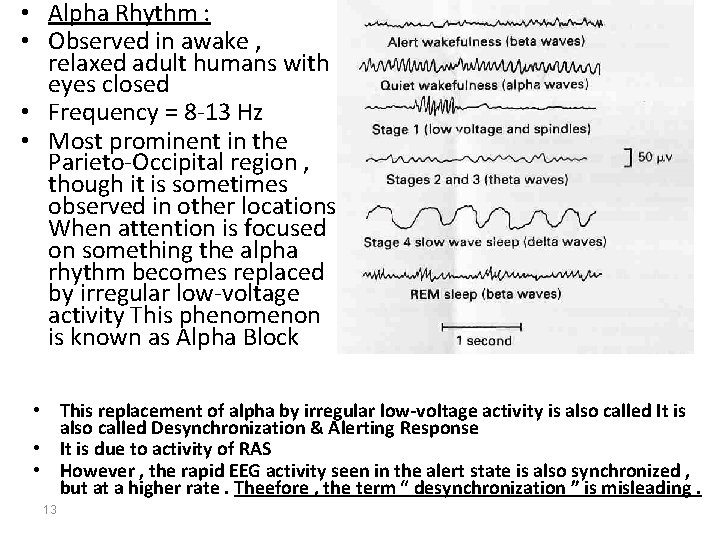
• Alpha Rhythm : • Observed in awake , relaxed adult humans with eyes closed • Frequency = 8 -13 Hz • Most prominent in the Parieto-Occipital region , though it is sometimes observed in other locations When attention is focused on something the alpha rhythm becomes replaced by irregular low-voltage activity This phenomenon is known as Alpha Block • This replacement of alpha by irregular low-voltage activity is also called It is also called Desynchronization & Alerting Response • It is due to activity of RAS • However , the rapid EEG activity seen in the alert state is also synchronized , but at a higher rate. Theefore , the term “ desynchronization ” is misleading. 13
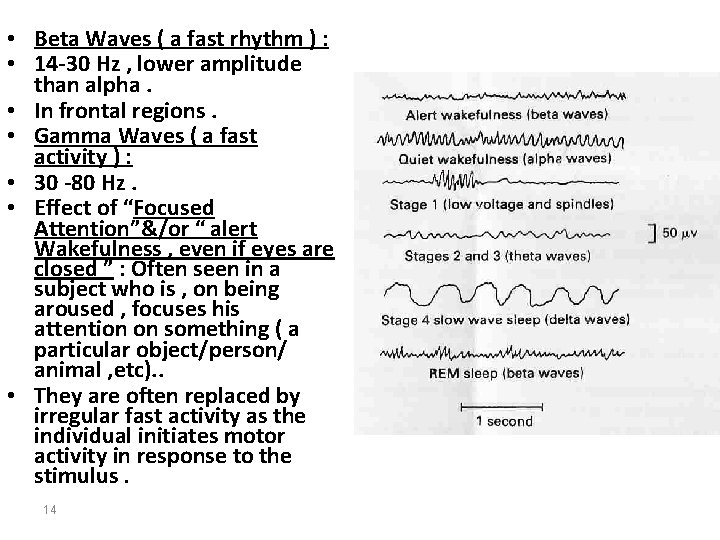
• Beta Waves ( a fast rhythm ) : • 14 -30 Hz , lower amplitude than alpha. • In frontal regions. • Gamma Waves ( a fast activity ) : • 30 -80 Hz. • Effect of “Focused Attention”&/or “ alert Wakefulness , even if eyes are closed ” : Often seen in a subject who is , on being aroused , focuses his attention on something ( a particular object/person/ animal , etc). . • They are often replaced by irregular fast activity as the individual initiates motor activity in response to the stimulus. 14
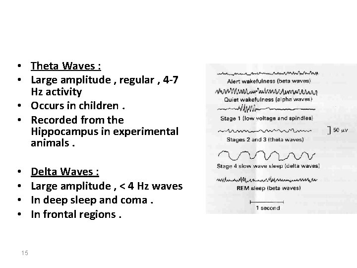
• Theta Waves : • Large amplitude , regular , 4 -7 Hz activity • Occurs in children. • Recorded from the Hippocampus in experimental animals. • • 15 Delta Waves : Large amplitude , < 4 Hz waves In deep sleep and coma. In frontal regions.
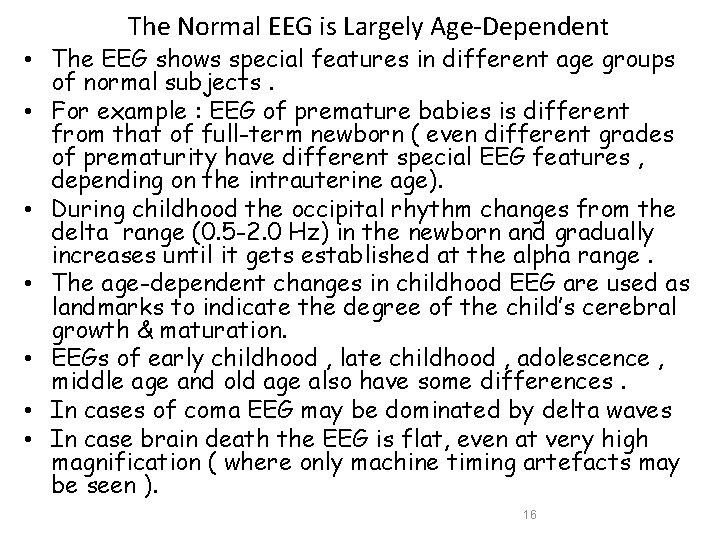
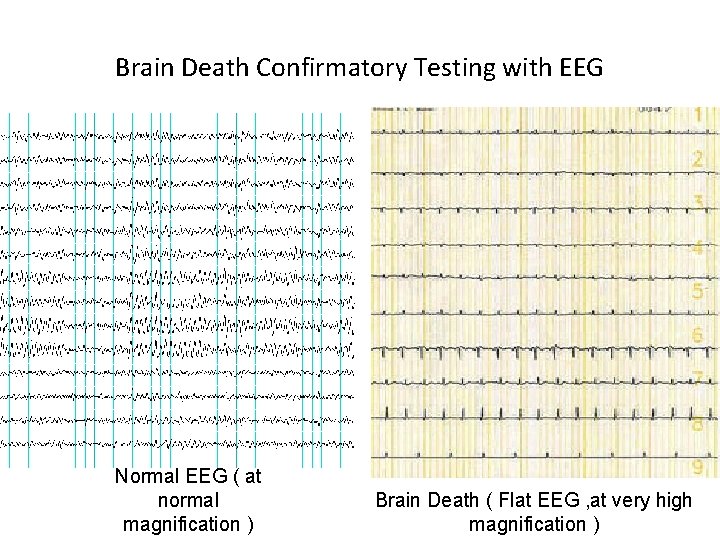
The Normal EEG is Largely Age-Dependent • The EEG shows special features in different age groups of normal subjects. • For example : EEG of premature babies is different from that of full-term newborn ( even different grades of prematurity have different special EEG features , depending on the intrauterine age). • During childhood the occipital rhythm changes from the delta range (0. 5 -2. 0 Hz) in the newborn and gradually increases until it gets established at the alpha range. • The age-dependent changes in childhood EEG are used as landmarks to indicate the degree of the child’s cerebral growth & maturation. • EEGs of early childhood , late childhood , adolescence , middle age and old age also have some differences. • In cases of coma EEG may be dominated by delta waves • In case brain death the EEG is flat, even at very high magnification ( where only machine timing artefacts may be seen ). 16
Brain Death Confirmatory Testing with EEG Normal EEG ( at normal magnification ) Brain Death ( Flat EEG , at very high magnification )
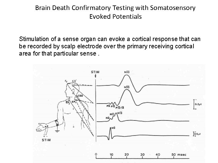
Brain Death Confirmatory Testing with Somatosensory Evoked Potentials Stimulation of a sense organ can evoke a cortical response that can be recorded by scalp electrode over the primary receiving cortical area for that particular sense.
• Thanks 19