Pelvic Fractures Author Walter Harrill Wray III MD

- Slides: 17
Pelvic Fractures Author: Walter Harrill Wray III, MD Academic Affiliation : Duke University Medical Center Department of Orthopaedic Surgery Chief Resident Level: Basic Version No: 1. 2 Submitted: May 2012 Editors’ Review: August 2012
2 Disclaimer/Liability • The information provided in the VAP is made available in good faith and is derived from sources believed to be reliable and accurate at the time of release. • The materials presented on the VAP may include links to external Internet sites. These external information sources are outside the control of Duke-NUS. The user of the Internet links is responsible for making his or her own decision about the accuracy, reliability and correctness of the information found. • In no event shall Duke-NUS be liable for any indirect, special, incidental, or consequential damages arising out of any use of reliance of any information contained in the VAP. Nor does Duke-NUS assume any responsibility for failure or delay in updating or removing the information contained in the VAP. • Moreover, information provided on the VAP does not constitute medical advice or treatment nor should it be considered as a replacement of the patient/physician relationship or a physician’s professional judgment. Duke-NUS expressly disclaims all liability for treatment, diagnosis, decisions and actions taken or not taken in reliance upon information contained in the VAP. This work is licensed under a Creative Commons Attribution-Non. Commercial-No. Derivs 3. 0 Unported License To view a copy of this license, visit [http: //creativecommons. org/licenses/by-nc-nd/3. 0/]
3 Financial Disclosures No Disclosures
4 Learning Objectives • Pelvis Anatomy • Mechanism of injury • Clinical Evaluation • Radiographic Evaluation • Classification • Treatment
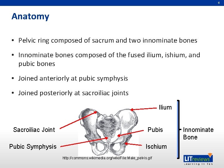
5 Anatomy • Pelvic ring composed of sacrum and two innominate bones • Innominate bones composed of the fused ilium, ishium, and pubic bones • Joined anteriorly at pubic symphysis • Joined posteriorly at sacroiliac joints Ilium Sacroiliac Joint Pubis Pubic Symphysis Ischium http: //commons. wikimedia. org/wiki/File: Male_pelvis. gif Innominate Bone
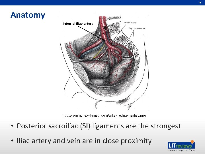
6 Anatomy http: //commons. wikimedia. org/wiki/File: Internaliliac. png • Posterior sacroiliac (SI) ligaments are the strongest • Iliac artery and vein are in close proximity
7 Mechanism of Injury • High energy injuries – MVC / motorcycle accident – Falls from a height – Pedestrian accidents • Anteroposterior (AP) force – Pelvis opens up like a book (“open-book” injury) • Lateral compression (LC) force – Side impact MVC
8 Clinical Evaluation • ABCDE – Airway, Breathing, Circulation, Disability, Exposure • Vitals – BP, heart rate, respiratory rate, temp, pulse ox • Address all life-threatening injuries – Head, chest, abdomen, spine • Full neurologic evaluation – Rectal exam • Orthopaedic evaluation – Observe any ecchymoses, urethral / rectal bleeding – Leg lengths – Pelvic compression for instability (performed only once)
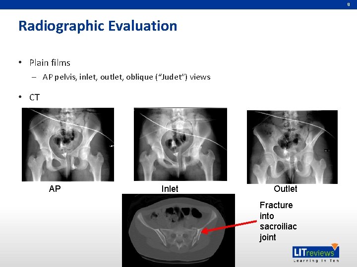
9 Radiographic Evaluation • Plain films – AP pelvis, inlet, outlet, oblique (“Judet”) views • CT AP Inlet Outlet Fracture into sacroiliac joint
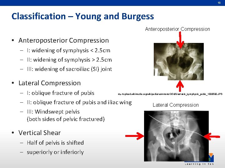
10 Classification – Young and Burgess Anteroposterior Compression • Anteroposterior Compression – I: widening of symphysis < 2. 5 cm – II: widening of symphysis > 2. 5 cm – III: widening of sacroiliac (SI) joint • Lateral Compression – I: oblique fracture of pubis – II: oblique fracture of pubis and iliac wing – III: Windswept pelvis (both sides of pelvic fractured) http: // upload. wikimedia. org/wikipedia/commons/3/3 d/Diastasis_symphysis_pubis_1300500. JPG • Vertical Shear – Half of pelvis is shifted – superiorly or inferiorly Lateral Compression
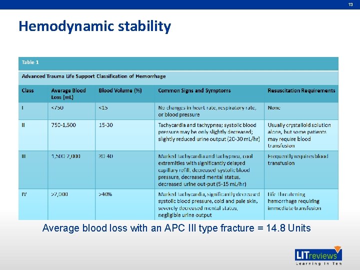
11 Initial Treatment • Resuscitation – Based on blood pressure, heart rate, urine output, lactate, base deficit, hematocrit, platelets, INR / PTT • Retroperitoneal hemorrhage may result in massive blood volume loss • Requires fluid and blood replacement – Initial 2 Liters of normal saline through large-bore IVs – Followed by units of packed red blood cells as needed
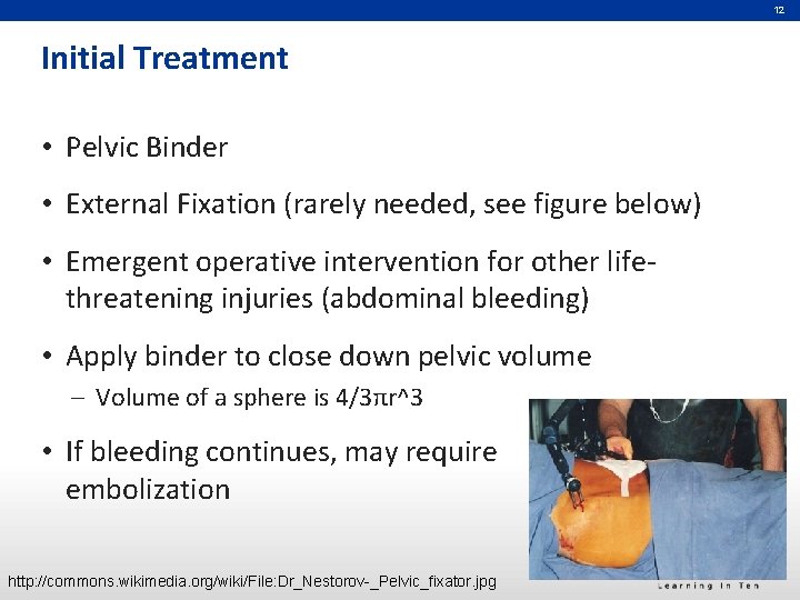
12 Initial Treatment • Pelvic Binder • External Fixation (rarely needed, see figure below) • Emergent operative intervention for other lifethreatening injuries (abdominal bleeding) • Apply binder to close down pelvic volume – Volume of a sphere is 4/3πr^3 • If bleeding continues, may require embolization http: //commons. wikimedia. org/wiki/File: Dr_Nestorov-_Pelvic_fixator. jpg
13 Hemodynamic stability Average blood loss with an APC III type fracture = 14. 8 Units
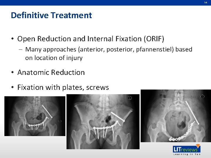
14 Definitive Treatment • Open Reduction and Internal Fixation (ORIF) – Many approaches (anterior, posterior, pfannenstiel) based on location of injury • Anatomic Reduction • Fixation with plates, screws
15 Post-operative management • Perioperative antibiotics • Early mobilization • Non-weight bearing • Anticoagulation – Sequential compression devices, TED hose, low molecular weight heparin (Lovenox®)
16 References • Hak DJ, Smith WR, Suzuki T. Management of hemorrhage in life-threatening pelvic fracture. JAAOS 2009 Jul; 17(7): 447 -57). • Sagi HC. Pelvic Ring Fractures. in Rockwood and Green’s Fractures in Adults. Bucholz et al (ed), 7 th ed. vol 2. pg 1415 -1462.
Partners in Academic Medicine