Pathology of Thyroid gland Hypo Hyperthyroidism and Hashimotos


 2.")

 Thyroiditis: • Subacute thyroiditis is believed to be")


 • • •")


- Slides: 22
Pathology of Thyroid gland Hypo, Hyperthyroidism and Hashimoto’s Thyroiditis Objectives: • Know the ways in which thyroid disorders present. • Know the major causes and manifestations of hypo, hyperthyroidism and thyroiditis. • Know the causes of the thyroid endemic goiter and its pathology. • Black: Doctors’ slides. • Red: Important! • Light Green: Doctors’ notes • Grey: Extra. • Italic black: New terminology.
Thyroid anatomy The thyroid gland consists of two bulky lateral lobes connected by a relatively thin isthmus, usually located below and anterior to the larynx. • � • The thyroid gland develops embryologically from an invagination of the developing pharyngeal epithelium that descends from the foramen cecum at the base of the tongue to its normal position in the anterior neck Histology
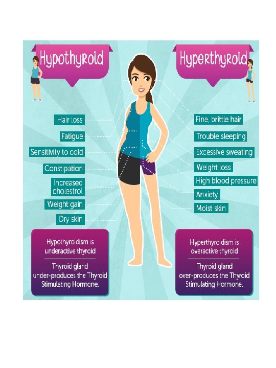
Thyroid Diseases Ø Ø Ø Clinical recognition of diseases of the thyroid is important, because most are amenable to medical or surgical management. Diseases include: Excessive release of thyroid hormones (hyperthyroidism). Thyroid hormone deficiency (hypothyroidism). Mass lesions. Hyperthyroidism : Ø Thyrotoxicosis 1 is a hypermetabolic state due to elevated circulating levels of free T 3 and T 4. Ø Because it is caused most commonly by hyperfunction of the thyroid gland, thyrotoxicosis often is referred to as hyperthyroidism. Ø In certain conditions, however, the oversupply either is related to excessive release of pre-formed thyroid hormone (e. g. , in thyroiditis) or comes from an extrathyroidal source, rather than a hyperfunctioning gland. The three most common causes of thyrotoxicosis: Primary Ø Ø Ø Diffuse hyperplasia of the thyroid associated with Graves disease (accounts for 85% of cases) Hyperfunctional multinodular goiter Hyperfunctional adenoma of the thyroid 1: Hyperthyrodisim is when a hyperfunctionig thyroid gland causes an increase in the blood levels of thyroid hormones , where as thyrotoxicosis is an increase in blood levels of thyroid hormones due to any other reason , ex: a teratoma that has a thyroid tissue in the ovaries (extra thyroidal cause).
Causes of Thyrotoxicosis Ø ASSOCIATED WITH HYPERTHYROIDISM q Primary • • • Diffuse toxic hyperplasia (Graves disease) Hyperfunctioning (“toxic”) multinodular goiter Hyperfunctioning (“toxic”) adenoma Iodine-induced hyperthyroidism Neonatal thyrotoxicosis associated with maternal Graves disease q Secondary TSH-secreting pituitary adenoma (rare) Ø NOT ASSOCIATED WITH HYPERTHYROIDISM • • Granulomatous (de Quervain) thyroiditis (painful) Subacute lymphocytic thyroiditis (painless) Struma ovarii (ovarian teratoma with ectopic thyroid) Factitious 2 thyrotoxicosis (exogenous thyroxine intake) • Clinical manifestation of thyrotoxicosis: v v v v Constitutional symptoms (weight loss, fever, sweating…) Gastrointestinal Cardiac Neuromuscular Ocular Thyroid storm (medical emergency) Apathetic 3 hyperthyroidism 2: Artificially created , from an exogenous source 3: No symptoms , silent.
Diagnosis of hyperthyroidism: • • The diagnosis of hyperthyroidism is based on clinical features and laboratory data. The measurement of serum TSH is the most useful single screening test for hyperthyroidism Free thyroid hormone assays Measurement of radioactive-iodine 4 uptake by the thyroid gland 4: Radioactive iodine uptake (RAIU) tests thyroid function. It measures how much radioactive iodine is taken up by your thyroid gland in a certain time period. Its increased in hyperthyrodisim.
Hypothyroidism • Hypothyroidism is caused by any structural or functional derangement that interferes with the production of adequate levels of thyroid hormone • Worldwide, the most common cause of hypothyroidism is dietary deficiency of iodine , while in most developed nations, autoimmune causes predominate Causes of hypothyroidism: Ø PRIMARY • Developmental (thyroid dysgenesis 5: PAX 8, FOXE 1, TSH receptor mutations) • • • Thyroid hormone resistance syndrome (THRB mutations) Postablative Surgery, radioiodine therapy, or external irradiation Autoimmune hypothyroidism (Hashimoto thyroiditis) Iodine deficiency[*] Drugs 6 (lithium, iodides, p-aminosalicylic acid) Congenital biosynthetic defect (dyshormonogenetic goiter) Ø Secondary (CENTRAL) • Pituitary failure • Hypothalamic failure (rare) 5: Embryological failure of development 6: They interfere with the de-Iodonation of T 4 to T 3
Clinical manifestations of hypothyroidism Cretinism refers to hypothyroidism developing in infancy or early childhood: Endemic cretinism, sporadic cretinism. Clinical features of cretinism include impaired development of the skeletal system and central nervous system, with severe mental retardation, short stature, coarse facial features, a protruding tongue, and umbilical hernia. • • Hypothyroidism developing in older children and adults results in a condition known as myxedema Manifestations of myxedema include : q generalized apathy and mental sluggishness that in the early stages of disease may mimic depression. q Cold intolerance, obesity. q Broadening and coarsening of facial features, enlargement of the tongue, and deepening of the voice. Constipation. Pericardial effusions are common q In later stages, the heart is enlarged, and heart failure may supervene. Laboratory evaluation • • • Measurement of serum TSH is the most sensitive screening test for this disorder The serum TSH is increased in primary hypothyroidism. Serum T 4 is decreased in patients with hypothyroidism of any origin.
Thyroiditis Clinically significant types of thyroiditis: 1. Hashimoto thyroiditis (or chronic lymphocytic thyroiditis) 2. Subacute: A. (granulomatous) thyroiditis, (de Quervain) B. Subacute Lymphocytic Thyroiditis 1 -Chronic Lymphocytic (Hashimoto) Thyroiditis : Ø Ø Ø Hashimoto thyroiditis is the most common cause of hypothyroidism in areas of the world where iodine levels are sufficient. it is characterized by gradual thyroid failure secondary to autoimmune destruction of the thyroid gland. It is most prevalent between the ages of 45 and 65 years and is more common in women than in men, with female predominance in a ratio of 10: 1 to 20: 1. the most common cause of hypothyroidism depends on iodine levels So if it’s insufficient, the cause is Dietary deficiency And if it’s sufficient, the cause is Autoimmunity (hashimoto thyroiditis) Ø A significant genetic component. It runs in families Ø Increased susceptibility to Hashimoto thyroiditis is associated with polymorphisms in multiple immune regulation–associated genes, the most significant of which is the linkage to cytotoxic T lymphocyte– associated antigen-4 gene (CTLA 4) Hashimoto Thyroiditis Pathogenesis: Ø Hashimoto thyroiditis is caused by a breakdown in self-tolerance to thyroid autoantigens. Thus, circulating autoantibodies against thyroid antigens are present in the vast majority of patients Multiple immunologic mechanisms that may contribute to thyrocyte damage: Ø CD 8+ cytotoxic T cell–mediated cell death (T killer) → thyrocyte destruction. Ø Cytokine-mediated cell death: Excessive T helper cell activation → production of inflammatory cytokines (interferon-γ) → recruitment and activation of macrophages and damage to follicles. Ø Binding of antithyroid antibodies (antithyroglobulin, and antithyroid peroxidase antibodies).
clinical features of Hashimoto Thyroiditis : Ø Ø Painless enlargement of the thyroid, usually associated with some degree of hypothyroidism. 7 It may be preceded by transient thyrotoxicosis caused by disruption of thyroid follicles, with secondary release of thyroid hormones (hashitoxicosis). As hypothyroidism supervenes, T 4 and T 3 levels progressively fall, accompanied by a compensatory increase in TSH. Patients with Hashimoto thyroiditis often have other autoimmune diseases and are at increased risk for the development of B cell non. Hodgkin lymphomas. Hashimoto Thyroiditis Morphology : • • Diffuse and symmetrical enlargement The cut surface is pale and gray-tan in appearance, and the tissue is firm and somewhat friable. Under the microscope : 1. infiltration of the parenchyma by a mononuclear inflammatory infiltrate containing small lymphocytes, plasma cells, and well-developed germinal centers. 2. The thyroid follicles are atrophic and are lined in many areas by epithelial cells distinguished by the presence of abundant eosinophilic, granular cytoplasm, termed Hürthle, or oxyphil, cells. 3. Interstitial connective tissue is increased and may be abundant. Less commonly, the thyroid is small and atrophic as a result of more extensive fibrosis (fibrosing variant) Unlike Reidel thyroiditis, the fibrosis does not extend beyond the capsule of the gland. 7: the transient hyperthyroidism is because of the damage of the follicular cells by the antibodies, so it releases the reservoir in it , which causes that increase. When the reservoir finishes and the follicular cells are already destroyed then the blood levels of hormones will drop.
2 -Granulomatous Subacute (Quervain de) Thyroiditis: • Subacute thyroiditis is believed to be caused by a viral infection or an inflammatory process triggered by viral infections. • A majority of patients have a history of an upper respiratory infection just before the onset of thyroiditis. • The onset of this form of thyroiditis often is acute, characterized by pain in the neck (particularly with swallowing), fever, malaise, and variable enlargement of the thyroid. • Transient hyperthyroidism may occur, as in other cases of thyroiditis, as a result of disruption of thyroid follicles. • The leukocyte count and erythrocyte sedimentation rates are increased. With progression of disease and gland destruction, a transient hypothyroid phase may ensue. • The condition typically is self-limited, with most patients returning to a euthyroid state within 6 to 8 weeks. Morphology • The gland is firm, with an intact capsule, and may be unilaterally or bilaterally enlarged. • Histologic examination reveals disruption of thyroid follicles, with extravasation of colloid leading to a polymorphonuclear infiltrate, which is replaced over time by lymphocytes, plasma cells, and macrophages. • The extravasated colloid provokes an exuberant granulomatous reaction with giant cells. Healing occurs by resolution of inflammation and fibrosis. •
Female slides 3 -Subacute lymphocytic thyroiditis • • • In a subset of patients the onset of disease follows pregnancy (postpartum thyroiditis). This disease is most likely to be autoimmune in etiology. Painless neck mass or features of thyroid hormone excess. The histologic features consist of lymphocytic infiltration and hyperplastic germinal centers within the thyroid parenchyma. From Robbins basic pathology: • This disease is though to be autoimmune in etiology, Because circulating anti thyroid antibodies are found in a majority of patients • The initial phase of thyrotoxicosis(which is likely to be secondary to thyroid tissue damage) is followed by euthyroid* state within few months. • In a minority of affected persons the condition eventually progress to hypothyroidism. Except for possible mild symmetric enlargement the thyroid appears normal on gross inspection. Euthyroid means having a normally functioning thyroid gland
Riedel thyroiditis • • • Rare disorder of unknown etiology, is characterized by extensive fibrosis involving the thyroid and contiguous neck structures. It spreads Clinical evaluation demonstrates a hard and fixed thyroid mass, simulating a thyroid neoplasm. From Robbins(it may be associated with idiopathic fibrosis in other sites of the body such as retroperitoneum ). Graves disease is the most common cause of endogenous hyperthyroidism. It is characterized by a triad of manifestations: 1 -Thyrotoxicosis, caused by a diffusely enlarged, hyperfunctional thyroid, is present in all cases. 2 -An infiltrative ophthalmopathy with resultant exophthalmos is noted in as many as 40% of patients. 3 -A localized, infiltrative dermopathy (sometimes designated pretibial myxedema) is seen in a minority of cases. • Graves disease has a peak incidence between the ages of 20 and 40, with women being affected up to seven times more commonly than men. • Genetic factors are important in the causation of Graves disease Pathogenesis • Graves disease is characterized by a breakdown in self-tolerance to thyroid autoantigens, of which the most important is the TSH receptor. The result is the production of multiple autoantibodies, including : • Thyroid-stimulating immunoglobulin: An Ig. G antibody that binds to the TSH receptor and mimics the action of TSH, relatively specific for Graves disease. • Thyroid growth-stimulating immunoglobulins: Also directed against the TSH receptor.
• TSH-binding inhibitor immunoglobulins: These anti-TSH receptor antibodies prevent TSH from binding to its receptor on thyroid epithelial cells and in so doing may actually inhibit thyroid cell function. • The coexistence of stimulating and inhibiting immunoglobulins in the serum of the same patient is not unusual—a finding that may explain why some patients with Graves disease spontaneously develop episodes of hypothyroidism. In Graves ophthalmopathy pathogenesis; a result of several causes Ø marked infiltration of the retroorbital space by mononuclear cells Ø inflammatory edema and swelling of extraocular muscles Ø accumulation of extracellular matrix components Ø increased numbers of adipocytes morphology • The thyroid gland is enlarged (usually symmetrically) due to diffuse hypertrophy and hyperplasia of thyroid follicular epithelial cells. The gland is usually smooth and soft, and its capsule is intact. • On microscopic examination, the follicular epithelial cells in untreated cases are tall, columnar, and more crowded than usual. This crowding often results in the formation of small papillae. so papillary thyroid carcinoma could be a deffrential and we have to exclude it. Such papillae lack fibrovascular cores, in contrast with those of papillary carcinoma. • The colloid within the follicular lumen is pale, with scalloped margins. • Lymphoid infiltrates, are present throughout the interstitium; germinal centers are common.
Microscopic views • Laboratory findings in Graves disease include elevated serum free T 4 and T 3 and depressed serum TSH. • Because of ongoing stimulation of the thyroid follicles by TSIs, radioactive iodine uptake is increased, and radioiodine scans show a diffuse uptake of iodine DIFFUSE AND MULTINODULAR GOITER • Goiter: Enlargement of the thyroid, is the most common manifestation of thyroid disease. • They reflect impaired synthesis of thyroid hormone, most often caused by dietary iodine deficiency • Pathogenesis of goiter : Impairment of thyroid hormone synthesis leads to a compensatory rise in the serum TSH, causes hypertrophy and hyperplasia of thyroid follicular cells and, ultimately, gross enlargement of the thyroid gland. • The compensatory increase in functional mass of the gland is enough to overcome the hormone deficiency, ensuring a euthyroid metabolic state in the vast majority of affected persons. • If the underlying disorder is sufficiently the compensatory responses may be inadequate to overcome the impairment in hormone synthesis, resulting in goitrous hypothyroidism.
• Goiters can be endemic or sporadic. • diffuse, symmetric enlargement of the gland (diffuse goiter): The follicles are lined by crowded columnar cells, which may pile up and form projections • colloid-rich gland (colloid goiter) • All long-standing diffuse goiters convert into multinodular goiters. • Multinodular goiters typically are hormonally silent, although a minority (approximately 10% over 10 years) can manifest with thyrotoxicosis secondary to the development of autonomous nodules that produce thyroid hormone independent of TSH stimulation. This condition, known as toxic multinodular goiter or Plummer syndrome. Goiter can arise in the following settings: 1. Endemic goiter due to iodine deficiency 2. ingestion of certain food e. g. cabbage 3. Rare inherited defect in thyroid hormone synthesis 4. Drug induced Multinodular goiter • Multinodular goiters are multilobulate, asymmetrically enlarged glands, which may attain massive size. • On cut surface, irregular nodules containing variable amounts of brown, gelatinous colloid are evident. Older lesions often show areas of fibrosis, hemorrhage, calcification, and cystic change.
• The microscopic appearance includes colloid-rich follicles lined by flattened, inactive epithelium and areas of follicular epithelial hypertrophy and hyperplasia Clinical features • The dominant clinical features of goiter are those caused by the mass effects of the enlarged gland. • In addition to the obvious cosmetic problem of a large neck mass, goiters also may cause airway obstruction, dysphagia, and compression of large vessels in the neck • a hyperfunctioning (toxic) nodule may develop within a long-standing goiter, resulting in hyperthyroidism. • The incidence of malignancy in long-standing multinodular goiters is low (less than 5%) but not zero, and concern for malignancy arises with goiters that demonstrate sudden changes in size or associated symptoms (e. g. , hoarseness).
Robbins Basic Pathology notes (these are the extra information from Robbins) • • • Clinical manifestations of thyrotoxicosis are duo to two main reasons hypermetabolic state and over activity of sympathetic nervous system. In Hyperthyroidism there is weight loss despite increased appetite Occular manifestation in hyperthyroidism : Staring gaze and lid lag are duo to over stimulation of the levator palpebrae superioris by the sympathatic system The difference in endemic and sporadic critinism is that in endemic cretinism is duo to areas that lack iodine example mountainous areas such as the Himalayas and the Andes. While sporadic is duo to enzyme defects that interfere with the thyroid hormone synthesis In hypothyroidism mucopolysaccharides rich edematous fluid accumulates in the skin , subcutaneous tissue and a number of visceral sites. Multiple autoimmune mechanisms account for Hashimoto disease. Cytotoxicity mediated by CD-8 T cells , cytokines : IFN-gamma, antithyroid antibodies. In Graves disease a T cell-mediated autoimmune phenomenon is in involved in the development of the infiltrative ophthalmopathy. Clinical features of graves disease: diffuse hyperplasia of the thyroid , ophthalmopathy, dermopathy. Increase flow of blood through the hyperactive gland often produces an audible bruit. Endemic goiter: occurs in geographic areas where the soil water and food contain little iodine. Sporadic goiter: occurs less commonly than endemic goiter, the condition is more common in females than males , with a peak incidence in puberty or young adulthood, when there is an increased physiological demand for T 4 Goiter: may result from hereditary enzymatic defects that interfere with thyroid hormone synthesis dyshormonogenetic goiter. In goiter airway obstruction dysphagia and compression of large vessels in the neck and upper thorax is referred as superior vena cava syndrome.
Summary
Questions 1. Which of the following is false regarding Thyrotoxicosis? A. Hypermetabolic state. B. Commonly caused by excessive release of pre-formed thyroxin. C. Elevated Thyroxin levels. D. Not always related with Exophthalmos. ANS: B 4. Plummer syndrome refer to which of the following? A. Autonomous thyroxine releasing nodules Independent of TSH. B. Colloid rich glands. C. Diffuse, symmetric enlargement of the gland. D. Silent Multinodular goiter. 2. Which of the following is the most useful screening test for hyperthyroidism? A. Serum TSH. B. Serum T 4 and T 3. C. Serum Free T 3. D. Measurement of radioactive iodine uptake. ANS: A 3. Exophthalmos and pretibial myxedema are likely to be found in which of the following? A. Thyrotoxicosis. B. Hypothyroidism. C. Hashimoto’s disease. D. Graves’ disease. ANS: D ANS: A
5. A 43 -year-old woman living in the US has had increasing lethargy and weakness over the past 3 years. She has cold intolerance and wears a sweater in the summer. She has difficulty concentrating, and her memory is poor. She has chronic constipation. She has alopecia (Hair loss affecting scalp), and her skin appears coarse and dry. Her face, hands, and feet appear puffy, with doughlike skin. Which of the following serologic test findings is most likely to be True in this woman? A. Elevated T 4. B. Low TSH. C. Positive Anti-thyroid peroxidase antibodies. D. Negative CTLA 4. ANS: C 6. A 2 -year-old child has failure to thrive since infancy. Physical examination shows that the child is short and has coarse facial features, a protruding tongue, and an umbilical hernia. As the child matures, profound intellectual disability becomes apparent. A deficiency of which of the following hormones is most likely to explain these findings? A. Growth hormone. B. Thyroxine. C. Somatostatin. D. Glycogen. ANS: B 7. Which of the following is true about Hashimoto’s Thyroiditis? A. Painful enlargement. B. Always preceded by transient thyrotoxicosis. C. Decrease in TSH. D. Associated with other autoimmune diseases ANS: D 8. Toxic diffuse goiter refers to which of the following? A. Subacute thyroiditis. B. Hashimoto’s disease. C. Graves’ disease. D. Iodine deficiency. ANS: C, Hashimoto’s is called chronic lymphocytic thyroiditis. 9. . Subacute granulomatous thyroiditis patients progress to an euthyroid state in 6 -8 weeks. What does that mean? A. The patient is in a fulminant stage. B. The condition is self-limiting. C. Sky-high elevation of Thyroid hormones. D. No thyroid hormone at all. ANS: B