Neurosonography Part Two Harry H Holdorf Frontal view


- Slides: 37

Neurosonography Part Two Harry H. Holdorf

Frontal view showing: Medial Longitudinal Fissure, Pons, Medulla Oblongata, and Cerebellum.

Corpus collasum p Two hemispheres are connected by mass of white matter called Corpus Callosum: n n n Corpus callosum is a bundle of commissural fibers Allows communications between hemispheres 10 cm long Anterior end = Genu Posterior portion= Splenium

Corpus Collasum

Corpus Collasum

Corpus Collasum

Cerebrum cont. color page 73 p Cerebrum is devided into five lobes: n n n Frontal- Center for motor and mental response, emotion and personality Parietal- Center for Sensory information; pain Temporal- Center for hearing, smell, memory and speech Occipital- Vision center Insula

Cerebral regions, Insula not exposed

Cerebral regions, Insula exposed

Cerebrum cont. Anteriorly Central Sulcus ( Sulcus of Rolando) seperates frontal and parietal lobes. p Posterorly Parieto-Occipital Sulcus seperates Parietal lfrom occipital p Laterally Sylvian Fissure (Lateral Fissure) seperates the temporal lobe from the parietal lobe p Located deep within the sylvian fissuer is the fifth lobe or Insula or Island of Reil. p

Cerebral sulcuses

Basal Ganglia, Color page 74 Regions of grey matter in the cerebrum. p Largest basal ganglias are; p n Caudate Nucleus Lateral to the LV p Has head, body and tail p Tapers from anterior to posterior p n Lentiform or Lenticular Nucleus Centerally located in each hemisphere p Divided into: p § Putamen- Located laterally § Globus Pallidus- Located medially

Caudate Nucleus p Head: n n n p Body: n n p Largest Most anterior Lies in concavity of the lat. Surface of the frontal horn of LVs Division of the head and body occurs @ the level of the Foramen of Monro Occupies the superior concavity of the lat. Wall of the body of the LVs Tail: n n In the roof of the Temporal horn of the LVs Rarely imaged in US or CT

Basal Ganglia

Basal Ganglia

The blue parts are the ventricular system, while the green part lining the lateral parts of the ventricular system is the caudate nucleus.

Basal Ganglia

Basal Ganglia in the coronal view

Parasagital view of the Caudate nucleus

Diancephalon; Color page 75 Located deep within the cerebral hemispheres. p Consist of the : p n n n p Epithalamus Thalamus Hypothalamus Surrounds the midline third ventricle

Thalamus Largest mass of grey matter p Large egg shaped paired stuctures p Forms lateral walls of the third ventricle p Connected by the “Massa Intermedia” p Occupy the inferior concavity of the lat. surface of the LVs p Bounded: p n n n Anteriorly-Foramen of Monro, head of the Caudate N. Posterioly- Trigone of the LVs Inferiorly- hypothalamus

Thalamus

Thalamus

Thalamus

Thalamus

Thalamus

Thalamus

Epithalamus Forms roof of the third ventricle p Has a midline projection that forms the Pineal Gland. p

Epithalamus

Hypothalamus Forms the floor of the third ventricle. p Inferiorly lies : p n n n The infundibulum or pituitary stalk Optic Chiasma Mammillary bodies (responsible for swallowing)

Hypothalamus

Hypothalamus

Hypothalamus

Brain Stem

Lateral view of the CNS

Brain Stem, Color page 76 p Subdivided into : 1. Midbrain 2. Pons 3. Medulla Oblonga

Brain Stem
 The digestive system and body metabolism chapter 14
 Human alimentary canal
 Full frontal view
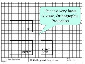
 Top view is directly above the front view
 Revolved section example
 Define a revolved section view?
 What are the section line symbols?
 Birds eye view vs worm's eye view
 What is end elevation
 Isometric orthographic drawing
 For the view create view instructor_info as
 Simple view and complex view
 Simple view and complex view
 Simple view and complex view
 Partial view
 Chest x ray lateral view positioning
 Supply chain cycle
 Components of operating systems
 Separatist view of ethics examples
 Multi-view drawing
 Front view top view
 Isometric drawing engineering
 For the view create view instructor_info as
 Selection tool in flash
 Part part whole addition
 Unit ratio definition
 Part part whole
 Technical description examples
 Parts of the back bar
 The part of a shadow surrounding the darkest part
 Part to part variation
 Convectional rainfall diagram
 Axial skeleton study guide
 Ejes corporales
 Frontal lobe stroke
 Surcos del lobulo frontal
 Cuerpo humano tronco
 Relief rainfall


