Necrotizing Fasciitis Clinical Features Systemic findings such as


, injection drug")


- Slides: 12
Necrotizing Fasciitis
Clinical Features • Systemic findings, such as fever, tachycardia, and hypotension • Typical signs and symptoms such as tense edema outside the involved skin, disproportionate pain, blisters/bullae, crepitus, and subcutaneous gas • Antibiotic therapy alone is associated with a mortality rate approaching 100 percent
Two Clinical Types • Type I - a mixed infection caused by aerobic and anaerobic bacteria and occurs most commonly after surgical procedures and in patients with DM and peripheral vascular disease. • Type II - a monomicrobial infection caused by group A streptococcus (GAS, Streptococcus pyogenes).
Type I • Cervical necrotizing fasciitis: "Ludwig's angina, " a rapidly expanding inflammation in the submandibular and sublingual spaces. • Fournier's gangrene: In the perineal area, penetration of the GI or urethral mucosa by enteric organisms can cause Fournier's gangrene, an aggressive infection.
Type II • Most cases were community-acquired but 20 % were nosocomial or acquired in a nursing home. • type II can occur in any age group and among patients who do not have complicated medical illnesses.
• Predisposing factors include a history of blunt trauma, varicella (chickenpox), injection drug use, a penetrating injury such as laceration, surgical procedures, childbirth, exposure to a "case, " burns, and perhaps NSAID.
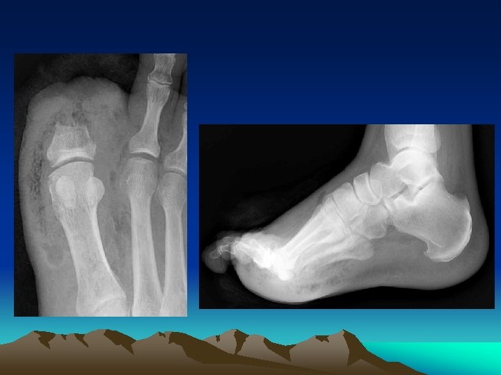
Diagnosis • Must be distinguished from gas gangrene, pyomyositis, and myositis. • Blood tests typically demonstrate a leukocytosis with a marked left shift, and elevations in the serum creatine kinase and creatinine concentrations. • Imaging - Soft tissue x-rays, CT scan and MRI
Treatment • Early and aggressive surgical exploration and debridement of necrotic tissue. The best indication for surgical intervention is severe pain, toxicity, fever and elevated CPK with or without radiographic findings. • Antibiotic therapy • Hemodynamic support as needed
Antibiotic therapy – Type I • Early empiric treatment could include ampicillin or ampicillin-sulbactam combined with either clindamycin or metronidazole. • G(-) coverage - substituting ticarcillinclavulanate or piperacillin-tazobactam for ampicillin-sulbactam or by adding a fluoroquinolone, an aminoglycoside, an extended spectrum cephalosporin, or a carbapenem.
Antibiotic therapy – Type II • penicillin G (4 million units IV every four hours in adults >60 kg in weight and with normal renal function) in combination with clindamycin
Mortality Rate • The mortality rates in different studies have included 21 % in type I necrotizing fasciitis , 30 and 34 % in type II necrotizing fasciitis in which streptococcal toxic shock syndrome is commonly associated with mortality in patients with GAS infection , 22 % in patients with cervical necrotizing fasciitis , and 22 to 40 % in those with Fournier's gangrene.