LOCOMOTION AND MOVEMENT Deleted portions Types of movement


- Slides: 18

LOCOMOTION AND MOVEMENT

Deleted portions • Types of movement • Skeletal System & its functions • joints • Disorders of muscular & skeletal system

MUSCLES • Properties of Muscle : • Excitability • Contractility • Extensibility • Elasticity

Types of Muscles : Skeletal muscles or striated muscles Closely associated with skeleton. They are striped appearance under the microscope and called Striated muscles. They are under voluntary control of nervous system, hence called voluntary muscles. These involved in locomotion and change of body postures. Unbranched and multinucleated.

Types of Muscle • Visceral muscles or smooth muscles • These are located in inner wall of hollow visceral organ. • Spindle shaped and uni-nucleated. • They do not exhibit any striation and are smooth in appearance. • They are called smooth muscles or nonstriated muscles. • Their activities are not under voluntary control of nervous system hence called as involuntary muscles. • They assist in transport of food through digestive tract and gametes through the genital tract.

Cardiac muscles Types of Muscles • The muscles of heart, involuntary in nature. • Cardiac muscle cells assemble in a branching pattern to form a cardiac muscle. • These are uni-nucleated with characteristic intercalated disc.

Structure of skeletal muscle : • Each organized skeletal muscle in our body is made of a number of muscle bundles called fascicles held together by common fibrous covering called fascia. • Each fascicle consists of a number of muscle fibres (cell) covered by a common fibrous perimysium. • Each muscle fibre is lined by the plasma membrane called sarcolemma, enclosing cytoplasm called sarcoplasm. • The sarcoplasm contain endoplasmic reticulum, called sarcoplasmic reticulum is the store house of calcium ion.

Structure of skeletal muscle :

• Muscle fibre is a syncitium as the sarcoplasm contain many nuclei. • Muscle fibres contain a large number of parallelly arranged filaments in the sarcoplasm called myofilaments or myofibrils. • There are two types of myofibrils are present in the sarcoplasm – • Thin filament – Actin • Thick filament – Myosin.

• The arrangement of thick and thin filament gives the characteristic striated appearance. • The light bands contain only actin filaments and are called I-band or isotropic band. • The dark band called ‘A’ or anisotropic band contains both actin and myosin.

• In the centre of each ‘I’ band is an elastic fibre called ‘Z’ line which bisects it. • The thin filaments or actin are firmly attached with the ‘Z’ line. • The thick filaments or myosin in the ‘A’ band are also held together in the middle by a thin fibrous membrane called ‘M’ line.

• The portion between two successive ‘Z’ lines is considered as the functional unit of the muscle called sarcomere. • Each ‘A’ band contains two overlap zone of thick and thin filament called ‘O’ band. • The central part of thick filament, not overlapped by thin filament is called ‘H’ band. • ‘A’ band = 2(O) + H.

Structure of Contractile proteins : • Thin filament or Actin : • Each actin filament is made of two ‘F’ actins helically wound to each other. • Each ‘F’ actin is made of polymer of monomeric ‘G’ (Globular) actin. • Each ‘F’ actin associated with another protein, tropomyosin also run throughout its length.

• Another complex protein, Troponin is distributed at regular intervals on the tropomyosin. • Each troponin has three component – • Troponin-C binds with calcium. • Troponin-M, binds with the tropomyosin. • Troponin T, masks the active site on the ‘G’ actin (thin filament) • • In the resting state a sub-unit of Troponin (Tn-T), masks the active binding sites on the thin filaments for myosin.

Thick filament • Each myosin (thick) filament is consists of many monomeric protein called Meromyosins. • Each meromyosin has two parts – • Heavy meromyosin (HMM) - A globular head with a short arm. • Light meromyosin (LMM) – a tail. • • The HMM component, i. e. the head and short arm projects outwards at regular distance and angle from each other from the surface of a polymerized myosin filament and is known as cross arm. • The globular head is an active ATPase enzyme and has binding sites for ATP and active sites for actin.

Mechanism of muscle contraction : • Mechanism of muscle contraction is explained by sliding filament theory which states that contraction of a muscle fibre takes place by the sliding of the thin filaments over the thick filaments. • Muscle contraction is initiated by a signal sent by the central nervous system via a motor neuron. • A motor neuron along with the muscle fibres connected to it constitutes a motor unit. • The junction between a motor neuron and the sarcolemma of the muscle fibre is called neuromuscular junction or motor-end plate. • Neurotransmitter releases here which generates an action potential in sarcolemma. • These causes release of Ca++ into sarcoplasm. • These Ca++ binds with troponin, thereby remove masking of active site. • Myosin head binds to exposed active site on actin to form a cross bridge, utilizing energy from ATP hydrolysis.

Mechanism of muscle contraction : • This pulls the actin filament towards the centre of ‘A’ band. • ‘Z’ lines also pulled inward thereby causing a shortening of sarcomere i. e. contraction. • ‘I’ band get reduced, whereas the ‘A’ band retain the length. • During relaxation, the cross bridge between the actin and myosin break. • Ca++pumped back to sarcoplasmic cisternae. • Actin filament slide out of ‘A’ band length of ‘I’ band increases. This returns the muscle to its original state. • Repeated muscle contraction causes accumulation of lactic acid, produced from anaerobic breakdown of glycogen leads to muscle fatigue. • Muscle contains red coloured oxygen storing pigment called myoglobin

Mechanism of muscle contraction : • Muscle with myoglobin called red muscle fibres, they are also contain large number of mitochondria which can utilize large amount of oxygen stored in them for ATP production also called aerobic muscle. • Some muscles possess very less quantity of myoglobin and less mitochondrion hence called white fibres. Amount of sarcoplasmic reticulum is high in these muscles. They depend on anaerobic process for energy
 Osteichthyes locomotion
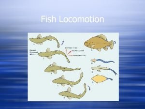
 Types of locomotion in fishes
 Explain miss maudie's cake portions
 Midsagittal plane
 Passion of the christ deleted scenes
 What is different about the deleted scene in the crucible
 Deleted objects container missing
 To kill a mockingbird page numbers
 Chapter 5 axial skeleton worksheet answers
 Chapter 5 the skeletal system figure 5-13
 Protection support and locomotion answer key
 Locomotion and support
 Locomotion and support
 Sporozoa locomotion
 Locomotion book summary
 Reptiles organs for locomotion
 Carl schissler
 Reptiles organs for locomotion
 Reptiles organs for locomotion
