Introduction a phenomenon termed crossmodal plasticity Sadato et


- Slides: 20
Introduction • a phenomenon termed cross-modal plasticity (Sadato et al. 1996; Cohen et al. 1997; Büchel et al. 1998; Bavelier and Neville 2002; Collignon et al. 2011; Watkins et al. 2013; Almeida et al. 2015). • arm amputees • the deaf individuals • the blind individuals • Even in such reorganization, the cortex typically retains elements of its original functions. • some “visual” cortex functions are preserved in congenital blindness (Striem-Amit, Cohen, et al. 2012; Striem-Amit, Dakwar, et al. 2012; Striem-Amit and Amedi 2014) • However, in congenital blindness, visual cortices take on entirely different functions from their typical role in visual perception. • nonvisual sensory responses • reflect higher-cognitive operations, such as language and mathematical processing (for review see Bedny 2017)
Introduction • However, in congenital blindness, visual cortices take on entirely different functions from their typical role in visual perception. • nonvisual sensory responses. • Recruitment of visual cortices for higher-cognitive functions is one of the • reflect higher-cognitive operations, such as language and mathematical mostprocessing extreme examples of cortical cognitive repurposing identified to date, (for review see Bedny 2017). since language and mathematics are cognitively and evolutionarily distant • Lateral occipital and ventral occipitotemporal regions from low-level vision. • the grammatical complexity of spoken sentences (Bedny et al. 2011; Lane et al. 2015). • Right middle occipital gyrus ( r. MOG) • The limits on such reorganization cortex? • the difficulty of mathcognitive equations (Kanjlia et al. 2016; Amalric et al. in 2017). the lifespan? sensitive periods of development? • TMS to the occipital cortex:reading Braille: generating semantically appropriate verbs to heard nouns. • congenitally blind × • sighted individuals √ • Even at rest, activity in visual cortices is synchronized with higher-cognitive frontoparietal networks in congenitally blind but not sighted individuals (Liu et al. 2007; Bedny et al. 2011; Watkins et al. 2012; Kanjlia et al. 2016).
Introduction • It is generally established that plasticity in the developing brain is enhanced relative to the mature brain. • monocular visual deprivation (Hubel and Wiesel 1970); • dense cataracts in one eye (Banks et al. 1975; Lewis and Maurer 2005). • the mouse model(Pizzorusso 2002; Hensch 2005 a, b; Bavelier et al. 2010) • Whether the capacity of cortex to take on novel cognitive functions similarly depends on sensitive period plasticity? • the functional plasticity observed in amputation——subtle; adult√ ? • functional repurposing of cortex——dramatic; circumscribed • auditory motion and spatial perception • congenital blindness vs adult-onset blindness (Haxby et al. 1991; Goodale and Milner 1992; Voss et al. 2006; Collignon et al. 2013).
Question • However, studies of higher-cognitive plasticity in the visual cortex of adult-onset blind individuals have thus far yielded mixed results. • V 1 responds more to sentences than nonverbal sounds only in those • Whether visual cortices sensitive to higher-cognitive who are congenitally blindare (Bedny et al. 2012). information in adult-onset blindness? • visual cortices appear to be active during higher-cognitive tasks, such as Braille reading, phonological judgments of spoken words and sentence comprehension, even in adult-onset blindness (Cohen et al. 1999; Burton et al. 2002; Burton and Mc. Laren 2006). • resting-state activity of visual cortices becomes synchronized with that of Broca’s area in adult-onset blindness, suggesting repurposing of visual cortices for language even in adulthood (Sabbah et al. 2016).
Question • Prior studies: • • comparing higher-cognitive tasks to a resting baseline or low-level perceptual control condition, making it difficult to determine what cognitive processes visual cortex activity truly reflects in the adult-onset blind population (Cohen et Whether such region-specific increases in functional al. 1999; Burton et al. 2002; Burton and Mc. Laren 2006). connectivity of visual cortex a sensitive and, if so, • The most compelling evidence for follow visual cortex involvementperiod in highercognitive functions in congenital blindness that whether this sensitive period alignscomes withfrom thatstudies of task-based manipulate fine-grained higher-cognitive information, such as the grammatical responses? complexity of sentences and difficulty of math equations (Röder et al. 2002; Bedny et al. 2011; Lane et al. 2015; Kanjlia et al. 2016). • A further open question concerns whether cognitive repurposing, as measured by task-based responses, follows a similar developmental time-course as changes in resting-state connectivity.
Question • Comparing task-based activation and resting-state functional connectivity across adult-onset blind (blind after 17 -years-of-age), congenitally blind and blindfolded sighted participants. • First, we asked whether visual cortices of adult-onset blind individuals show regional specialization for math as opposed to language processing and whether they show load dependent responses during higher-cognitive tasks—in particular, during symbolic mathematical reasoning. • Second, we tested whether adult-onset blind individuals, like the congenitally blind group, show higher resting-state functional connectivity between the r. MOG and the frontoparietal number network and higher functional connectivity between a language-responsive visual cortex area (ventral occipitotemporal cortex or VOT) and prefrontal language areas.
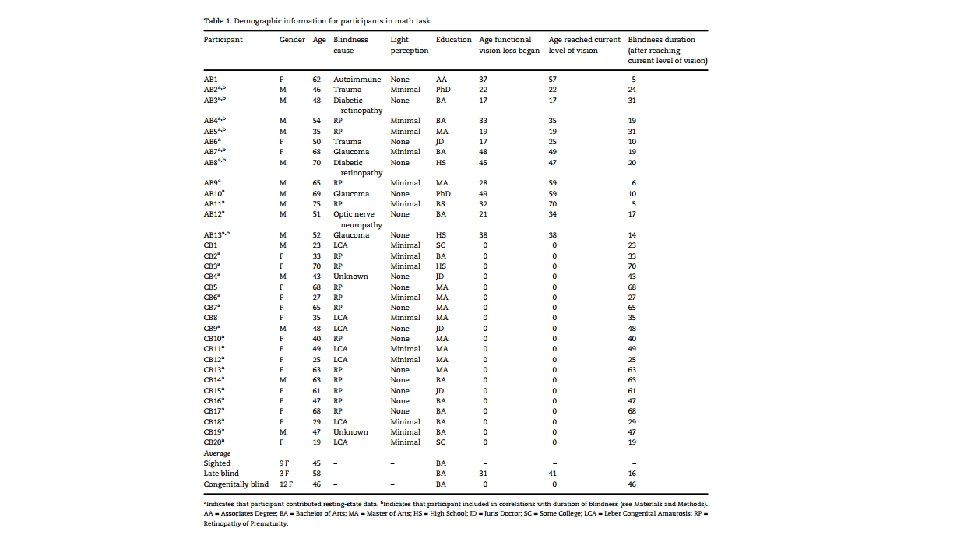
Materials and Methods • Task-based f. MRI experiment • 19 blindfolded sighted (age = 21. 45– 75. 49 years, mean = 45. 61, SD = 16. 03; 9 females) • 13 adult-onset blind (age = 34. 74– 74. 72, mean = 57. 18, SD = 11. 77; 3 females); blind for an average of 16. 11 years after reaching their current level of vision (SD = 8. 99, min = 4. 72, max = 31. 35) • 20 congenitally blind (age = 19. 34– 70. 12, mean age = 46. 08 years, SD = 16. 80; 15 females) • 7 additional participants were scanned but excluded(5 CB) • Resting-state • A total of 43 blindfolded sighted (mean age = 34. 12 years, SD = 14. 33, min = 18. 88, max = 63. 19; 25 females), • 12 adult-onset blind (mean age = 56. 79, SD = 12. 21, min = 34. 74, max = 74. 75; 2 females) • 25 (mean age = 46. 63, SD = 16. 91, min = 18. 81, max = 72. 98; 18 females) congenitally blind individuals
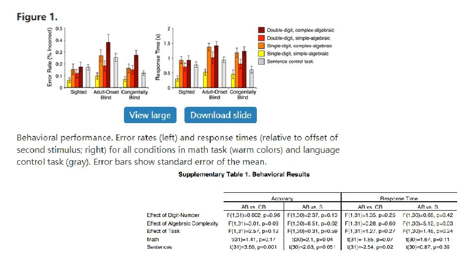
Behavioral Task • math trials • e. g. , 7− 2 = x 1( lasted 3. 5 s ) 2. 75 s delay • e. g. , 9− 6 = x 2( lasted 3. 5 s ) • Press button to identify if X 1=X 2 • 4 s to respond, respond at any point after the onset of the second math equation or sentence. e. g. , • 7− 2 = x (algebraically simple) • 27− 12 = x • x− 2 = 7 (algebraically complex) • x− 12 = 27 • 6 runs each with 24 trials (16 math trials and 8 language trials) • 96 unique math trials and 48 unique language trials in total
f. MRI Data Analysis • Task-based f. MRI data were analyzed using a standard general linear model (GLM). • Within each participant, each run was modeled separately and then combined using a fixed-effects model. Data across participants (within-group and between-group) were analyzed using a random-effects model. • Math-responsive regions of interest (ROIs) in the intraparietal sulcus (IPS)——within an anatomical IPS search-space——leave-one-run-out——the top 20 verticesthe greatest math > language effect (Destrieux et al. 2010). • We then looked for an effect of digit-number and algebraic complexity. And also tested for selectivity for math over language by comparing the math and sentence conditions. • Math-responsive regions of interest (ROIs) visual cortex——taking the r. MOG cluster that responded to the math > language contrast in CB > S (P < 0. 0001, uncorrected). • Each congenitally blind and sighted participant’s search-space was created based on functional data from the remaining subjects. • The adult-onset blind group used the same search-space. • Additionally, we looked at responses in V 1 because this is the first cortical stage of visual processing (Van Essen et al. 2012). • Paired t-tests were used to compare means within a group and unpaired t-tests were used when comparing means across groups. • Correlations with duration of blindness were conducted by including only adult-onset blind participants who lost their vision abruptly (within 2 years, n = 7; see Table 1)
Resting-State Functional Connectivity Analysis • ROI-to-ROI resting-state functional connectivity analyses were conducted in the right hemisphere, since task-based effects were right-lateralized. • We used data from all 13 adult-onset blind participants, the first 13 congenitally blind and first 13 sighted participants to define search-spaces. • Within these broad regions, math- and language-responsive prefrontal ROI’s were defined separately for each group (using all participants for that group) by taking the top 250 vertices with the greatest response to the math > language and language > math contrast, respectively. • Math-responsive IPS ROI’s were defined for each group by taking the top 250 vertices with the greatest math > language effect within anatomically defined IPS search-space (Destrieux et al. 2010). • Math- and language-responsive ROIs in the visual cortex could only be defined in the congenitally blind group and thus CB ROIs were used for all groups.
Similar Frontoparietal Responses in Adult-Onset Blind, Congenitally Blind and Sighted Groups • The IPS of adult-onset blind showed the same sensitivity to digit-number (hemisphere by digit-number by algebraic complexity repeated-measures ANOVA; • main effect of digit-number in AB group: F[1, 12] = 14. 38, P= 0. 003; • digit-number by group [AB vs. S] interaction: F[1, 30] = 0. 95, P = 0. 34; • digit-number by group [AB vs. CB] interaction: F[1, 31] = 0. 002, P = 0. 96). • The adult-onset blind group did not show an effect of algebraic complexity (AB group: F[1, 12] = 0. 20, P = 0. 66) in the IPS. • The effect of algebraic complexity was not different across adult-onset blind and congenitally blind groups; • slightly larger in the sighted group compared with the adult-onset blind group (algebraic complexity by group [AB vs. CB] interaction: F[1, 31] = 0. 84, P = 0. 37; algebraic complexity by group [AB vs. S] interaction: F[1, 30] = 3. 18, P = 0. 09).
Different Visual Cortex Sensitivity to Higher-Cognitive Functions in Congenitally Blind as Opposed to Adult-Onset Blind and Sighted Groups • In whole-cortex analyses, relative to the sighted, congenitally blind but not adult-onset blind participants‘ r. MOG was more active for math than language while the r. VOT and right lateral occipital cortex (r. LO) were more active for language than math (Fig. 2). • Although some visual cortex activity was observed in the within-group analysis of the adult-onset blind group, this activity was focused around the location of the so-called visual number-form area (VNFA).
Different Visual Cortex Sensitivity to Higher-Cognitive Functions in Congenitally Blind as Opposed to Adult-Onset Blind and Sighted Groups • In ROI analyses, overall response to all math and language conditions in r. MOG was greater in both congenitally and adult-onset blind groups compared with the sighted group (CB vs. S: t[37] = 6. 30, P < 0. 001; AB vs. S: t[30] = 4. 73, P < 0. 001; Fig. 3). • The r. MOG of the adult-onset blind group was not different from that of the sighted in its sensitivity to either math difficulty manipulation (digit-number by group interaction: F[1, 30] = 2. 88, P = 0. 10; algebraic complexity by group interaction: F[1, 30] = 0. 004, P = 0. 95). • • • In V 1, selectivity for mathematical stimuli over sentence stimuli • the congenitally blind > the adult-onset blind group • the sighted > the adult-onset blind group Notably, selectivity for math in the r. MOG and V 1 was not predicted by duration of blindness among adult-onset blind participants with abrupt vision loss (see Materials and Methods) or congenitally blind participants (i. e. , age) No correlation between blindness duration and the size of the math difficulty effect in either the r. MOG or V 1 of the adult-onset blind or congenitally blind groups
Functional Connectivity Between “Visual” Cortices and Frontoparietal Cortices in Adult-Onset Blindness • In congenital blindness, visual cortices become more correlated at rest with parietal and prefrontal cortices (Kanjlia et al. 2016). Math-responsive r. MOG and language-responsive r. VOT were more correlated with the r. IPS, r. DLPFC, and r. IFG in the congenitally blind group as opposed to sighted group • As previously reported, we found that increases in functional connectivity among congenitally blind individuals are network-specific. • Math-responsive r. MOG but not language responsive r. VOT shows elevated resting-state correlations with math-responsive r. IPS (seed [r. MOG vs. r. VOT] by group [CB vs. S] interaction: F[1, 65] = 5. 32, P = 0. 02). • Both for the congenitally blind and for the sighted, withinnetwork correlations (math visual cortex to math prefrontal cortex) are higher than between network correlations (math visual cortex to language prefrontal cortex) • Among the adult-onset blind group, resting-state functional connectivity of visual cortices show an intermediate pattern between that of the sighted and congenitally blind groups (Fig. 4). • Selectivity of functional connectivity across number and language networks in adult-onset blindness did not differ from either the congenitally blind or sighted groups. • Notably, among adult-onset blind individuals with abrupt vision loss (see Materials and Methods), resting-state functional connectivity between r. MOG and r. IPS but not r. PFC was significantly correlated with blindness duration
Discussion • Sensitive Period for Cognitive Repurposing in Visual Cortex • The capacity of cortex to take on novel cognitive functions narrows in adulthood. • congenital blindness vs adult-onset blindness • Differences in the functional profile of visual cortex cross the adultonset and congenitally blind groups do not appear to be related to the blindness duration. • Why might the recruitment of visual cortex for higher-cognitive functions be limited to a sensitive period during development? • Cognitive specialization of cortex requires circuit-internal structural changes that are uniquely possible during sensitive periods in development. • Alternatively, establishing one set of representations (e. g. , visual) could block cortex from representing other content (e. g. , number).
Discussion • Sensitive Period for Cognitive Repurposing in Visual Cortex • Future work should test the generalizability of the present findings to tactile tasks, such as Braille reading, and ask whether the capacity of visual cortex to specialize for specific cognitive operations declines gradually over childhood or abruptly after birth. • The visual cortex of adult-onset blind individuals may take on nonvisual cognitive functions that are different from those it takes on in congenital blindness. Adult cortex can repurpose but only within a narrow cognitive range. • Blindness in adulthood does, in fact, change the function of the visual cortex, but not in the same way or to the same degree as blindness at birth. • Exactly what defines the cognitive potential of cortex in adulthood and what distinguishes it from the cognitive range of developing cortex remains an open question.
Discussion • Functional Connectivity of Visual Cortices Changes, Even in Adult-Onset Blindness • resting-state correlations between visual cortices and the frontoparietal number network increase. • resting-state correlations between visual cortex and higher-cognitive networks are lower in those who are adult-onset as compared with congenitally blind (Bedny et al. 2010; Butt et al. 2013). • A dissociation between long-range resting-state connectivity and local functional properties. • The “visual” cortex of adult-onset and congenitally blind participants show similar resting-state functional connectivity yet different taskbased responses.