IN THE NAME OF GOD THE COMPASSIONATE THE


: chondrocytes show mitotic figure and")


- Slides: 35
IN THE NAME OF GOD THE COMPASSIONATE THE MERCIFUL
Histology Lecture 8 Bone Formation and Joints
2 Types of Bone Tissue 'Bone histogenesis may be of two types intramembranous or endochondral 'First bone that is formed by either of the two methods is the same and is called primary bone which is later replaced by secondary bone 'Immature, primary or woven bone where collagen bundles have random orientation 'Mature, secondary or lamellar bone where collagen bundles are oriented in bone lamellae
Primary Bone OFirst to be formed during embryonic period and during bone repair in adults OReplaced by secondary except near sutures in flat bones of skull, tooth sockets and insertion areas of some tendons OCollagen bundles are irregular and random in orientation ODecreased mineral content and more osteocytes per unit volume than secondary bone
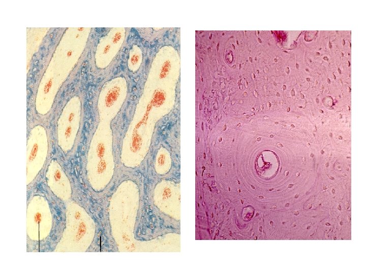
Secondary Bone ñSecondary bone is mature form of primary bone ñIt is composed of parallel or concentric lamellae ñOsteocytes are dispersed at regular intervals ñCanaliculi connect neighboring lacunae ñMatrix is more calcified than primary bone ñCollagen fibers are arranged so that they parallel each other within a given lamellae
2 Types of Bone Histogenesis ìIntramembranous ossification occurs in a layer of condensed mesenchymal connective tissue ìEndochondral ossification occurs within a cartilaginous template, cartilage precursor is gradually replaced by ossified tissue differentiating from the surrounding periosteum
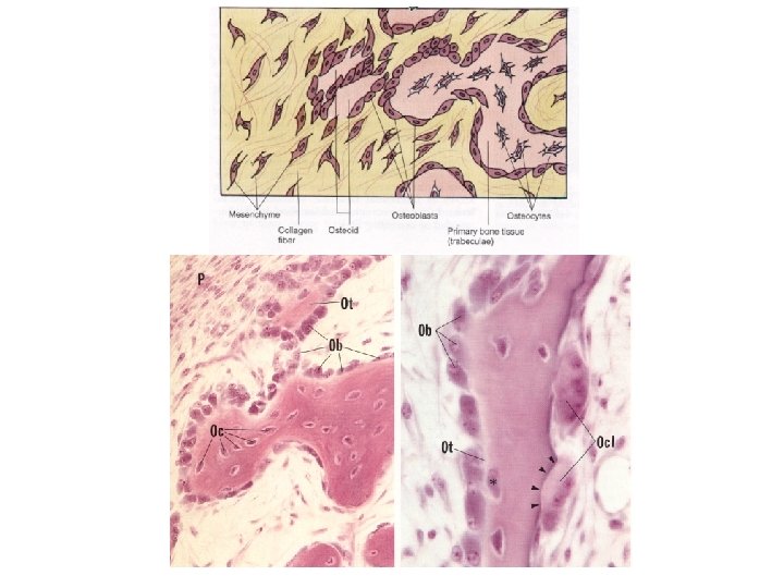
Intramembranous Ossification y. It is the source of most flattened bones such as skull, mandible and maxilla y. Intramembranous ossification starts with a mesenchymal connective tissue y. Primary ossification centers form when mesenchymal cells differentiate into osteoblasts around growing vessels y. Islands of developing bone are spicules and trabeculae of primary bone whose surface is lined by these cells y. Following fusion of spicules the mesenchymal connective tissue in their interstices transformed into bone marrow y. Uncalcified mesenchymal tissue on the surface of trabeculae differentiate into periosteum y. In skull, internal and external plates of compact bone are separated by a layer of spongy bone form in this way
Endochondral Ossification ö Endochondral Ossification Occurs in hyaline cartilage template ö It forms most of the long and short bones of the body ö Endochondral Ossification has 2 Phases • Formation of a hyaline cartilage template • Growing of cartilage followed by resorbtion and replacement by bone
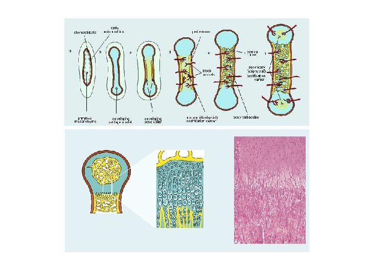
Primary Center Events YFormation and development of a hyaline cartilage model in the region where bone should be formed YIn the middle of cartilage chondrocytes accumulate glycogen, hypertrophied, and vacuolated YThe lacunae enlarge and intervening matrix reduced and calcified YPerichondrium at the middle of the diaphysis of cartilage model becomes vacularized and chondrogenic cells replaced by osteoprogenitor cells YOsteoprogenitor cells forming chondroblasts and perichondrium becomes periosteum YBy intramembranous ossification osteoblasts forming subperiosteal bone collar on the surface of the cartilage model
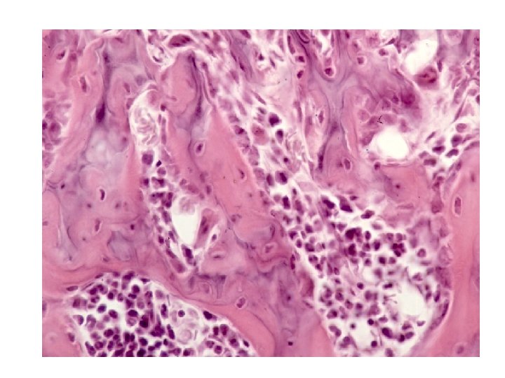
Primary Center Events ÔBone collar stops diffusion of nutrients to chondrocytes in cartilage template and causing them to die ÔThe lacunae becomes empty, confluents and forming large cavities ÔOsteoclasts makes holes in bone collar and permit osteogenic bud enter the cavities in cartilage model ÔOsteogenic bud is composed of osteoprogenitor cells, hemopoietic cells and blood vessels ÔOsteoblasts secrete bone matrix on the surface of calcified cartilage forming calcified cartilage – bone complex ÔCalcified cartilage is basophilic (blue) and new bone is acidophilic (pink)
Primary Center Events 0 Collar bone becomes thicker and grows in each direction toward epiphysis 0 As this happen osteoclasts resorbs the calcified cartilage bone complex enlarging bone marrow 0 In this way the cartilage of the diaphysis replaced by bone except for the epiphysial plate 0 Later secondary ossification centers form at each epiphysis
Secondary Centers Events è Secondary center forms epiphysis è The process of bone formation is identical to diaphysis except that a bone collar is not present è The cartilage of epiphysis replaced by bone except at the articular surface and epiphysial plate è As epiphyseal plate growth continues, matrix produced is processed into bone by expanding diaphyseal ossification center
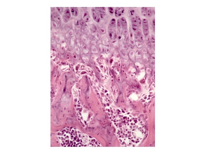
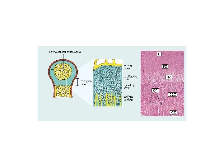
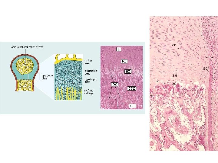
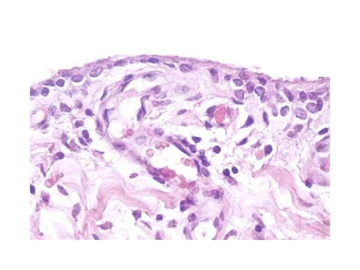
Zones of Epiphyseal Plate Û Resting zone (Reserve zone): chondrocytes show mitotic figure and randomly distributed in hyaline cartilage Û Proliferative zone: rapidly dividing chondrocytes form columns of stacked cells Û Hypertrophied cartilaginous zone: chondrocytes mature accumulate glycogen in cytoplasm, and hypertrophied Û Calcified cartilage zone: chondrocytes dying, deposition of hydroxy apatite in matrix Û Ossification zone: endochondral bone appears, vessels invade from periosteum, osteoblasts deposit calcified bone matrix on calcified cartilage followed by resorption of this complex
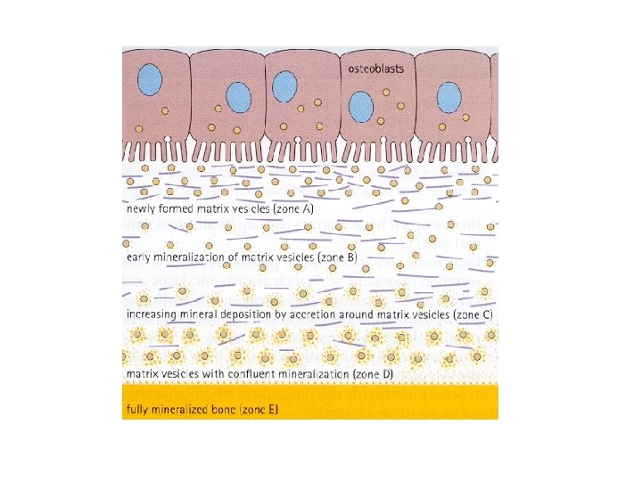
Mechanisms of Calcification v. Not completely understood v. Alkaline phosphatase causes accumulation of phosphate for precipitation of hydroxy apatite crystals v. Calcium binding glycoproteins secreted v. Vesicles in osteoblasts accumulate calcium and phosphate for calcification
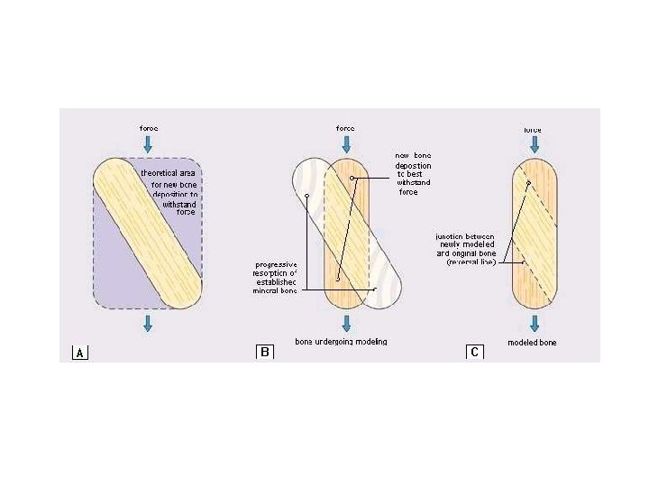
Remodeling and Growth of Bone * Resorption of bone matrix and deposition of new bone remain active throughout life * As bone matrix is added to outer bone surfaces marrow cavity enlarges leading to increase in bone diameter * Bone length increases due to epiphyseal plate growth * Bone diameter increases as periosteal cells differentiate into new osteoblasts and deposit new bone on the surface (Appositional growth)
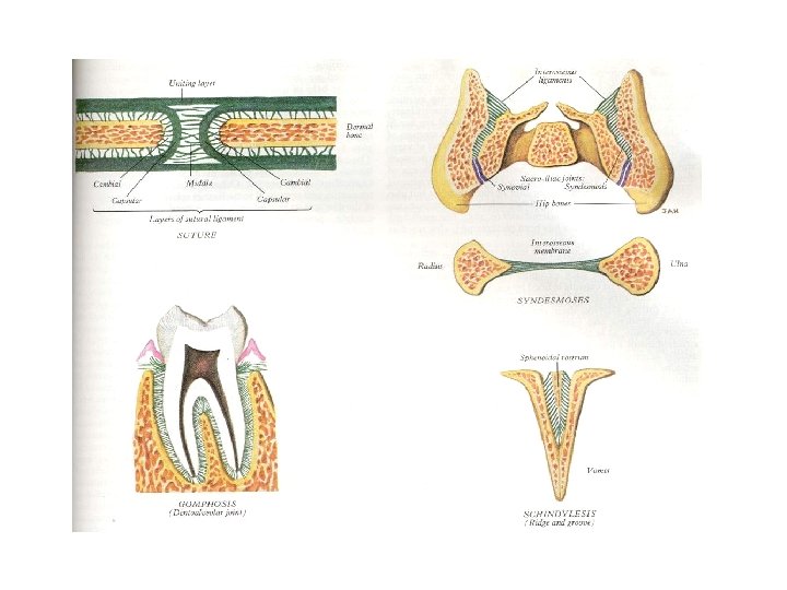
Joints Bones are connected to each other by joints Joints classification: - Permit limited movement - Permit free movement ome joints are formed by fibrocollagenous S (Syndesmoses in skull) or cartilaginous tissue (Synchondroses between ribs and sternum ) In old age will replace by bone (Synostosis )
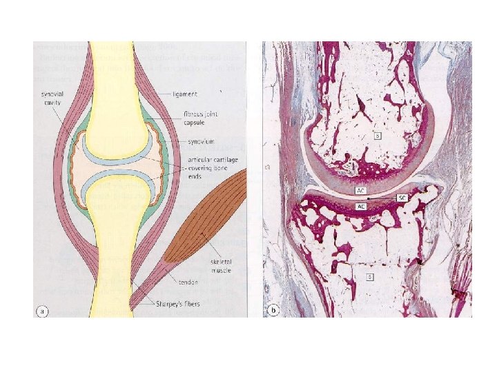
Synovial joints ¶Allow free movement ¶The bone held together by collagenous tissue (Joint capsule and Ligaments) ¶Bone ends are coated by a smooth, friction-free layer of hyaline cartilage (articular cartilage) ¶Joint capsule is composed of two layers an outer fibrous layer, and an inner cellular synovial layer ¶The internal lining of the joint capsule is a specialized secretory epithelium, the synovium
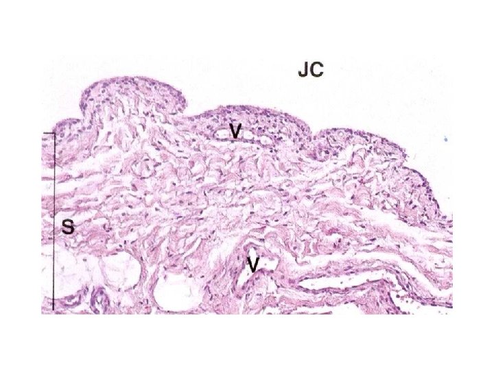
Synovial joints ØSynovium produce synovial fluid and composed of 1 -4 layers of cells ØCells vary from flat to cuboidal or polyhedral in shape ØTwo cell types defined: -Type A are phagocytic and have numerous lysosomes - Type B have abundant RER and produce protein ØThese layers of cells supported by a loose fibrocollagenous tissue containing adipocytes, mast cells, fibroblasts and macrophages
Synovial joints öThe synovial membrae has an abundant blood , lymph, and nerve supply running in the loose fibrocollagenous tissue öSynovial fluid contains of hyaluronic acid and glycoprotein(Lubricin) combined with a filtrate of plasma öThe synovial membrane supported by dense fibrocollagenous tissue ( Fibrous layer) öThe joint supported by some internal and external ligaments
SOMATIC CONNECTIVE TISSUES Articular cartilage Dermis Marrow Hypodermis Bone Joint capsule Ligament & Tendon Periosteum Muscle
Department of Histology F. Rajaei