Hymenolepis diminuta Disease Hymenolepiasis or Rat tapeworm infection


. Control:")
- Slides: 19
Hymenolepis diminuta Disease: Hymenolepiasis or Rat tapeworm infection Parasite of rats, mice & other rodents also it has been reported from human, usually children.
Epidemiology n World wide. n Common among children due to ingestion of infected grain beetles, dried fruits, flour and cereals. n Prevalence of H. diminuta in Philippine rats is about 8%.
Common parasite of rats and mice. ♣ Accidental human infections. ♣ Differs from Hymenolepis nana in morphology and life cycle because it requires an intermediate host. ♣ 2 Hosts: § Larval stage: cysticercoid is passed in fleas. § Adult stage: in rats and mice and accidentally in humans especially children who accidentally ingest infected ♣
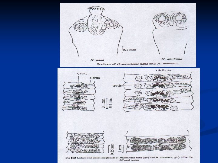
Morphology: v Adult larger than Hymenolepis nana. v Measure 20 -60 cm. X 3. 54. 0 mm. It contain 1000 or more proglottids. v Scolex is provided with 4 - small suckers and a rudimentary unarmed rostellum. v The neck is short and stout.
Scolex of Hymenolepis diminuta
v Mature segment: wider than long testes 3 globes mid-lateral genital pore. v Gravid proglottids usually disintegrate while still attached to the strobila, liberating fully embryonated ovoid to subspherical eggs.
MP of H. diminuta GP of H. diminuta segments of H. diminuta Note : just larger than H. nana
v Egg: there is a space between the tanned outer membrane and the inner one. The latter one is provided with a pair of polar thickenings, but lacks the polar filaments the characteristic of H. nana eggs.
Iodine stain Ova of H. diminuta, stool smear 201
Cysticercoid of Hymenolepis diminuta: This larva has no bladder, but instead has a tail. n The dark body within the spherical portion is the scolex. n The longer the tail, the more mature cysticercoid. n This stage is found inside an insect such as the mealworm. n
Life cycle: | Eggs of Hymenolepis diminuta are passed out in the feces of the infected definitive host (rodents & man). | The mature eggs are ingested by an intermediate host (various arthropod adults or larvae). | Oncospheres are released from the eggs and penetrate the intestinal wall of the host, which develop into cysticercoid larvae.
| The cysticercoid larvae persist through the arthropod's morphogenesis to adulthood. | H. diminuta infection is acquired by the mammalian host after ingestion of an intermediate host carrying the cysticercoid larvae. | Humans can be accidentally infected through the ingestion of insects in precooked cereals, or other food items, and directly from the environment (e. g. , oral exploration of the environment by children).
| After ingestion, the tissue of the infected arthropod is digested releasing the cysticercoid larvae in the stomach and small intestine. | Eversion of the scoleces occurs shortly after the cysticercoid larvae are released. | Using the four suckers on the scolex, the parasite attaches to the small intestine wall.
| Maturation of the parasites occurs within 20 days. | Eggs are released in the small intestine from gravid proglottids that disintegrate after breaking off from the adult worms. | The eggs are expelled to the environment in the mammalian host's feces.
Hymenolepis diminuta life cycle
Pathogenesis & Symptoms: H. diminuta infection usually produce no symptoms. Worm burden in rodents is relatively low.
Diagnosis: Stool examination Diagnostic stage: egg.
Treatment: Similar to H. nana infection. (Praziquantel 25 mg/kg body weight single dose). Control: Eradicate rats around the home.