GIT secretions hormones This include Digestive enzymes mouthileum
: based on location")


 a")


 • 5. water inside the oxyntic cell dissociates into hydrogen and hydroxyl")
 ,")
 stimulation of the peptic cells by acetylcholine released")


 cells in response to")


 long vagovagal")


")


- Slides: 53
GIT secretions & hormones • This include • Digestive enzymes (mouth-ileum): based on location and type of food • Mucus (mouth - anus)
Glands • They are the structures on the surface of the epithelium or invaginations of the epithelium that produce alimentary tract secretions • Examples are: mucous glands, crypts of lieberkuhn, tubular glands and complex glands (lying outside the wall of the tract) like salivary glands, pancreas, liver…. .
Structure of a glandular cell & gland
Functions of mucous secretions • They have adherent qualities • Prevent direct contact of food with the mucosa • Provides a low resistance for slippage • Causes fecal particles to adhere to one another • Its resistant to digestion by GIT enzymes • The glycoproteins of mucus have amphoteric or buffering properties
Stimulation of the glands • The glands are stimulated by: • Mechanical presence of food in a part of the GIT • Local epithelial stimulation that activates the ENS eg tactile stimulation, chemical irritation, gut distension…. . • Parasympathetic stimulation • Sympathetic stimulation has a dual effect • Regulation: majorly hormonal
Saliva secretion • Glands
Salivary secretions Glands parotid, submandibular, sublingual glands; small buccal glands. Secretion rate: 800 -1500 mls/day (ave=1000) • Ph = 6. 0 – 7. 0 • • •
• Composition • contains two major types of protein secretion • (1) a serous secretion that contains ptyalin (an a -amylase), which is an enzyme for digesting starches, and • (2) mucus secretion that contains mucin for lubricating and for surface protective purposes. • Parotid: majorly serous • Submandibular/sublingual: serous and mucus • Buccal: mucus
Mechanism for secretion of saliva
Mechanism • Salivary secretion is in two stages: • the first stage involves the acini, and • The second, the salivary ducts. • The acini secrete a primary secretion that contains ptyalin and/or mucin in a solution of ions in concentrations not greatly different from those of typical extracellular fluid. • As the primary secretion flows through the ducts, two major active transport processes take place that markedly • modify the ionic composition of the fluid in the saliva.
• First, sodium ions are actively reabsorbed from all the salivary ducts and potassium ions are actively secreted in exchange for the sodium. • Therefore, the sodium ion concentration of the saliva becomes greatly reduced, whereas the potassium ion concentration becomes increased. • However, there is excess sodium reabsorption over potassium secretion, and this creates electrical negativity of about -70 millivolts in the salivary ducts; this in turn causes chloride ions to be reabsorbed passively. • Therefore, the chloride ion concentration in the salivary fluid falls.
• Second, bicarbonate ions are secreted by the ductal epithelium into the lumen of the duct. • partly caused by passive exchange of bicarbonate for chloride ions, and • active secretory process. • Sodium conc (m. Eq/L) 145 -15 • Potassium conc (m. Eq/L) 4. 5 -30 • Bicarbonate ions (m. Eq/L) ? ? ? - 50 -70 • Increased rate of production: saliva rich in sodium is produced (copious) • Decreased rate of production: saliva rich in potassium is produced (sticky)
Functions of saliva • For oral hygiene • Wash away pathogenic bacteria and food particles • Destroy bacteria: proteolytic enzyme and thiocyanate
Regulation of salivary secretion • The salivary glands are controlled mainly by parasympathetic nervous signals all the way from the superior and inferior salivatory nuclei in the brain stem.
Secretion is stimulated by • Taste: acid • Tactile: smooth objects • Higher centres: appetite area close to anterior hypothalamus, amygdala and cerebral cortex • Gastric and small intestinal reflexes • Sympathetic stimulation: dual • Blood vasodilators: kallikrein and bradykinin
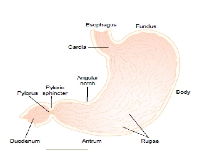
Gastric secretion • Glands • 1. oxyntic / gastric glands: HCl, pepsinogen, intrinsic factor and mucus • location: inside surfaces of the body and fundus of the stomach • 2. pyloric glands: mucus and gastrin • Location: antral portion and distal part of stomach
Oxyntic/ Gastric glands • • Is composed of three types of cells Mucus neck cells: mucus Peptic or chief cells: pepsinogen Parietal/oxyntic cells: HCl and intrisic factor
Mechanism of acid secretion by the oxyntic cells
Mechanism of acid secretion • 1. Chloride ion is actively transported from the cytoplasm of the parietal cell into the lumen of the canaliculus • 2. sodium ions are actively transported out of the canaliculus into the cytoplasm of the parietal cell • 3. a negative potential of -40 to -70 millivolts is created in the canaliculus • 4. the negative potential causes diffusion of small quantities of sodium and potassium ions back into the lumen
Mechanism (cont’d) • 5. water inside the oxyntic cell dissociates into hydrogen and hydroxyl ions • 6. hydrogen ions are actively secreted into the lumen in exchange for potasium ions using H+-K+ ATPase • 7. sodium ions are reabsorbed back into the ECF using Na+-K+ATPase. • 8. water moves downhill from the ECF through the cell to the lumen • 9. CO 2 in the cell combines with OH- in the presence of CA to form HCO 3 - which is reabsorbed back into the ECF • Final secretion is thus: HCl and water, conc= 150 to 160 m. Eq/L, KCl= 15 m. Eq/L
Secretion of pepsinogen by peptic cells • pepsinogen is secreted (no digestive activity) , • comes in contact with HCl • it is activated to form active pepsin. • functions • an active proteolytic enzyme • necessary for protein digestion in the stomach
Regulation of pepsinogen secretion • (1) stimulation of the peptic cells by acetylcholine released from the VN or from the gastric ENS • (2) stimulation of peptic cell secretion in response to acid in the stomach. Applied Physiology • In people who have lost the ability to secrete normal amounts of acid, secretion of pepsinogen isalso decreased, even though the peptic cells may otherwise appear to be normal.
Secretion of intrinsic factor by parietal cells • Intrinsic factor is essential for absorption of vitamin B in the ileum, • it is secreted by the parietal cells along with the secretion of HCl. 12 • Applied physio • Destruction of parietal cells caused by acute or chronic gastritis can cause: 1. achlorhydria (lack of stomach acid secretion) 2. pernicious anemia because of failure of maturation of the RBCs in the absence of vitamin B stimulation of the bone marrow 12
Secretion of mucus and gastrin • • The pyloric gland secretes mucus and gastrin Mucus Its produced by surface mucous cells Coats the stomach mucosa Protects the stomach wall Lubricates food for easy transport Alkaline in nature • Gastrin to be discussed later
Stimulation of Gastric Acid Secretion • the parietal cells operate in close association with enterochromaffin-like cells (ECL cells), the primary function of which is to secrete histamine. • The rate of secretion of HCl by the parietal cells is directly related to the amount of histamine secreted by the ECL cells. • ECL is stimulated/regulated by 1. 2. 3. Gastrin Acetylcholine Hormones from the ENS
Stimulation by gastrin • Gastrin is produced by Gastrin (G) cells in response to presence of meat or other proteincontaining foods in the stomach • Gastrin stimulates the ECL cells to produce histamine • Histamine stimulates the parietal cells to produce HCl
Phases of Gastric Secretion • a cephalic phase, • a gastric phase, and • an intestinal phase.
Cephalic phase • The cephalic phase occurs even before food enters the stomach • It results from the sight, smell, thought, or taste of food, and the greater the appetite, the more intense is the stimulation. • Neurogenic signals that cause the cephalic phase of gastric secretion originate in the cerebral cortex and in the appetite centers of the amygdala and hypothalamus. • They are transmitted through the dorsal motor nuclei of the vagi and thence through the vagus nerves to the stomach. • This phase of secretion normally accounts for about 20% of the gastric secretion associated with eating a meal.
Gastric phase • Once food enters the stomach, it excites • (1) long vagovagal reflexes from the stomach to the brain and back to the stomach, • (2) local enteric reflexes, and • (3) the gastrin mechanism • The gastric phase accounts for about 70% of the total gastric secretion associated with eating a meal
Intestinal phase • The presence of food in the upper portion of the small intestine, particularly in the duodenum inhibits gastric secretion by 1. Initiation of a reverse enterogastric reflex 2. Release of inhibitory hormones eg secretin, GIP, VIP and somatostatin in the presence of acid, fat, protein breakdown products, hyperosmotic or hypo-osmotic fluids, or any irritating factor in the upper small intestine 3. Reduction of gastric motility • purpose: to slow passage of chyme from the stomach when the small intestine is already filled or already overactive.
Pancreatic secretion
Phases of pancreatic secretion • Cephalic: vagal signals, 20% of total secretion • Gastric: vagal signals fire on, 5 -10%, chyme in stomach • Intestinal: chyme in the small intestine: 70 -75% caused by secretin
Pancreatic secretion • The pancreas is a large compound gland • Its pancreatic duct joins the hepatic duct immediately before it empties into the duodenum through the papilla of Vater, surrounded by the sphincter of Oddi. • Its acini produce digestive enzymes • The epithelial cells of the ductules secrete bicarbonate and water
Pancreatic secretion • Stimulated by: • Presence of chyme in the duodenum • Its islets of Langerhans secrete insulin directly into the blood • Total amount is 1 liter/day
Pancreatic digestive enzymes • These include: • Proteins: trypsin & chymotrypsin (proteins to peptides) , carboxypolypeptidase (peptides to amino acids) • CHO: pancreatic amylase (CHO, glycogen to tri n di saccharides) • Fat: pancreatic lipase (neutral fat to f. a. & monoglycerrides), cholesterol esterase, phospholipase
Activation of the enzymes • The enzymes become activated only in the intestine. • Trypsinogen is converted to trypsin in the presence of enterokinase • Chymotrypsinogen is activated to form chymotrypsin in the presence of trypsin • Note: trypsin inhibitor is formed in the cytoplasm of the glandular cells, and it prevents activation of trypsin both inside the secretory cells and in the acini and ducts of the pancreas. • trypsin inhibitor prevents activation of the others as well.
Applied physiology • Acute pancreatitis: occurs when the effect of trypsin inhibitor is overwhelmed eg when a duct is blocked • Thus the pancreatic secretions rapidly become activated and can literally digest the entire pancreas within a few hours,
Bicarbonate ion secretion • Are secreted by epithelial cells of the ductules and ducts of the pancreas along with water • Normal conc is 29 m. Eq/L (can rise to 145 m. Eq/L, to neutralize acidic chyme) • Steps involved: 3 2 1 4
Regulation of pancreatic • ACh: more enzymes, less water & electrolytes • CCK: secreted by I cells of the mucosa of duodenum and jejunum. more enzymes, less water & electrolytes • Secretin: secreted by S cells of the mucosa of duodenum and jejunum. Causes production of large water & bicarbonate, less enzyme all in response to acidic chyme. • An important protective mechanism to prevent the development of duodenal ulcer. HO • HCl +Na. HCO 3 Na. Cl +H 2 CO 3 2 CO 2
CCK • Secreted by the I cells of the mucosal of the duodenum and jejunum • Release is potentiated by presence of proteoses, peptones and long chain f. a in the chyme
Regulation
Bile secretion by the liver The liver produces 600 -1000 ml of bile per day. Functions of bile 1. fat digestion and absorption 2. means of excretion of waste products like bilirubin Secretion of bile: secreted in two stages 1. hepatocytes secrete large amounts of bile acids, cholesterol, other organic constituents into bile canaliculi • 2. bile flows in the system of ducts through the cystic duct into the gall bladder or empties directly into the duodenum: these ducts add Na+ & HCO 3 - to the secretion (secretin effect) • • •
Gallbladder: for storage and concentrating bile • water, sodium, chloride, and most other small electrolytes are continually absorbed through the gallbladder mucosa, concentrating the remaining bile constituents that contain the bile salts, cholesterol, lecithin, and bilirubin. • Most of this gallbladder absorption is caused by active transport of sodium through the gallbladder epithelium, and this is followed by secondary absorption of chloride ions, water, and most other diffusibleconstituents. • Bile is normally concentrated in this way about 5 -fold, but it can be concentrated up to a maximum of 20 -fold.
Composition of bile
CCK stimulates gallbladder emptying • Stimulus: food digestion in the stomach and presence of fatty foods in the duodenum, Ach & ENS • Rhythmical contraction of the bladder wall begins under the influence of CCK • Sphincter of Oddi relaxes • gallbladder empties its store of concentrated bile into the duodenum
Regulation
Role of bile salts in fat digestion and absorption • Emulsifying or detergent function: to decrease the surface tension of the particles • Help in the absorption of fatty acids, monoglycerrides, cholesterol and other lipids from the tract • They form physical complexes called micelles, through which the lipids are ferried into the blood
Applied physio: Gallstone • Bile salts are formed in the hepatic cells from cholesterol in the blood plasma. • Under abnormal conditions, the cholesterol may precipitate in the gallbladder causing gallstone formation • Other causes include ü Too much absorption of water from bile ü Too much absorption of bile acids from bile ü Too much cholesterol in bile ü Inflammation of the epithelium
Formation of gallstone
Small intestinal secretions • • A. Brunner’s glands secrete alkaline mucus in response to: (1) tactile or irritating stimuli on the duodenal mucosa; (2) vagal stimulation: increased; and SNS inhibits (3) gastrointestinal hormones, especially secretin. • Functions • to protect the duodenal wall from digestion by the highly acid gastric juice emptying from the stomach • Neutralizes the HCl entering the duodenum from the stomach.
Intestinal secretions contd • B. Crypts of Lieberkühn with villi: • contains goblet cells that secrete mucus and enterocytes which secrete water and electrolytes • Mainly to aid digestion and absorption
Large intestinal secretions Crypts of Lieberkühn without villi: The epithelial cells secrete only mucus Regulation Tactile stimulation Functions protects the intestinal wall against excoriation provides an adherent medium for holding fecal matter together • protects the intestinal wall from the great amount of bacterial activity that takes place inside the feces • provides a barrier to keep acids formed in the feces from attacking the intestinal wall. • Local reflexes: vagal stimulation through pelvic nerves increases mucus production • •