Genes and Genetic Conditions Genetic Terminology Inheritance terms


 contained in")


")


 Trisomy 18 (Edwards Syndrome)")


")


 Normal male genitalia but small testes Sparse body hair Some")


 to make melanin Cystic fibrosis")

 Recessive Diseases • Virtually all sex liked")


- Slides: 57
Genes and Genetic Conditions
Genetic Terminology Inheritance terms to know: Gene Locus Phenotype Homozygous Heterozygous Allele Genotype Carrier Expressivity Penetrance Dominant traits vs recessive traits
Genetics • In the 19 th century, scientists still only suspected that inheritance was contained in the nucleus of the cell. • Mid 1800’s, Gregor Mendel experiments describe the outward expression of heritable traits without yet knowing where in cells the information was contained. • Not until 1953 do we discover DNA and start to understand the real nature of chromosomes.
Genetics: The Central Dogma DNA RNA Protein Transcription & Translation DNA makes RNA makes Protein
Genetics: The Central Dogma DNA RNA Protein Transcription & Translation • DNA to RNA is called transcription • A friend asks to borrow your class notes but you only have a recording so they will have to transcribe them. • Same language, different format • RNA to Protein is called translation • Your German friend want to borrow your class notes. Since they are in English they will have to translate them. • Different language
Genetics: The Central Dogma DNA RNA Protein Transcription & Translation • Is the Central Dogma ever violated? • Does Protein ever make RNA? • Does RNA ever make DNA?
The Central Dogma - Exceptions DNA RNA Protein Transcription & Translation • Some viruses use RNA as their genetic material • These carry the code for an enzyme called reverse transcriptase that converts RNA to DNA • Because they work in a reverse process, they are called retroviruses. • When a retrovirus invades a cell, it inserts DNA into the host cell • When a retrovirus makes a foreign insertion like this, it is a destructive process for the host cell, damage to the host DNA is common and diseases such as cancer can be the result.
Genes and Genetic Diseases What is DNA? What is a Gene? What is a Chromosome?
Genes and Genetic Diseases What is DNA? What is a Gene? What is a Chromosome? DNA (deoxyribonucleic acid) 1. Double helix, strings of components which carry codes for proteins 2. Two nitrogenous base pairs between each sugar Adenine with Thymine Guanine with Cytosine 3. 4. Linked by weak hydrogen bonds Order of molecules: Sugar, phosphorus, sugar, phosphorus (bases in-between sugars) DNA is made up of thousands of segments called Genes. Specific Genes are located on Specific Chromosomes.
The Human Genome Project
DNA Replication • DNA strands separate • Two new DNA strands are constructed using the original strands as a template. • Same base-paring rules allow new strands to be made with exactness
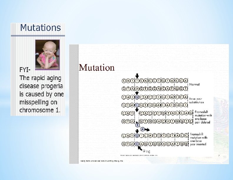
Mutation Alteration of order of the base pair • A change in the sequence of DNA will alter the m. RNA thus changing what is manufactured. This is called a gene polymorphism. • Most polymorphisms are silent, that is they do not change the protein sequence. • Some polymorphisms cause noticeable changes in the organism, these are called mutations.
Mutagens Agents known to increase the frequency of mutations • • Ionizing Radiation Chemical agents UV Radiation Viruses
Somatic Gene therapy using an Adenovirus vector. A new gene is inserted into an adenovirus vector, which is used to introduce the modified DNA into a human cell. If the treatment is successful, the new gene will make a functional protein
Transgenic fluorescent green pigs in Taipei January 13, 2006. Taiwan, home to the world's first transgenic glowing fish, has success
Two Kinds of Human Cells There are two basic types of human cells • Somatic Cells are almost all of 10 trillion cells that make up our body • They are diploid cells • • • 23 pairs of chromosomes for a total of 46 chromosomes Formed by mitosis Gametes • • Specialized reproductive cells • Sperm and ovum • These are haploid cells (23 total chromosomes) • When a sperm and an ovum join they make a diploid cell Formed by the process of meiosis
Structure and Function of Chromosomes Twenty-three pairs • Autosomes (1 through 22) contained in all somatic cells – Each pair is identical (homologous to one another) • Sex chromosomes are 23 rd pair – In females, chromosomes are homologous – In males, they are non-homologous Characteristics of genes • Chromosomes contain all genes • 50, 000 to 100, 000 genes that encode proteins in humans, many of which are duplicated or seem to have to no function.
Chromosomes are long strands of genes in linear order • Each gene has a definite locus on each paired set • Alleles are genes for the same characteristic • Somatic genes are located on chromosomes 1 -22 • Karotype • Displayed set of 23 chromosomes arranged according ot size (largest to smallest) • Pair # 1 has more genetic material
Karyotype
Polyploidy • Cells that have a multiple of the normal number / set (23) chromosomes are euploid. • Is a haploid cell euploid? Yes 23 x 1 • Is a diploid cell euploid? Yes 23 x 2 • When a euploid cell has more than the diploid number, it is called a polyploid cell (they still contain a multiple of 23). • Triploid fetuses have 3 copies of each chromosome (69 total) • Tetraploid fetuses have 4 copies of each chromosome (92 total) • Neither triploid or tetraploid fetuses can survive
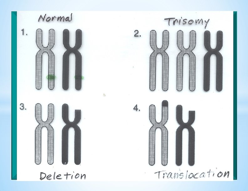
Aneuploidy • A Somatic cell that does not contain an exact multiple of 23 chromosomes is an aneuploid cell. • A cell containing 3 copies of one of the chromosome pairs is said to be trisomic (trisomy). • Monosomy is the presence of only one of any of the chromosome pairs. • Monosomy is more often lethal, but some infants can survive with trisomy of certain chromosomes. • “It is better to have extra than less. ”
Disjunction Normal separation of chromosomes during cell division. Non-disjunction One daughter cell receives both chromosomes and the other receives neither.
Non Disjunction Either in Meiosis I or Meiosis II Every cell in offspring will be affected (Three)
TRISOMIES Specific chromosomes are affected: The larger the chromosome, the greater the number of defects. Trisomy 9 – Incompatible with life – Often cyclops (one eye) Trisomy 13 (Pateau Syndrome) – Mental deficiency, deafness, cleft palate, cardiac anomalies - Incompatible with life Trisomy 18 ( Edwards Syndrome) – Rocker bottom feet – Low set ears – Multiple physical defects – Extremely profound mental retardation – Survivable with multiple surgeries and constant care
Trisomy 13 (Pateau Syndrome) Trisomy 18 (Edwards Syndrome)
Downs Syndrome Occurrence: < 1: 770 in U. S. Result of non-disjunction of 21 st Chromosome. 47 chromosomes instead of 46
Occurrence Downs correlated to maternal age 3/1000 if 20 y/o 100/1000 if 45 y/o 90 -95% is nondisjunction of mother’s egg
CLINICAL ASPECTS OF DOWN’S SYNDROME Physical Features: Short limbs and fingers Enlarged, protruding tongue Slanted eyes with skin fold over inner corners Flattened facial features and back of head Print ridges: – Ulnar loops fingertips – Tibial arch Single “simian” palmar crease Mentally challenged Moderate to severe (can usually get to 8 th grade) Leukemia (20 X)and heart defects common (50%)
MOSAICISM *Result of non-disjunction in the first or second mitotic division after fertilization (cleavage) *Some cells (about half) will be normal 46 chromosomes, others will be 47 (those of 45 number will die, but the 47 s will keep dividing) *Abnormalities less severe
Chromosome Structure Abnormalities • Not a change in chromosome number • Chromosome alterations at the gene level • Breakage • Deletions • Inversions • Duplications • Translocations
PROBLEMS OF DELETION Broken chromosomes and loss of DNA. Example: Cri du chat syndrome, deletion of part of 5 th chromosome: – “Cry of the cat” – Low birth weight – Severe mentally challenged – Microcephaly – Heart defects – Endocardial cushion defects most common in chromosomal abnormalities
From Jorde LB, Carey JC, Bamshad MJ: Medical genetics, ed 4, Philadelphia, 2010, Mosby.
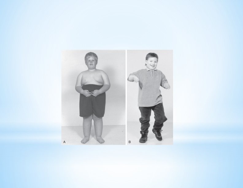
*Prader-Willi and Angelman syndromes * Deletion of 4 million base pairs of long arm of chromosome 15 * Manifestations of Prader-Willi syndrome if inherited from father * Short stature, hypotonia, small hands and feet, obesity, mentally challenged * Manifestations of Angelman syndrome if inherited from mother * Severely mentally challenged, seizures, ataxic gait * Indistinguishable at DNA sequence level
Sex Chromosome Aneuploidy Turner’s Syndrome Females with only one X chromosome Characteristics Absence of ovaries (sterile) Short stature (~ 4'7") Webbing of the neck Edema Underdeveloped breasts Widely spaced nipples High number of fetuses with a single X are aborted. The single X chromosome is usually inherited from mother
Klinefelter Syndrome (XXY, XXXXY) Normal male genitalia but small testes Sparse body hair Some female breast development Slight mentally challenged Male appearance Small testes Long limbs The abnormalities Will increase With each “extra” X chromosome
Genotype vs. Phenotype Heterozygous vs. Homozygous When for a single locus, more than two alternative alleles exist in the population Classic example - blood types Genes: O, A, B Genotypes – OO – AA – AO – BB – BO – AB Questions: What gene is recessive? What genes are codominant? Phenotypes O A A B B AB
Errors in Genetic Inheritance AUTOSOMAL DOMINANT INHERITANCE Allele that carries the gene that encodes for the disease will take precedence over the normal allele: A = Affected a = Normal Criteria: 50% chance for each pregnancy Unaffected persons do not transmit Not influenced by sex
EXAMPLES OF AUTOSOMAL DOMINANT Achondroplasia Skeletal disorder of short-limbed dwarfism and large head size, bulging forehead, and “scooped out” nose bridge Normal intelligence Marfan Syndrome Defect in elastic fibers of connective tissue Tall, thin, long tapering fingers, poorly developed musculature Defects: aortic aneurysm, dislocation of optic lens, winged scapula
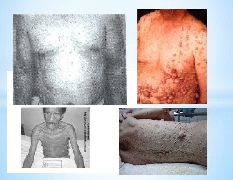
AUTOSOMAL DOMINANT - continued Neurofibromatosis Defect in neural cells Café-au-lait spots, multiple fibromas, neuro and opthalmologic defects, skeletal anomalies Osteogenesis imperfecta Defect in procollagen Fragile, short, bowed bones, blue sclera, hyperextensible joints, hearing loss – Note: Most autosomal dominant diseases affect the structure of the individual
Osteogenis Imperfecta
AUTOSOMAL RECESSIVE INHERITANCE Allele that carries the defective gene will not be expressed if other allele is normal Criteria Males and females affected equally Affected person’s parents will be unaffected (but carriers) 25% change of each pregnancy occurrence if both parents have normal gene Affected individuals mated to normal persons will have all normal but carrier children Often no evidence in family history of disease
AUTOSOMAL RECESSIVE DISORDERS Albinism No tyronase (tyrosine to dopa) to make melanin Cystic fibrosis Defect in pancreatic enzymes Galactosemia Galactose build-up in liver Maple syrup disease Defective metabolism of branched amino acids Phenylketonuria Defect in phenylalanine metabolism
Tay-Sachs disease Deficiency of hexosaminidase: progressive neurologic, cherry red spot in macula, early death (Ashkenazi Jews) Thalassemias Defect in synthesis of globin, severe anemia Wilson disease Liver failure due to copper build-up – Note: Most recessive disease affect metabolism or lack of protein synthesis for enzymes
X - LINKED INHERITANCE Sex Linked (X-linked) Recessive Diseases • Virtually all sex liked disorders are recessive • Affected males cannot transmit the genes to sons, but they can to all daughters, who will likely be carriers. • People often say these disorders “skip” a generation. • Sons of female carriers have a 50% risk of getting the disease. • These disorders are only expressed by males because females have another X chromosome to mask the abnormal gene. • Examples of this are hemophilia A and redgreen colorblindness.
X - LINKED INHERITANCE - continued Disorders of X - linked recessive Hemophilia A Rarely, can be X - linked dominant Rare incidence Expression in both sexes but with a greater incidence in females due to the greater number of X chromosomes.
Exercises in Types of Inheritance Using Pedigrees PEDIGREE 1
*Scenario A child is born with a Duchene muscular dystrophy (an x-linked recessive form of muscular dystrophy) a short time later, the mother is tested for the gene but she does not have the genetic anomaly. The child definitely has the gene. How can this happen? Is this really her baby? Was there a mix-up at birth? *
Germ Line Mosaicism Very early in fetal development, cells that will eventually become the future sperm or egg cells separate form the rest of the embryo. This is called the germ line As the fetus develops and continuing into childhood the germ line cells divide and multiply. Sperm cells don’t complete their development until the child becomes an adolescent. Eggs complete part of their development during fetal development and the other part at puberty. At any stage, from fetus through puberty or even later, mutations in the germ cells can occur. If the mutation occurs early, they affect many sperm or eggs. If they occur late, they affect very few sperm of eggs.
Germ Line Mosaicism cont. When a germ line mutation occurs it does not affect the other cells of the body because they were separated early in the fetal development. The condition of having some germ cells affected and some not, is germ line mosaicism, derived from the idea of a mosaic pattern. If a child is conceived with a sperm or egg carrying the mutation, they can inherit the disease, even though the DNA tests of the parents won’t show the mutation.
Multifactorial Inheritance Multifactorial inheritance is responsible for the greatest number of individuals that will need special care or hospitalization because of genetic diseases. • Atopic reactions, Diabetes, Cancer, Spina Bifida, pyloric stenosis, cleft lip, cleft palate, congenital hip dysplasia, club foot, and a host of other diseases all result from multifactorial inheritance. • Some of these diseases occur more frequently in males. Others occur more frequently in females.
Threshold Model • Qualitative traits are not measured on a numeric scale • Multifactorial traits/diseases like cardiovascular disease are best explained by the threshold model • …you either have them or you don’t • There is a qualitative threshold at which the disease manifests
Threshold Model • Each factor that contributes to the disease moves a person closer to the disease threshold • Factors that push you toward the threshold are: • Genetic • Environment
Threshold Model Example • Pyloric Stenosis – narrowing or obstruction of the pyloric sphincter • 1/200 white males • 1/1000 white females • Which group has the lower threshold?