Gametogenesis SOME TERMS Gamete egg or sperm Gametogenesis


. Spermatogenesis Means formation of sperm ;")


 • Contributes Milky Alkaline Fluid that assists Sperm Activation")


- Slides: 34
Gametogenesis
SOME TERMS Gamete: egg or sperm Gametogenesis: production of eggs or sperm Oogenesis: production of eggs Spermatogenesis: production of sperm Spermiogenesis: differentiation of sperm morphology Follicle: where eggs mature in the ovary Ovulation: release of egg from follicle Polar body: nonfunctional product of meiotic divisions in oogenesis Zygote: Fertilized egg
SOME MORE TERMS Oogonia: mitotically dividing cells in the ovary, will become Oocytes Primary oocyte: decision has been made to undergo meiosis, cell has grown. Cells are arrested at this stage until puberty. Secondary oocyte: has completed first meiotic division the division was unequal in terms of cytoplasm Ovum: Ovulated egg, ready to be fertilized. If fertilized, the second meitoic division will occur, another polar body will be given off.
Gametogenesis The creation of highly specialized sex cells through a process called MEIOSIS
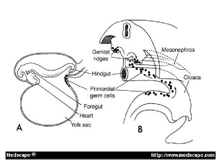
Primordial germ cells • Appears in the wall of the endodermal layer of the yolk sac due to their large size and high content of alkaline phosphatase , and migrate by amoeboid movement toward the hind gut epithelium and then through dorsal mesentery reach to the primordia of the gonads ( primitive sex glands). • Become recognizable at 24 days post-fertilization. • Invading the genital ridges in the 6 th week of development. • The primordia of the gonads give a chemostatic attractant for the germ cells.
• Also may be the germ cells directed toward the gonads by the mature of cellular and non-cellular microenvironment that surround them. • Or by tissue rearrangement that occur in the early embryos. • If they fail to reach the ridges, the gonads will not develop. • So, it have the inductive influence on the development of gonads into ovary and testes. • Determine sex of the embryo. • They will form different stages of spermatogenesis and oogenesis in the future. • Found in adult in gonads (testes and ovary)
MEIOSIS • The process where one diploid germ cells divides in order to create either 1 or 4 haploid cells ( 23 chromosomes).
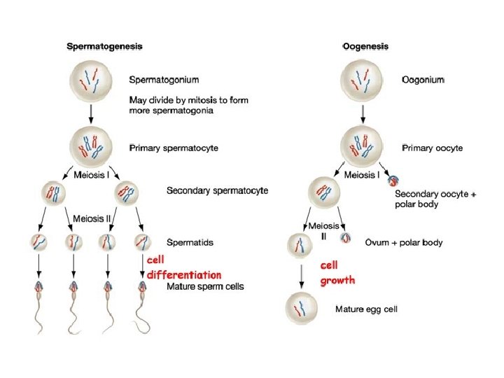
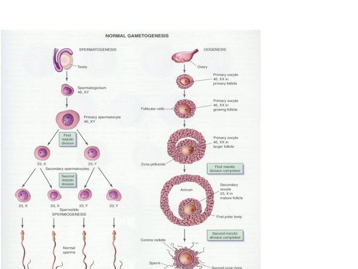
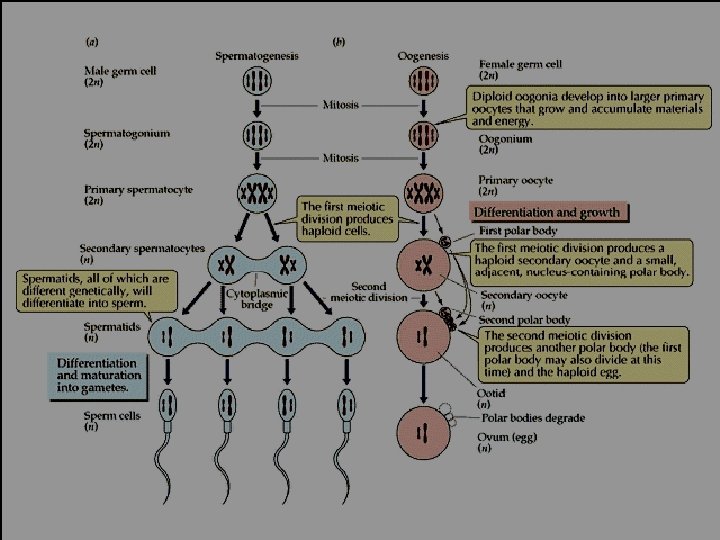
Gametogenesis means formation of gametes (Sperm & Ova). Spermatogenesis Means formation of sperm ; male gametes. Oogenesis Means formation of ovum ; female gametes. Sperm and ovum are highly specialized sex cells. Both types possess three main phases: 1. Period of multiplication: The primordial germ cells multiply by mitotic cell division giving rise to oogonia in case of females and spermatogonia in case of males. 2. Period of growth: During these phase both oogonia (female gamete) and spermatogonia (male gamete) grow into primary oocyte or primary spermatocyte.
3. Period of maturation: In case of female, the primary oocyte undergoes two meiotic cell division, the first gives rise to secondary oocyte and primary polar body. The secondary undergoes second meiotic division giving rise to mature ova and secondary polar body (mature ova /each oogonia). In case of male, each primary spermatocyte divides meiotically into secondary spermatocytes and intern to spermatid (4 spermatids / each primary spermatocyte).
Male Genital System
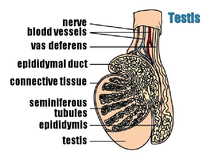
Overview of Gross Anatomy of Male Reproductive System: – Testis • Male sex gland, Located in the Scrotum. • Produce Sperm and Androgens. • Have also the interstitial cells (leydeg cells) that produce male sex hormone (testosterone) • Seminiferous tubules is the structural unit of testes. , it have the developmental phases of sperms in addition to Sertoli cells. – Epididymis • Sperm Storage • Complete of sperm maturation. – Vas Deferens • Duct that transports the Sperm from the Scrotum to the Prostate Gland – Seminal Vessicle • Secrets Fluid, rich in Fructose, to Semen
– Prostate Gland (Ejaculatory Duct) • Contributes Milky Alkaline Fluid that assists Sperm Activation – Cowpers Gland [Bulbourethral Gland] • Contributes Mucous to Semen – Urethra (Penis) • Organ of Copulation
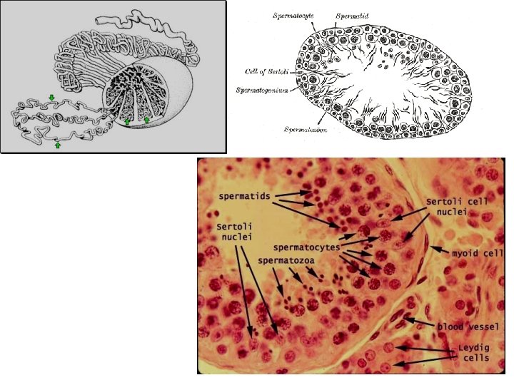
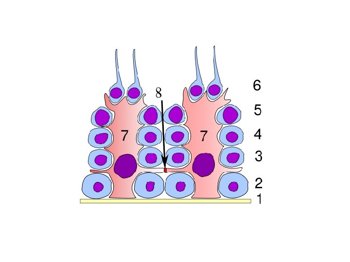
Sertoli Cells • At all stages of differentiation, the spermatogenic cells are in close contact with Sertoli cells which are provide structural and metabolic support to the developing sperm cells. • A single Sertoli cell extends from the basement membrane to the lumen of the seminiferous tubule. • Sertoli cells serve a number of functions during spermatogenesis like providing the cells with nourishment and molecular signals. , they support the developing gametes in the following ways:
1. Maintain the environment necessary for development and maturation via the blood-testis barrier. 2. Secrete substances initiating meiosis. 3. Secrete supporting testicular fluid. 4. Secrete androgen-binding protein, which concentrates testosterone in close proximity to the developing gametes. 5. Secrete hormones affecting pituitary gland control of spermatogenesis, particularly the polypeptide hormone, inhibin. 6. Phagocytose residual cytoplasm left over from spermiogenesis.
SPERMATOGENESIS • • • The process of development of spermatids from the male primordial germ cells and their differentiation into spermatozoa. Under stimulation of anterior pituitary gonadotropic hormones. PROCESS : 1. The Primordial germ cells develop into spermatogonia. There are two types of spermatogonia: Type A: that divide by mitosis to provide a continuous reserve of type B. Type B: that enter spermatogenesis.
2. This occurs after puberty, and they remain in the wall of the Seminiferous Tubule. 3. Spermatogonia form primary spermatocytes. 4. They remain in the prophase of 1 st meiotic division for 16 days. 5. Each contains 22 pairs of autosomes and one pair of sex chromosome XY. 6. Then divides into two secondary spermatocytes. (Meiotic div. completed). 7. Each secondary spermatocyte has equal chromosomes. (22+X) or (22+Y). 8. Each of these divides again(2 nd meiotic div), thus forming 4 Spermatids. 9. Each containing equal cytoplasm and, HAPLOID chromosomes…. TWO with 23 X & TWO with 23 Y
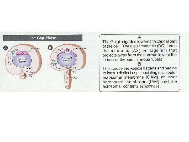
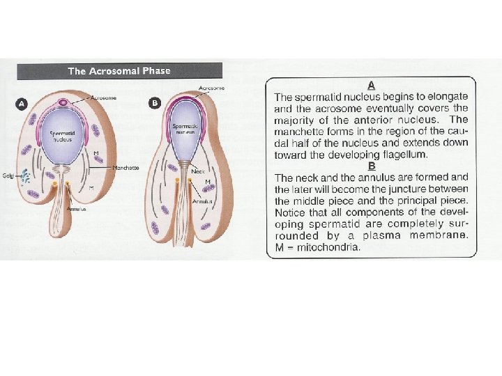
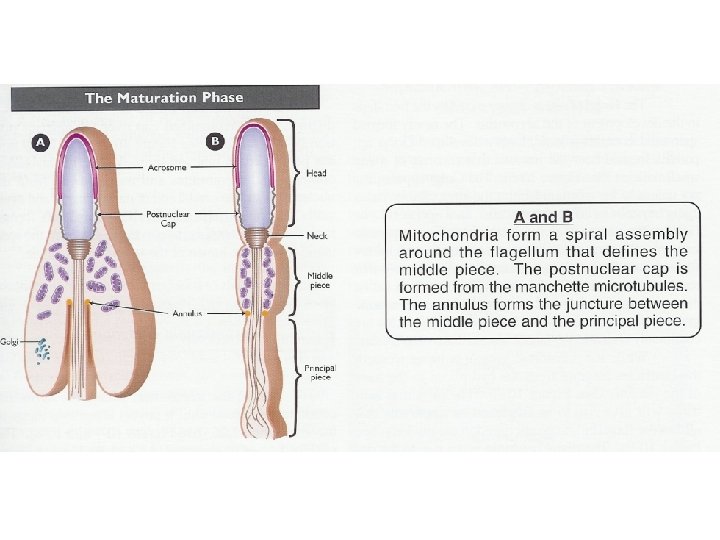
Spermiogenesis Spermatids undergo morphological changes to form spermatozoa
Spermiation • The mature spermatozoa released from the protective Sertoli cells into the lumen of the seminiferous tubule and a process called spermiation then takes place, which removes the remaining unnecessary cytoplasm and organelles. • The resulting spermatozoa are now mature but lack motility, rendering them sterile. The non-motile spermatozoa are transported to the epididymis in testicular fluid secreted by the Sertoli cells with the aid of peristaltic contraction. • In the epididymis they acquire motility and become capable of fertilization. However, transport of the mature spermatozoa through the remainder of the male reproductive system is achieved via muscle contraction rather than the spermatozoon's recently acquired motility.
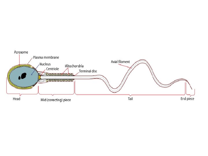
Structure Of The Sperm The Head • has two important features. 1. The acrosome (derived from Golgi apparatus) contains hydrolytic enzymes which are released when the sperm reaches an ovum. These enzymes digest the outer membrane of the egg (proteins and complex sugars) , allowing penetration of the sperm. 2. The nucleus (haploid) contains a single set of chromosomes derived from the male. This will include either an 'X' or 'Y' chromosome, because of the way the XY separate during meiosis. In many species, a region of globular actin molecules lies between 1&2 that used to extend a finger-like process during early stages of Fertilization.
The Middle Section • behind the head, contains numerous mitochondria. These respire sugars in the semen to generate ATP in order to provide the energy for movement of the tail. The Tail • (Flagellum) contains microfilaments running the length of the tail. Rhythmic contraction of the filaments causes the tail to wave and move against the fluid environment, providing forward motion.
Abnormalities of sperms 1. Morphological Abnormalities: • The head & tail may be abnormal, they may be: a) giants. b) Dwarfs. c) Joined in head or in tail. • Lack motility and don’t fertilize the egg. 2. Numerical Abnormalities: • Oligospermia: few number of sperms in semen. • Aspermia: no sperms at all in semen. • Necrospermia: sperms found dead.