Fluorescence Polarization Immunoassay FPIA Fluorescence polarization is ideal


 is secreted from the anterior pituitary gland in both men")


, it")


- Slides: 26
Fluorescence Polarization Immunoassay FPIA
• Fluorescence polarization is ideal for the study of small molecule fluorescent ligands binding to receptors. • In fluorescent polarization method of assay, polarized light excites a fluorescent label. As this label is bound, the degree of polarization of the fluorescent emission from the label increases.
disadvantages • Disadvantages of this method are nonlinear assay when the concentration of the test compound is related to polarization of the fluorescent emission. • Also, this type of assay has a limiting assayed can be accurately measured. • FPIA is utilized to provide accurate and sensitive measurement of small toxicology analytes such as therapeutic drugs, and drugs of abuse, toxicology and some hormones
FPIA reagent components • S: Antibody Reagent: Antiserum to analyte; • T: Tracer: Fluorescein-labeled analyte; • P: Pretreatment detergent: facilitate release of drug from serum proteins.
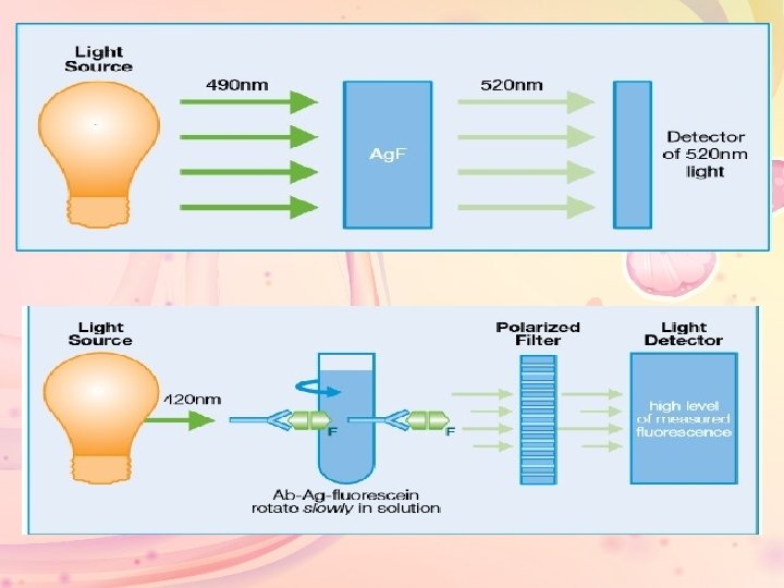
• FPIA utilizes three key concepts to measure specific analytes in a homogeneous format: fluorescence, rotation of molecules in solution, and polarized light. • Fluorescence: Fluorescein is a fluorescent label. It absorbs light energy at 490 nm and releases this energy at a higher wavelength (520 nm) as fluorescent light
• FPIA results in an inverse dose response curve such that lower levels of patient analyte result in a higher signal (in this case, the signal is polarized light). High signal at low patient analyte levels results in a highly sensitive Assay.
• fluorescence polarization immunoassays combine two technologist to determine analyte concentration: – competitive protein binding – fluorescence polarization • competitive binding requires two antigen or analyte systems: Ø Analyte from the sample Ø Tracer-labeled analyte provided by the assay reagents • The analyte from the sample competes with the analyte tracer for binding sites on the antibody. Each analyte system attaches to binding sites based on its relative concentration.
• If high concentrations of the sample analyte are present more specimen analyte binds to the antibody, leaving the analyte tracer unbound. • If low concentrations of the sample analytes are present less specimen analyte binds to the antibody leaving analyte tracer bound.
• Polarized light can be used to produce a polarized flurorescent emission from the analyte-tracer. • The average polarization of the emitted fluorescence is related to the speed of rotation of the molecule. • The rate of molecule rotation in liquid is related to the size of the molecule. Small, unbound analytes rotate more rapidly than the larger analyteantibody complex. • Axsym system undergo to this priniciple.
FPIA reaction sequence: • A typical FPIA reaction would occur as follows: – Sample, pretreatment reagent and line diluent are read as a blank. – Sample, pretreatment reagent, and line diluent are combined and incubated at reaction temperature. An initial read is taken by the FPIA optics system as a blank for the assay.
• Competitive binding occurs: • Antibody, tracer, line diluent and more sample are added to the reaction mixture and incubated. • The analyte from the sample competes with the analyte-tracer for binding sites on the antibodies. • If the sample contains a high concentration of the analyte, the sample analyte binds to the antibodies, leaving the analyte-tracer unbound. • If the sample contains a low concentration (or no concentration) of the analyte, then few or no sample analyte molecules bind to the antibodies, leaving the antibodies open for the analyte tracer.
• Change in polarization of emitted fluorescence indicates analyte concentration. • FPIA optics detect and measure the change in polarization of emitted fluorescence, which is inversely rotational to the concentration of the analyte in the specimen.
• If the sample contains a high concentration of the analyte, then many analyte-tracer molecules are left unbound. When excited by polarized vertical light, these small fluorescent molecules rotate rapidly, emitting light in many different planes. The result is a decrease in polarization. • If the sample contains a low concentration (or no concentration ) of the analyte, then the analyte-tracer binds to the antibodies, resulting in few unbound analyte-tracer molecules. These large molecules (complexes of analyte-tracer bound to antibody) rotate slower. Rotation is slowed enough so that light is emitted in a single plane. The result is an increase in polarization.
• Prolactin (PRL) is secreted from the anterior pituitary gland in both men and woman. Women normally have slightly higher basal prolactin levels than men. During and following pregnancy, prolactin, in association with other hormones, stimulates breast development and milk production.
• Hypersecretion of prolactin can be caused by pituitary tumors, hypothalamic diseases, hypothyroid, renal failure, acute exercise and several medications. Hyperprolactinemia inhibits hypogonadism in men and women with accompanying low FSH and LH levels. • Human Prolactine is simillar in structure to human growth hormone, and both are good lactogenes.
• Human prolactin is a single chain polypeptide hormone with a molecular weight of approximately 23. 000 daltons. • The primary biological action of the hormone is on the mammary gland where it is involved in the growth of the gland in the induction and maintenance of milk production. • There is evidence to suggest that prolactin may involved in steroidogenesis in the gonad, acting synergistically with luteinizing hormone. High levels of prolactin appear to inhibit steroidogenesis as well as inhibiting LH and FSH synthesis at the pituitary gland.
• Prolactinomas occur in both men and women but are more commonly diagnosed in women who are less than 50 years than in older women or men. • Clinical usefulness of the measurement of prolactin hormone in ascertaining the diagnosis of hyperprolactemia and for subsequent monitoring the effectiveness of the treatment has been well stablished.
• For the interpretation of the finding of an increased plasma (prolactin), it is important to enquire about the intake of drug. Many centrally acting drugs (phenothiazines, methyldopa, imipramine) inhibit the release of prolactin release-inhibiting hormone. Oestrogens and oral contraceptive may also cause raised plasma prolactin
• Measurement of plasma prolactin has been used as an index of response to the injection of TRH, which stimulates release of prolactin in addition to stimulating the release of TSH. (clinical notes of clinical chemistry). • If you are taking medicine for a prolactinoma, you will have your hormone levels checked at least once or twice a year.
• Surgery is often the first treatment for many types of pituitary adenomas. If you had a functional (hormone-making) pituitary adenoma, hormone measurements can often be done within days or weeks after surgery to see if the treatment was successful. Blood tests will also be done to see how well the remaining normal pituitary gland is functioning. If the results show that the tumor was removed completely and that pituitary function is normal, you will still need periodic visits with your doctor. Your hormone levels may need to be checked again in the future to check for recurrence of the adenoma.
• Blood should be drawn using standard venipuncture techniques and serum separated from blood cells as soon as possible. Samples should be allowed to clot for one hour at room temperature, centrifuged for 10 minutes (4°C) and serum extracted. This kit is for use with serum samples without additives only. • Avoid grossly hemolytic, lipidic or turbid samples. • Serum samples to be used within 24 -48 hours may be stored at 2 -8°C otherwise samples must be stored at 20°C to avoid loss of bioactivity and contamination. Avoid freeze-thaw cycles.
• When performing the assay slowly bring samples to room temperature. • It is recommended that all samples be assayed in duplicate. • DO NOT USE HEAT-TREATED SPECIMENS. • This test cannot be made for mother at lactation period. • Patient should be avoid Emotional stress and strenuous exercise.
The End