Fluorescence Polarization Immunoassay FPIA Fluorescence polarization is ideal


- Slides: 14
Fluorescence Polarization Immunoassay FPIA
• Fluorescence polarization is ideal for the study of small molecule fluorescent ligands binding to receptors. • In fluorescent polarization method of assay, polarized light excites a fluorescent label. As this label is bound, the degree of polarization of the fluorescent emission from the label increases.
disadvantages • Disadvantages of this method are nonlinear assay when the concentration of the test compound is related to polarization of the fluorescent emission. • Also, this type of assay has a limiting assayed can be accurately measured. • FPIA is utilized to provide accurate and sensitive measurement of small toxicology analytes such as therapeutic drugs, and drugs of abuse, toxicology and some hormones
FPIA reagent components • S: Antibody Reagent: Antiserum to analyte; • T: Tracer: Fluorescein-labeled analyte; • P: Pretreatment detergent: facilitate release of drug from serum proteins.
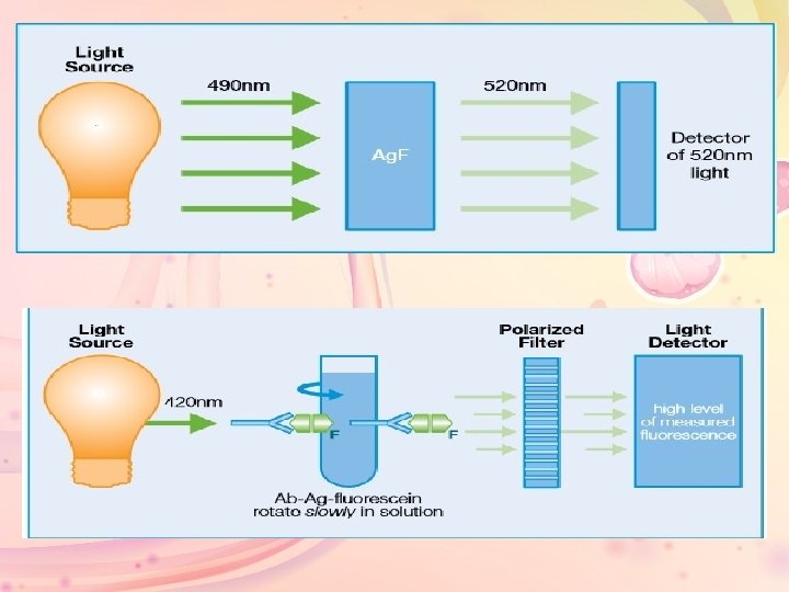
• FPIA utilizes three key concepts to measure specific analytes in a homogeneous format: fluorescence, rotation of molecules in solution, and polarized light. • Fluorescence: Fluorescein is a fluorescent label. It absorbs light energy at 490 nm and releases this energy at a higher wavelength (520 nm) as fluorescent light
• FPIA results in an inverse dose response curve such that lower levels of patient analyte result in a higher signal (in this case, the signal is polarized light). High signal at low patient analyte levels results in a highly sensitive Assay.
• fluorescence polarization immunoassays combine two technologist to determine analyte concentration: – competitive protein binding – fluorescence polarization • competitive binding requires two antigen or analyte systems: Ø Analyte from the sample Ø Tracer-labeled analyte provided by the assay reagents • The analyte from the sample competes with the analyte tracer for binding sites on the antibody. Each analyte system attaches to binding sites based on its relative concentration.
• If high concentrations of the sample analyte are present more specimen analyte binds to the antibody, leaving the analyte tracer unbound. • If low concentrations of the sample analytes are present less specimen analyte binds to the antibody leaving analyte tracer bound.
• Polarized light can be used to produce a polarized flurorescent emission from the analyte-tracer. • The average polarization of the emitted fluorescence is related to the speed of rotation of the molecule. • The rate of molecule rotation in liquid is related to the size of the molecule. Small, unbound analytes rotate more rapidly than the larger analyteantibody complex. • Axsym system undergo to this priniciple.
FPIA reaction sequence: • A typical FPIA reaction would occur as follows: – Sample, pretreatment reagent and line diluent are read as a blank. – Sample, pretreatment reagent, and line diluent are combined and incubated at reaction temperature. An initial read is taken by the FPIA optics system as a blank for the assay.
• Competitive binding occurs: • Antibody, tracer, line diluent and more sample are added to the reaction mixture and incubated. • The analyte from the sample competes with the analyte-tracer for binding sites on the antibodies. • If the sample contains a high concentration of the analyte, the sample analyte binds to the antibodies, leaving the analyte-tracer unbound. • If the sample contains a low concentration (or no concentration) of the analyte, then few or no sample analyte molecules bind to the antibodies, leaving the antibodies open for the analyte tracer.
• Change in polarization of emitted fluorescence indicates analyte concentration. • FPIA optics detect and measure the change in polarization of emitted fluorescence, which is inversely rotational to the concentration of the analyte in the specimen.
• If the sample contains a high concentration of the analyte, then many analyte-tracer molecules are left unbound. When excited by polarized vertical light, these small fluorescent molecules rotate rapidly, emitting light in many different planes. The result is a decrease in polarization. • If the sample contains a low concentration (or no concentration ) of the analyte, then the analyte-tracer binds to the antibodies, resulting in few unbound analyte-tracer molecules. These large molecules (complexes of analyte-tracer bound to antibody) rotate slower. Rotation is slowed enough so that light is emitted in a single plane. The result is an increase in polarization.