Fig 3 Parallel application of targeting methods and


- Slides: 55
Fig. 3. Parallel application of targeting methods and fluorophores B. N. G. Giepmans et al. , Science 312, 217 -224 (2006) Published by AAAS
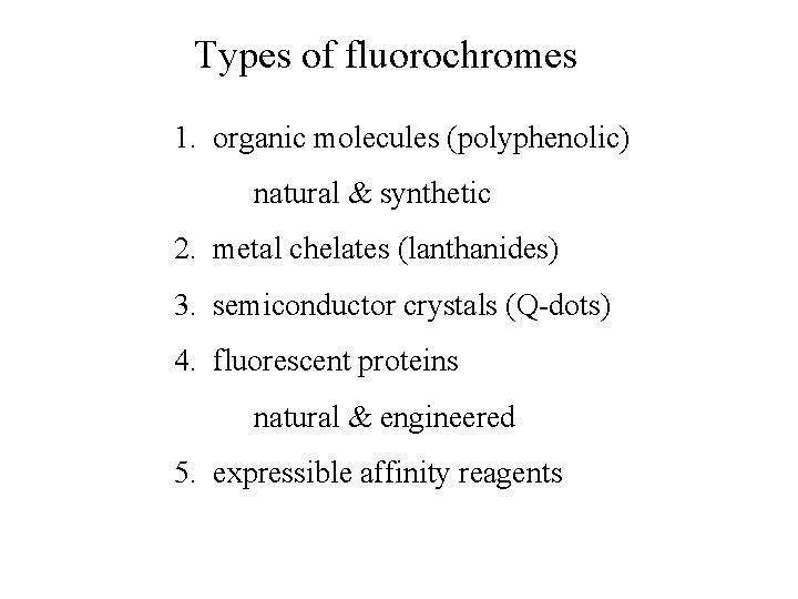
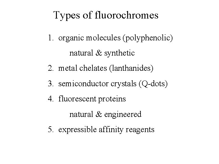
Types of fluorochromes 1. organic molecules (polyphenolic) natural & synthetic 2. metal chelates (lanthanides) 3. semiconductor crystals (Q-dots) 4. fluorescent proteins natural & engineered 5. expressible affinity reagents
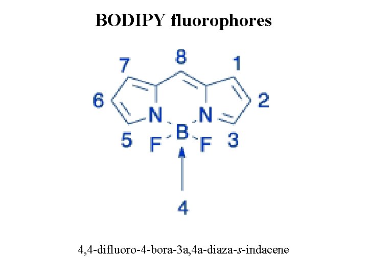
BODIPY fluorophores 4, 4 -difluoro-4 -bora-3 a, 4 a-diaza-s-indacene
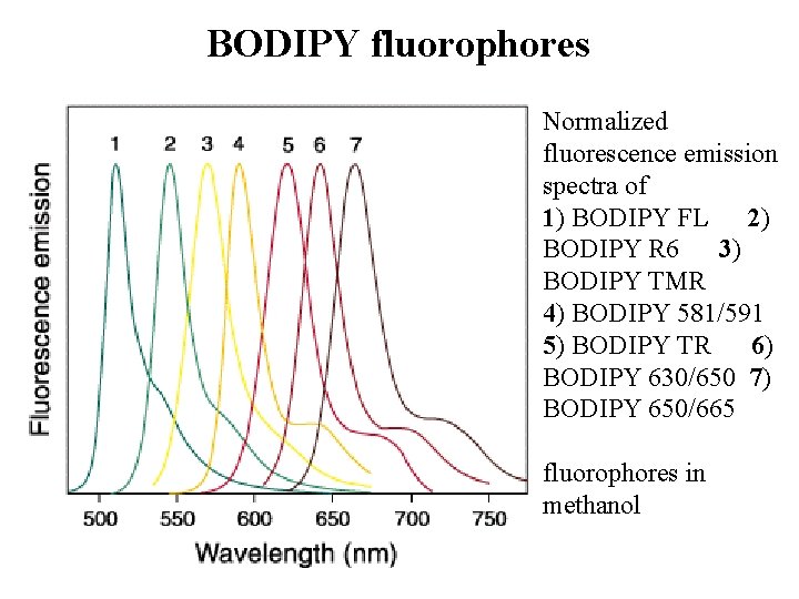
BODIPY fluorophores Normalized fluorescence emission spectra of 1) BODIPY FL 2) BODIPY R 6 3) BODIPY TMR 4) BODIPY 581/591 5) BODIPY TR 6) BODIPY 630/650 7) BODIPY 650/665 fluorophores in methanol
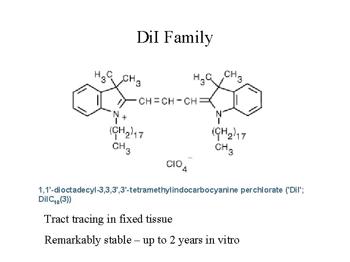
Di. I Family 1, 1'-dioctadecyl-3, 3, 3'- tetramethylindocarbocyanine perchlorate ('Di. I'; Di. IC 18(3)) Tract tracing in fixed tissue Remarkably stable – up to 2 years in vitro
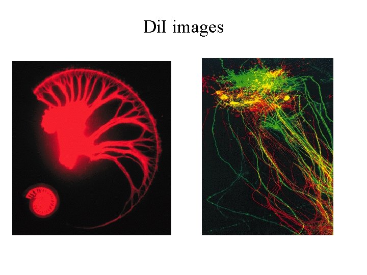
Di. I images
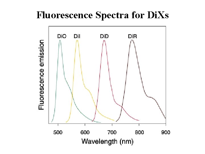
Fluorescence Spectra for Di. Xs
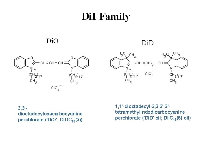
Di. I Family Di. O 3, 3'dioctadecyloxacarbocyanine perchlorate ('Di. O'; Di. OC 18(3)) Di. D 1, 1'-dioctadecyl-3, 3, 3'tetramethylindodicarbocyanine perchlorate ('Di. D' oil; Di. IC 18(5) oil)
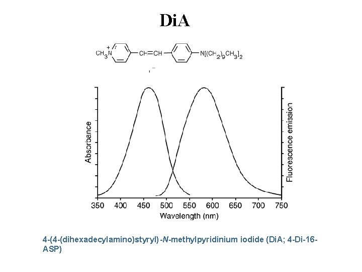
Di. A 4 -(4 -(dihexadecylamino)styryl) -N-methylpyridinium iodide (Di. A; 4 -Di-16 ASP)
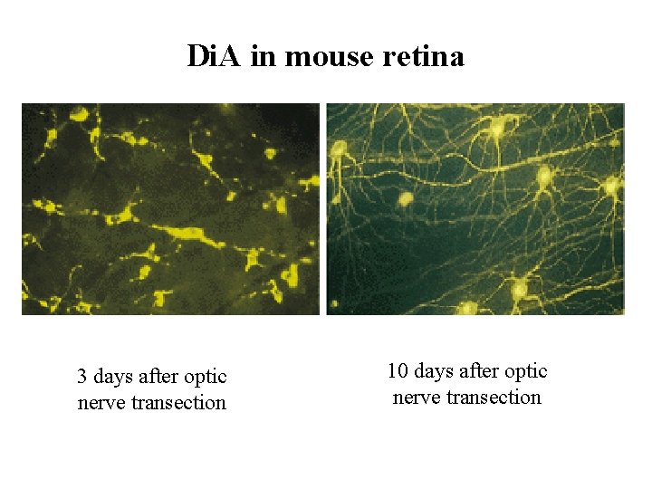
Di. A in mouse retina 3 days after optic nerve transection 10 days after optic nerve transection
Dextrans Substituted dextran polymers Poly-( a -D-1, 6 -glucose) linkages, which render them resistant to cleavage by most endogenous cellular glycosidases Wide molecular weight range (3000 – 300 K) Multiple fluorophores Biotin Lysine Fluoro-Ruby
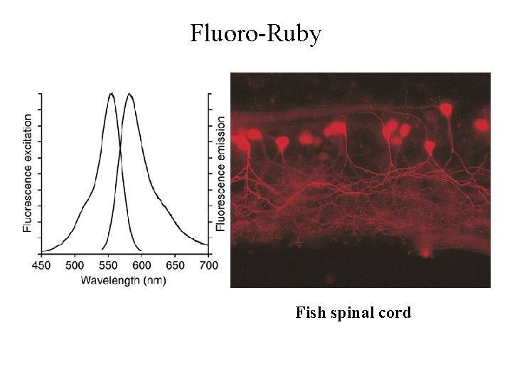
Fluoro-Ruby Fish spinal cord
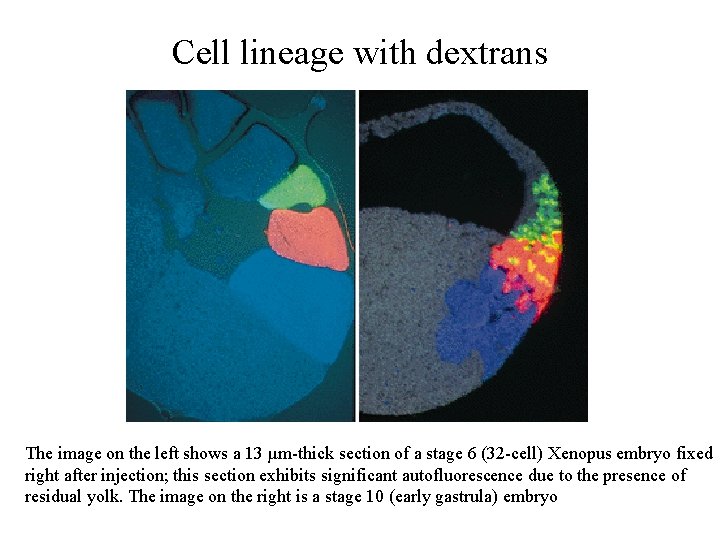
Cell lineage with dextrans The image on the left shows a 13 µm-thick section of a stage 6 (32 -cell) Xenopus embryo fixed right after injection; this section exhibits significant autofluorescence due to the presence of residual yolk. The image on the right is a stage 10 (early gastrula) embryo
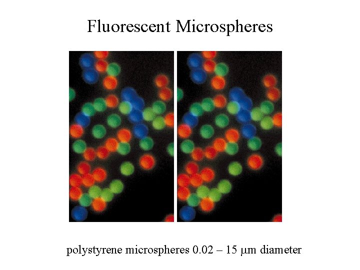
Fluorescent Microspheres polystyrene microspheres 0. 02 – 15 mm diameter
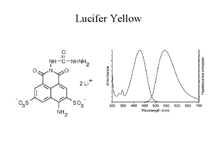
Lucifer Yellow
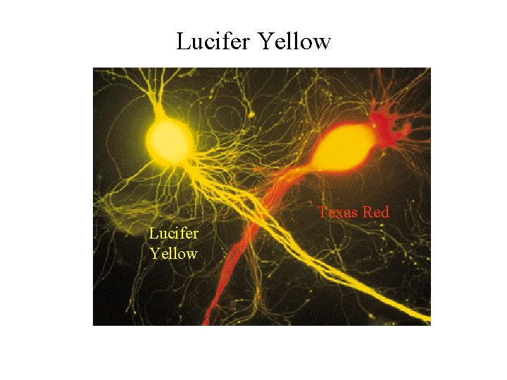
Lucifer Yellow Texas Red Lucifer Yellow
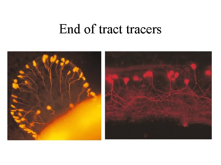
End of tract tracers
Types of fluorochromes 1. organic molecules (polyphenolic) natural & synthetic 2. metal chelates (lanthanides) 3. semiconductor crystals (Q-dots) 4. fluorescent proteins natural & engineered 5. expressible affinity reagents
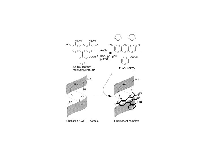
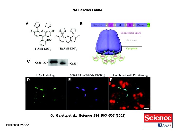
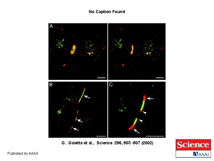
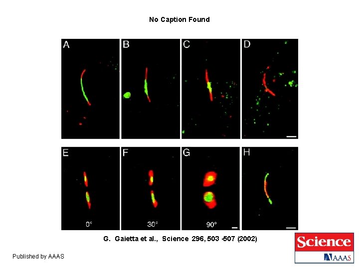
No Caption Found G. Gaietta et al. , Science 296, 503 -507 (2002) Published by AAAS
No Caption Found G. Gaietta et al. , Science 296, 503 -507 (2002) Published by AAAS
No Caption Found G. Gaietta et al. , Science 296, 503 -507 (2002) Published by AAAS
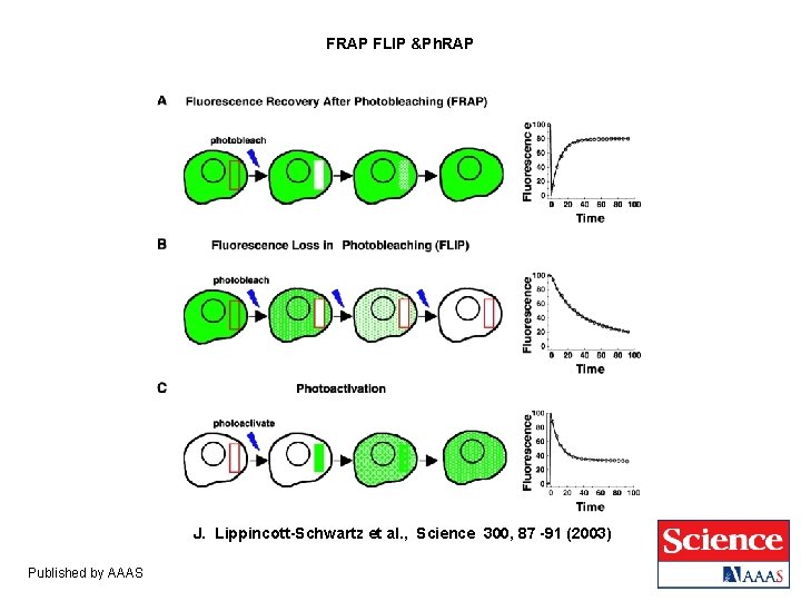
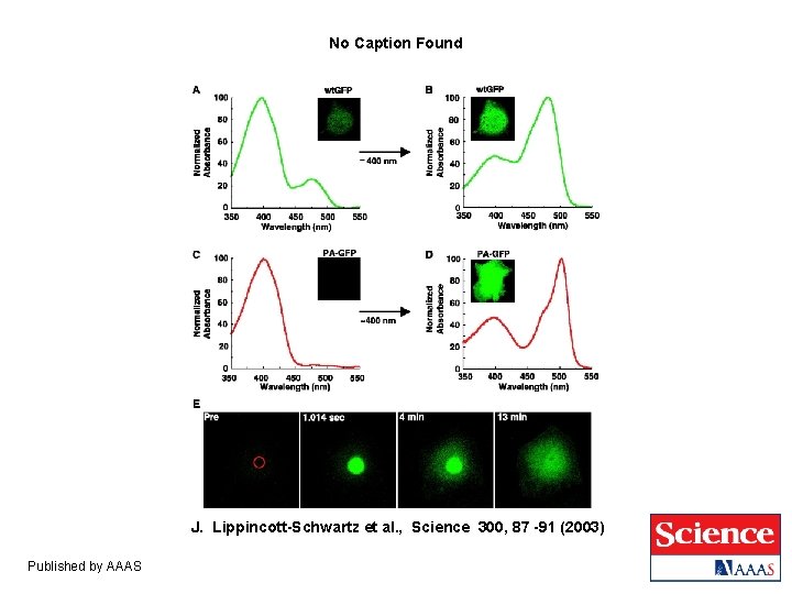
FRAP FLIP &Ph. RAP J. Lippincott-Schwartz et al. , Science 300, 87 -91 (2003) Published by AAAS
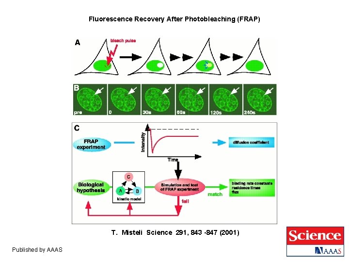
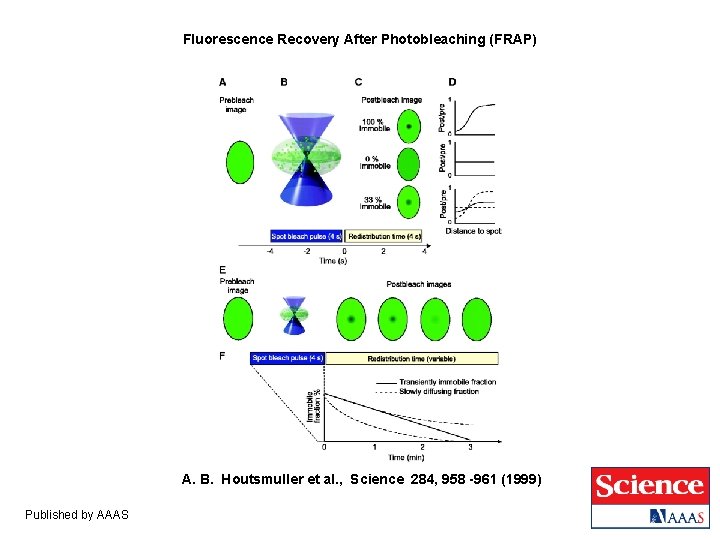
Fluorescence Recovery After Photobleaching (FRAP) T. Misteli Science 291, 843 -847 (2001) Published by AAAS
Fluorescence Recovery After Photobleaching (FRAP) A. B. Houtsmuller et al. , Science 284, 958 -961 (1999) Published by AAAS
No Caption Found J. Lippincott-Schwartz et al. , Science 300, 87 -91 (2003) Published by AAAS
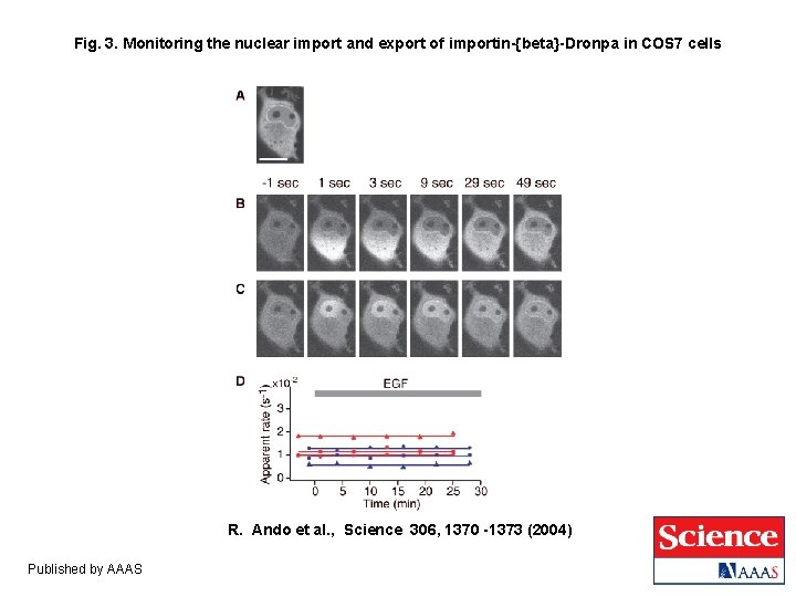
Fig. 3. Monitoring the nuclear import and export of importin-{beta}-Dronpa in COS 7 cells R. Ando et al. , Science 306, 1370 -1373 (2004) Published by AAAS
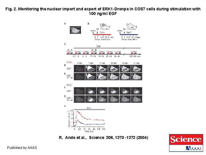
Fig. 2. Monitoring the nuclear import and export of ERK 1 -Dronpa in COS 7 cells during stimulation with 100 ng/ml EGF R. Ando et al. , Science 306, 1370 -1373 (2004) Published by AAAS
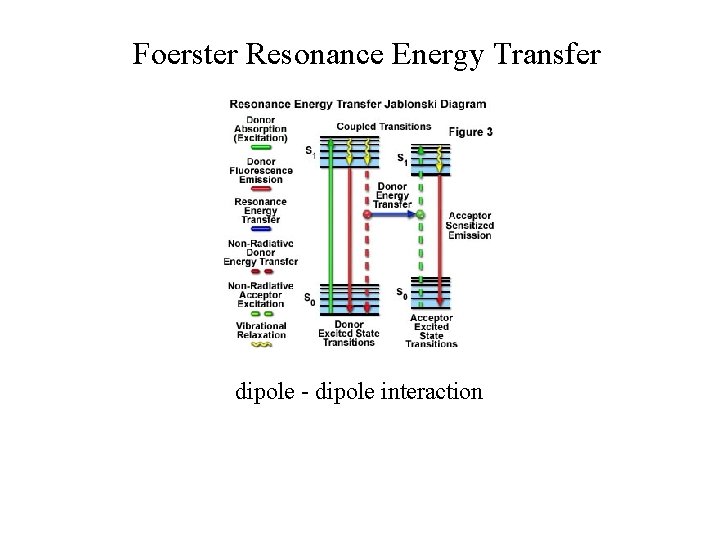
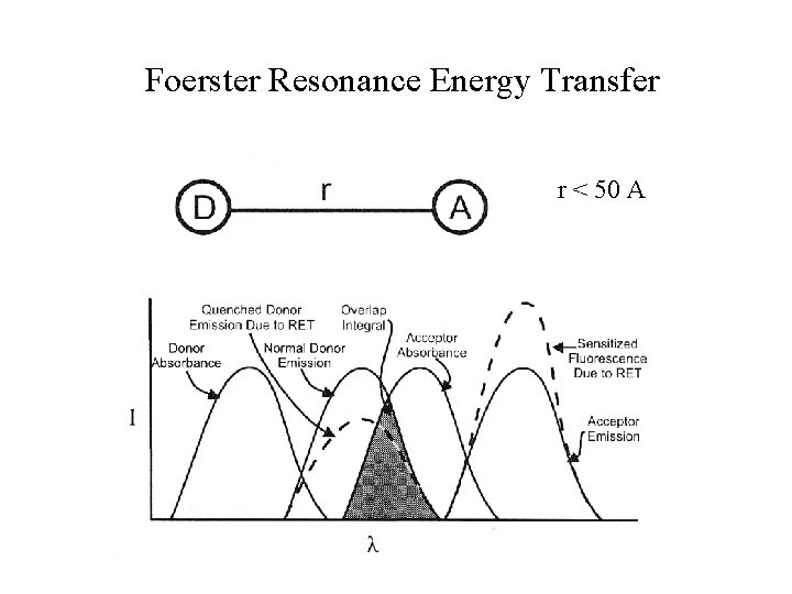
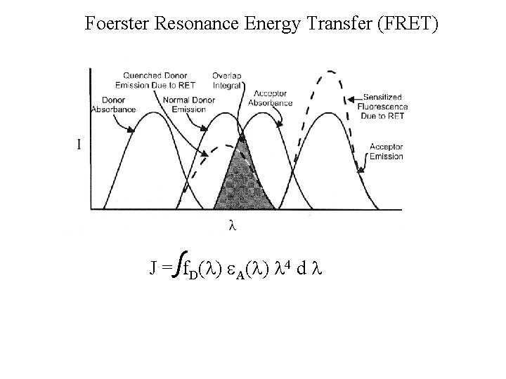
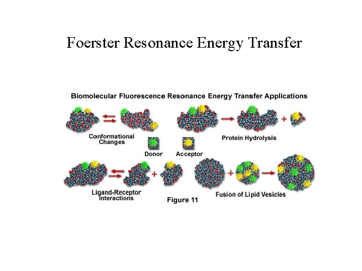
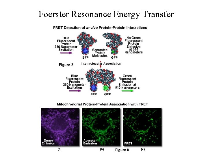
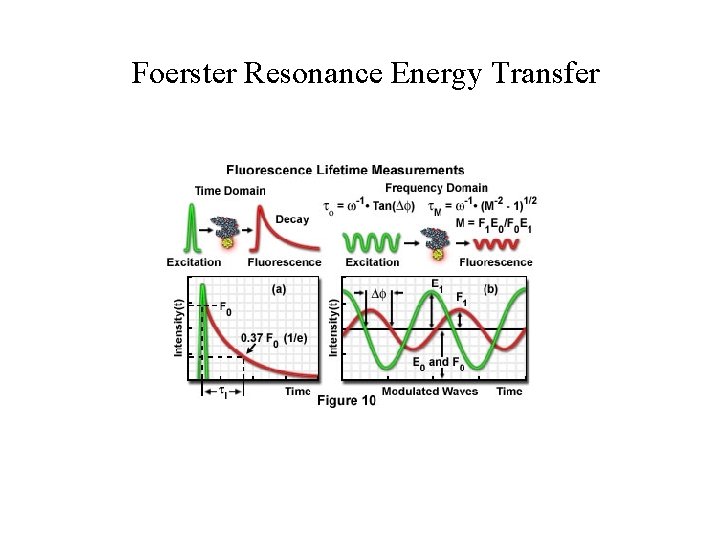
Foerster Resonance Energy Transfer dipole - dipole interaction
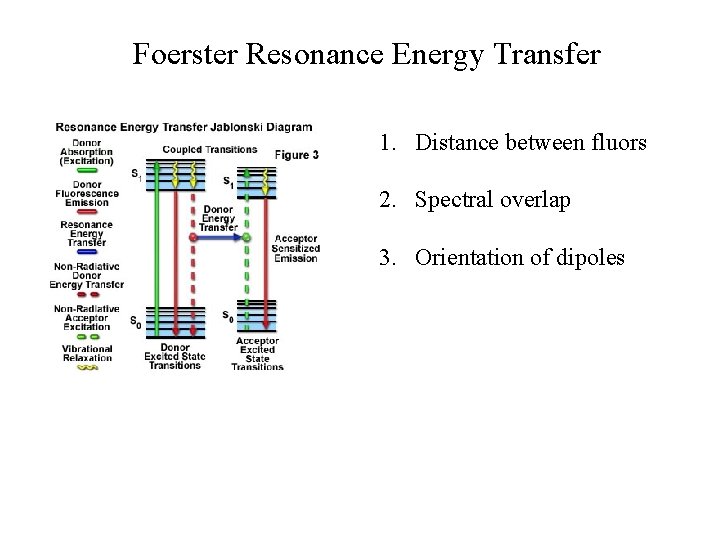
Foerster Resonance Energy Transfer 1. Distance between fluors 2. Spectral overlap 3. Orientation of dipoles
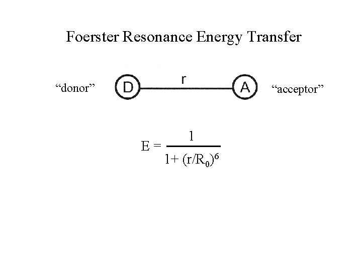
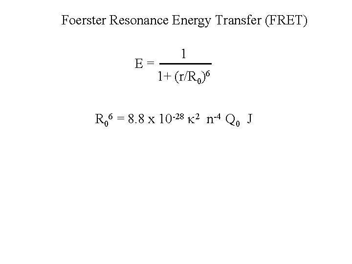
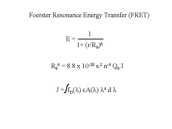
Foerster Resonance Energy Transfer “donor” “acceptor” 1 E = 1+ (r/R 0)6
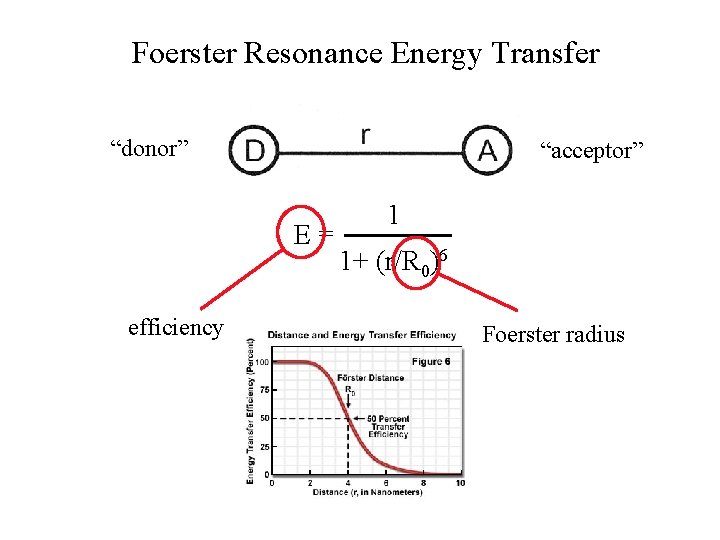
Foerster Resonance Energy Transfer “donor” “acceptor” 1 E = 1+ (r/R 0)6 efficiency Foerster radius
Foerster Resonance Energy Transfer r < 50 A
Foerster Resonance Energy Transfer (FRET) 1 E = 1+ (r/R 0)6 R 06 = 8. 8 x 10 -28 2 n-4 Q 0 J
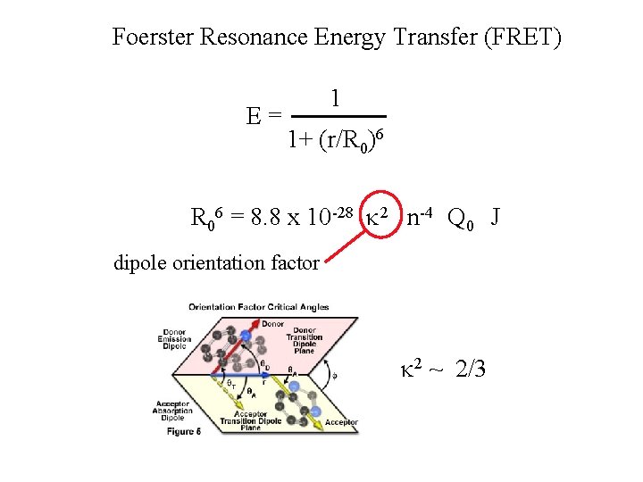
Foerster Resonance Energy Transfer (FRET) 1 E = 1+ (r/R 0)6 R 06 = 8. 8 x 10 -28 2 n-4 Q 0 J dipole orientation factor 2 ~ 2/3
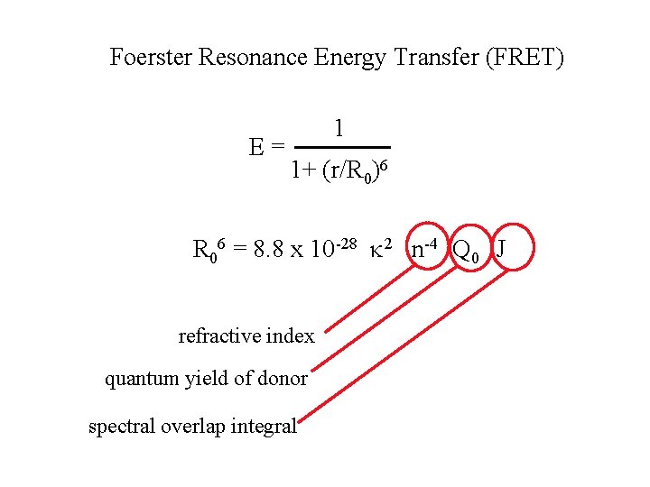
Foerster Resonance Energy Transfer (FRET) 1 E = 1+ (r/R 0)6 R 06 = 8. 8 x 10 -28 2 n-4 Q 0 J refractive index quantum yield of donor spectral overlap integral
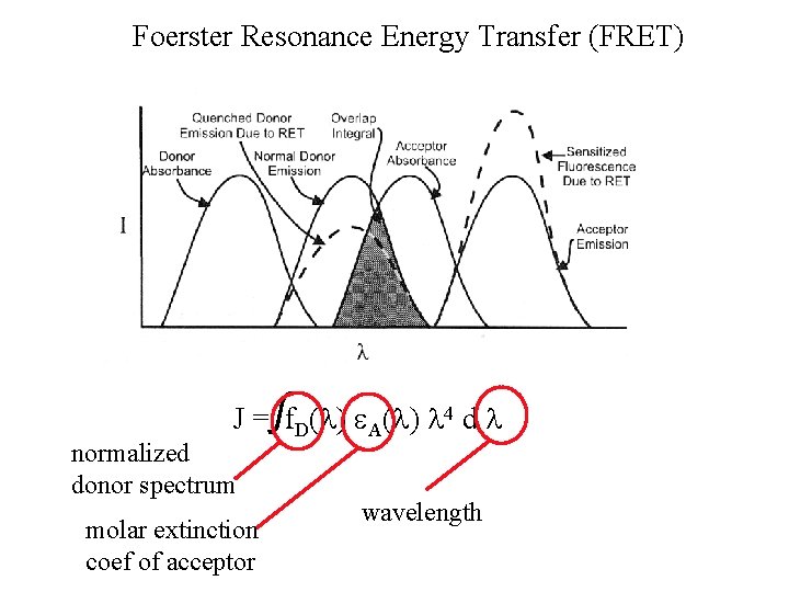
Foerster Resonance Energy Transfer (FRET) J = f. D( ) A( ) 4 d
Foerster Resonance Energy Transfer (FRET) J = f. D( ) A( ) 4 d normalized donor spectrum molar extinction coef of acceptor wavelength
Foerster Resonance Energy Transfer (FRET) 1 E = 1+ (r/R 0)6 R 06 = 8. 8 x 10 -28 2 n-4 Q 0 J J = f. D( ) A( ) 4 d
Foerster Resonance Energy Transfer
Foerster Resonance Energy Transfer
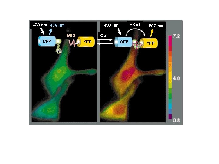
433 nm 476 nm M 13 CFP 527 nm Ca 2+ YFP Ca. M FRET 433 nm CFP YFP
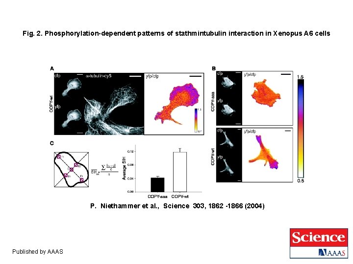
Fig. 2. Phosphorylation-dependent patterns of stathmintubulin interaction in Xenopus A 6 cells P. Niethammer et al. , Science 303, 1862 -1866 (2004) Published by AAAS
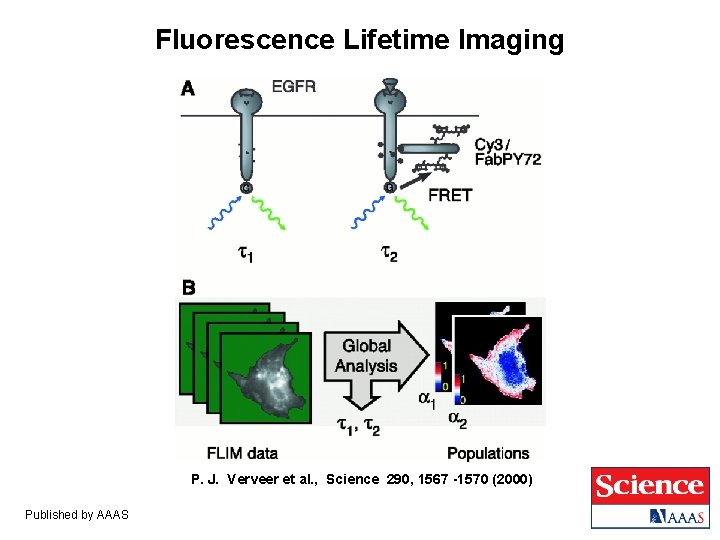
Fluorescence Lifetime Imaging P. J. Verveer et al. , Science 290, 1567 -1570 (2000) Published by AAAS
Foerster Resonance Energy Transfer
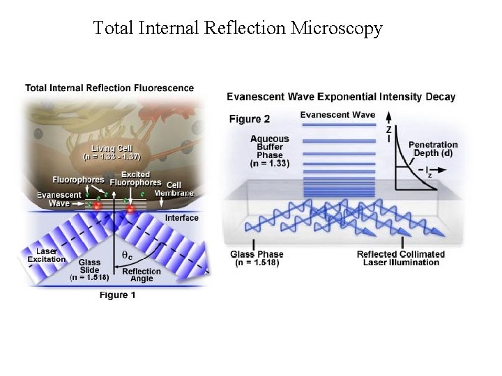
Total Internal Reflection Microscopy
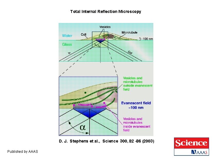
Total Internal Reflection Microscopy D. J. Stephens et al. , Science 300, 82 -86 (2003) Published by AAAS
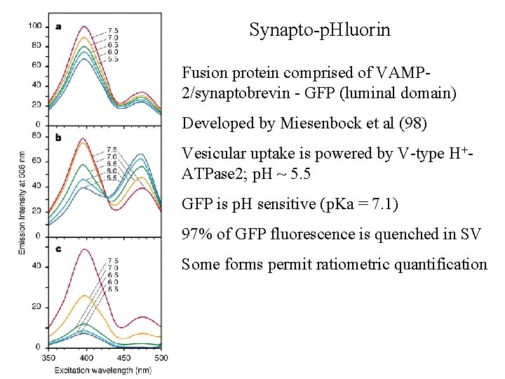
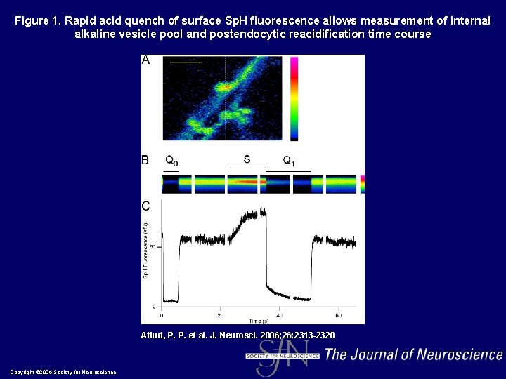
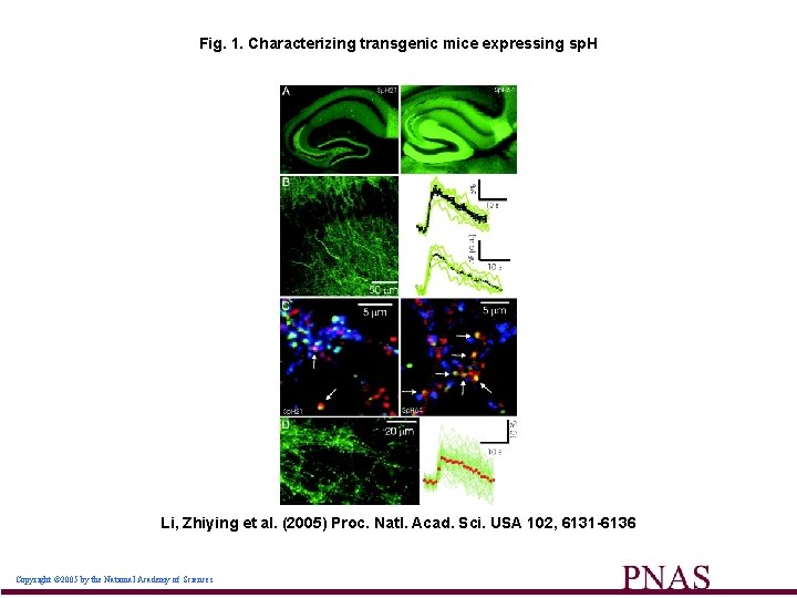
Synapto-p. Hluorin Fusion protein comprised of VAMP 2/synaptobrevin - GFP (luminal domain) Developed by Miesenbock et al (98) Vesicular uptake is powered by V-type H+ATPase 2; p. H ~ 5. 5 GFP is p. H sensitive (p. Ka = 7. 1) 97% of GFP fluorescence is quenched in SV Some forms permit ratiometric quantification
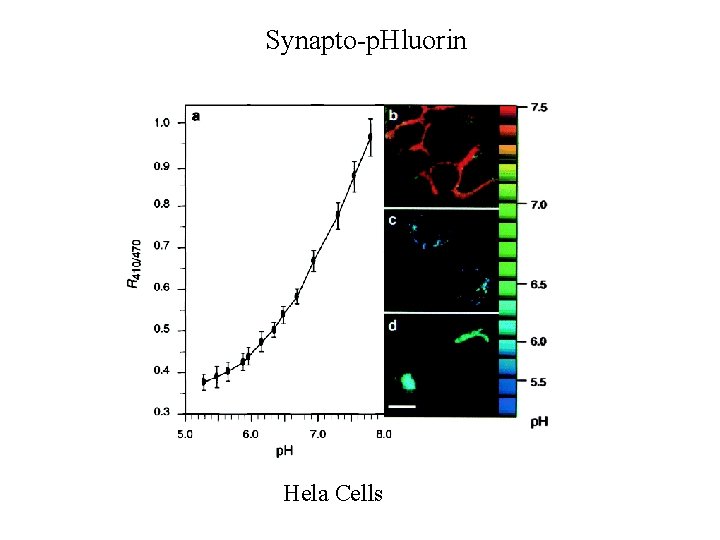
Synapto-p. Hluorin Hela Cells
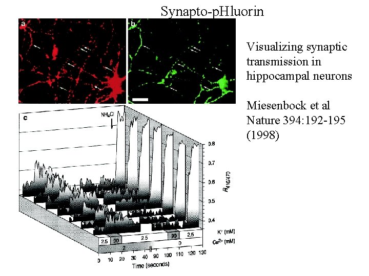
Synapto-p. Hluorin Visualizing synaptic transmission in hippocampal neurons Miesenbock et al Nature 394: 192 -195 (1998)
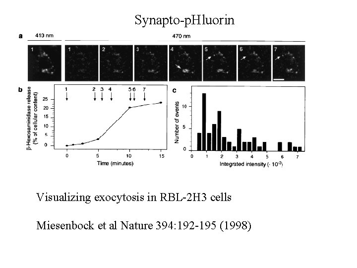
Synapto-p. Hluorin Visualizing exocytosis in RBL-2 H 3 cells Miesenbock et al Nature 394: 192 -195 (1998)
Figure 1. Rapid acid quench of surface Sp. H fluorescence allows measurement of internal alkaline vesicle pool and postendocytic reacidification time course Atluri, P. P. et al. J. Neurosci. 2006; 26: 2313 -2320 Copyright © 2006 Society for Neuroscience
Fig. 1. Characterizing transgenic mice expressing sp. H Li, Zhiying et al. (2005) Proc. Natl. Acad. Sci. USA 102, 6131 -6136 Copyright © 2005 by the National Academy of Sciences