e Ed E108 Treatment Tipping Points in Head

- Slides: 26
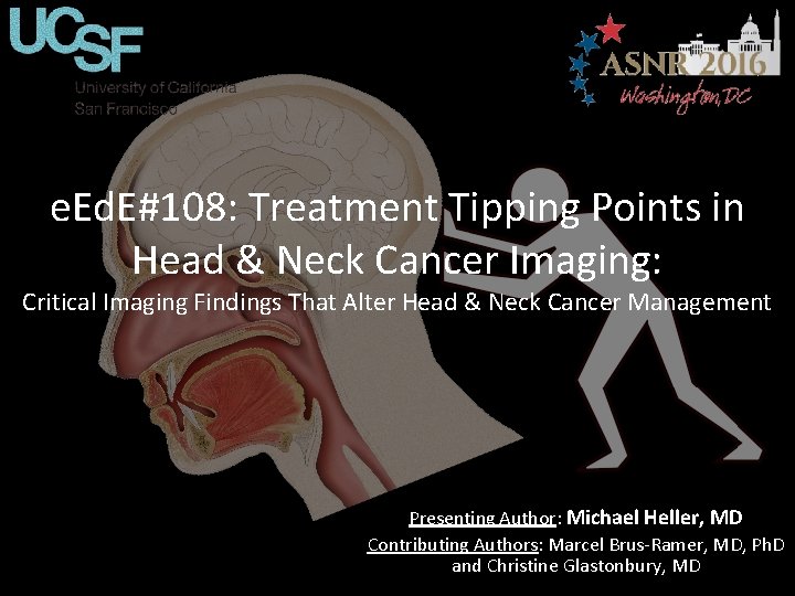
e. Ed. E#108: Treatment Tipping Points in Head & Neck Cancer Imaging: Critical Imaging Findings That Alter Head & Neck Cancer Management Presenting Author: Michael Heller, MD Contributing Authors: Marcel Brus-Ramer, MD, Ph. D and Christine Glastonbury, MD
Disclosures The authors have nothing to disclose.
Introduction Anatomy Case Vignettes Summary Conclusion Introduction When performing and reading staging scans for Head & Neck (H&N) cancers there are often very specific imaging findings which either alter the stage of the tumor or radically alter the management plan. These are the diagnostic and treatment tipping points; where radiology has a significant impact on patient care. Here, we present 8 clinical cases, highlighting the imaging ‘Tipping Points’ and clarifying the importance of careful evaluation of H&N cancer scans.
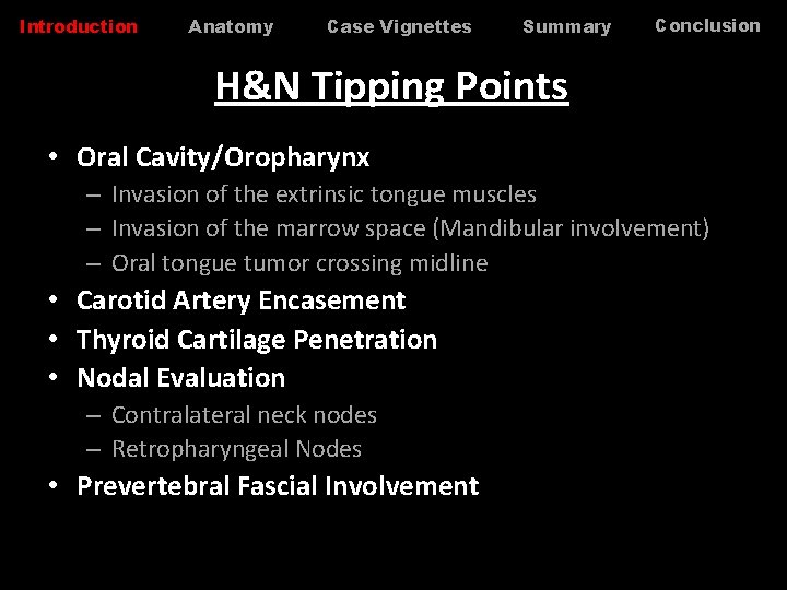
Introduction Anatomy Case Vignettes Summary Conclusion H&N Tipping Points • Oral Cavity/Oropharynx – Invasion of the extrinsic tongue muscles – Invasion of the marrow space (Mandibular involvement) – Oral tongue tumor crossing midline • Carotid Artery Encasement • Thyroid Cartilage Penetration • Nodal Evaluation – Contralateral neck nodes – Retropharyngeal Nodes • Prevertebral Fascial Involvement
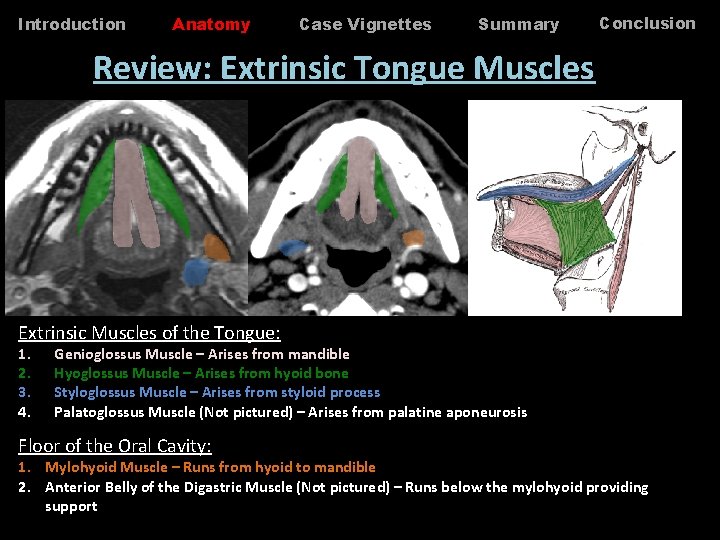
Introduction Anatomy Case Vignettes Summary Conclusion Review: Extrinsic Tongue Muscles Extrinsic Muscles of the Tongue: 1. 2. 3. 4. Genioglossus Muscle – Arises from mandible Hyoglossus Muscle – Arises from hyoid bone Styloglossus Muscle – Arises from styloid process Palatoglossus Muscle (Not pictured) – Arises from palatine aponeurosis Floor of the Oral Cavity: 1. Mylohyoid Muscle – Runs from hyoid to mandible 2. Anterior Belly of the Digastric Muscle (Not pictured) – Runs below the mylohyoid providing support
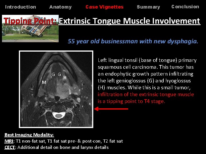
Anatomy Introduction Case Vignettes Summary Conclusion Tipping Point: Extrinsic Tongue Muscle Involvement 55 year old businessman with new dysphagia. G G M H Left lingual tonsil (base of tongue) primary squamous cell carcinoma. This tumor has an endophytic growth pattern infiltrating the left genioglossus (G) and hyoglossus (H) muscles. While this is a small tumor, infiltration of the extrinsic tongue muscle is a tipping point to T 4 stage. Best Imaging Modality: MRI: T 1 non-fat sat, T 1 fat sat pre- & post-con, T 2 fat sat CECT: Additional detail on bone and larynx details
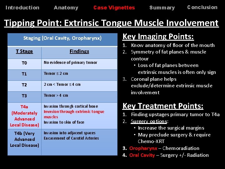
Introduction Anatomy Case Vignettes Summary Conclusion Tipping Point: Extrinsic Tongue Muscle Involvement Staging (Oral Cavity, Oropharynx) T Stage Findings T 0 No evidence of primary tumor T 1 Tumor ≤ 2 cm T 2 2 cm < Tumor ≤ 4 cm T 3 Tumor > 4 cm T 4 a (Moderately Advanced Local Disease) Invasion through cortical bone Invasion through extrinsic tongue muscles Invasion to skin of face Invasion into adjacent spaces T 4 b (Very Encasement of Carotid Arteries Advanced Local Disease) Key Imaging Points: 1. Know anatomy of floor of the mouth 2. Symmetry of fat planes & muscle contour • Loss of fat planes between extrinsic muscles is often only sign 3. Coronal plane helps exclude/determine extrinsic muscle involvement Key Treatment Points: 1. Finding upstages primary tumor to T 4 a 2. Surgery options: • Increase the surgical margins • May preclude surgery & require Chemo-XRT 3. Oropharynx – Chemoradiation 4. Oral Cavity – Surgery +/- Radiation
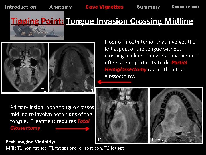
Anatomy Introduction Case Vignettes Summary Conclusion Tipping Point: Tongue Invasion Crossing Midline Floor of mouth tumor that involves the left aspect of the tongue without crossing midline. Unilateral involvement offers the opportunity to do Partial Hemiglossectomy rather than total glossectomy. T 1 Primary lesion in the tongue crosses midline to involve both sides of the tongue. Treatment requires Total Glossectomy. T 1 + C Best Imaging Modality: MRI: T 1 non-fat sat, T 1 fat sat pre- & post-con, T 2 fat sat T 1 + C
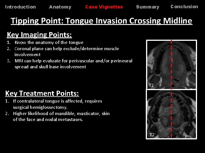
Introduction Anatomy Case Vignettes Summary Conclusion Tipping Point: Tongue Invasion Crossing Midline Key Imaging Points: 1. Know the anatomy of the tongue 2. Coronal plane can help exclude/determine muscle involvement 3. MRI can help evaluate for perivascular and/or perineural spread and skull base involvement Key Treatment Points: T 2 1. If contralateral tongue is affected, requires surgical hemiglossectomy. 2. Higher likelihood of mandible, masticator, skin of the face and nodal metastases. T 2
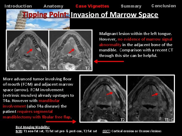
Introduction Anatomy Case Vignettes Summary Conclusion Tipping Point: Invasion of Marrow Space Malignant lesion within the left tongue. However, no evidence of marrow signal abnormality in the adjacent bone of the mandible. Comparison with a recent CT through this site can be helpful. T 1 T 2 More advanced tumor involving floor of mouth (FOM) and adjacent marrow space (arrow). FOM involvement (extrinsic muscles) already upstages to T 4 a. However with mandibular involvement (also T 4 a disease) the patient requires segmental mandiblectomy with fibular free flap. Best Imaging Modality: MRI: T 1 non-fat sat, T 1 fat sat pre- & post-con, T 2 fat sat T 2 CECT: Cortical erosion or Osseous lesions T 1+C
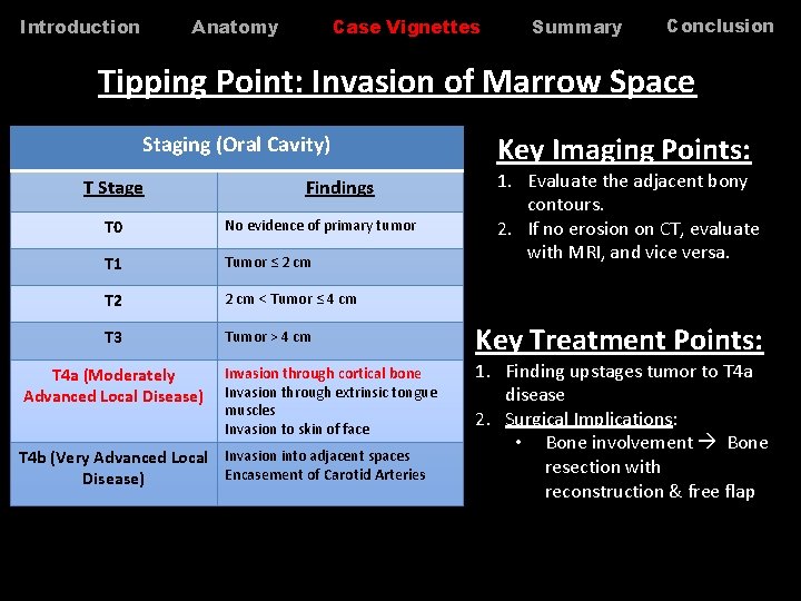
Anatomy Introduction Case Vignettes Summary Conclusion Tipping Point: Invasion of Marrow Space Staging (Oral Cavity) T Stage Findings T 0 No evidence of primary tumor T 1 Tumor ≤ 2 cm T 2 2 cm < Tumor ≤ 4 cm T 3 Tumor > 4 cm T 4 a (Moderately Advanced Local Disease) Invasion through cortical bone Invasion through extrinsic tongue muscles Invasion to skin of face T 4 b (Very Advanced Local Invasion into adjacent spaces Encasement of Carotid Arteries Disease) Key Imaging Points: 1. Evaluate the adjacent bony contours. 2. If no erosion on CT, evaluate with MRI, and vice versa. Key Treatment Points: 1. Finding upstages tumor to T 4 a disease 2. Surgical Implications: • Bone involvement Bone resection with reconstruction & free flap
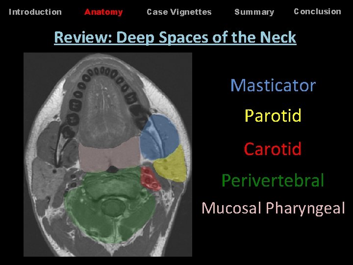
Introduction Anatomy Case Vignettes Summary Conclusion Review: Deep Spaces of the Neck Masticator Parotid Carotid Perivertebral Mucosal Pharyngeal
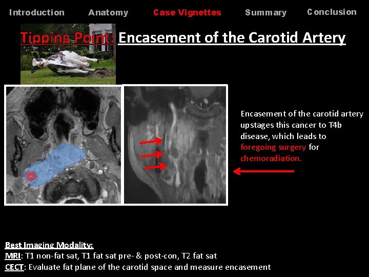
Introduction Anatomy Case Vignettes Summary Conclusion Tipping Point: Encasement of the Carotid Artery Encasement of the carotid artery upstages this cancer to T 4 b disease, which leads to foregoing surgery for chemoradiation. Best Imaging Modality: MRI: T 1 non-fat sat, T 1 fat sat pre- & post-con, T 2 fat sat CECT: Evaluate fat plane of the carotid space and measure encasement
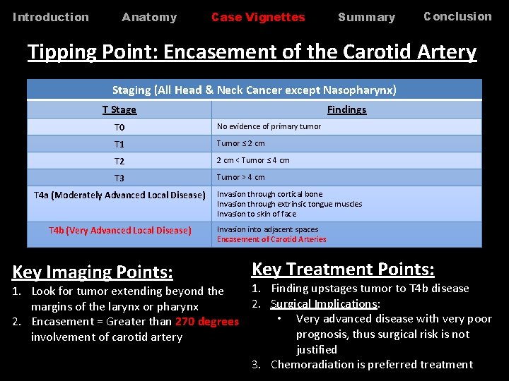
Introduction Anatomy Case Vignettes Summary Conclusion Tipping Point: Encasement of the Carotid Artery Staging (All Head & Neck Cancer except Nasopharynx) T Stage Findings T 0 No evidence of primary tumor T 1 Tumor ≤ 2 cm T 2 2 cm < Tumor ≤ 4 cm T 3 Tumor > 4 cm T 4 a (Moderately Advanced Local Disease) T 4 b (Very Advanced Local Disease) Key Imaging Points: Invasion through cortical bone Invasion through extrinsic tongue muscles Invasion to skin of face Invasion into adjacent spaces Encasement of Carotid Arteries Key Treatment Points: 1. Finding upstages tumor to T 4 b disease 1. Look for tumor extending beyond the 2. Surgical Implications: margins of the larynx or pharynx • Very advanced disease with very poor 2. Encasement = Greater than 270 degrees prognosis, thus surgical risk is not involvement of carotid artery justified 3. Chemoradiation is preferred treatment
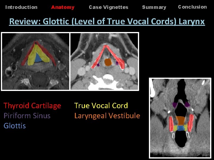
Introduction Anatomy Case Vignettes Summary Conclusion Review: Glottic (Level of True Vocal Cords) Larynx Thyroid Cartilage Piriform Sinus Glottis True Vocal Cord Laryngeal Vestibule
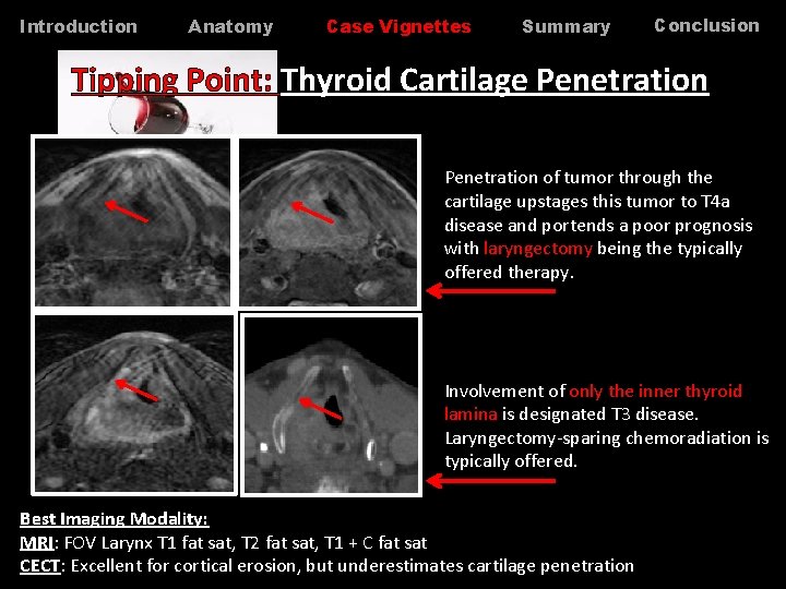
Introduction Anatomy Case Vignettes Summary Conclusion Tipping Point: Thyroid Cartilage Penetration of tumor through the cartilage upstages this tumor to T 4 a disease and portends a poor prognosis with laryngectomy being the typically offered therapy. Involvement of only the inner thyroid lamina is designated T 3 disease. Laryngectomy-sparing chemoradiation is typically offered. Best Imaging Modality: MRI: FOV Larynx T 1 fat sat, T 2 fat sat, T 1 + C fat sat CECT: Excellent for cortical erosion, but underestimates cartilage penetration
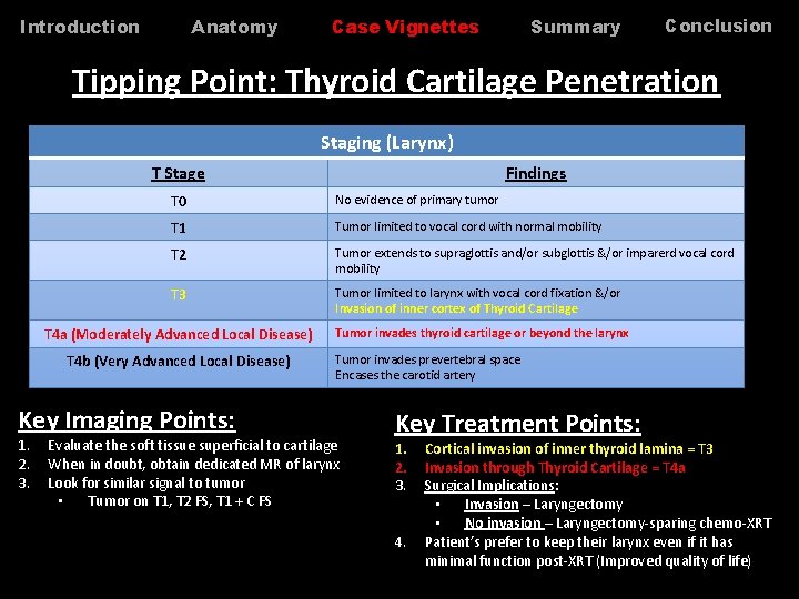
Anatomy Introduction Summary Case Vignettes Conclusion Tipping Point: Thyroid Cartilage Penetration Staging (Larynx) T Stage T 0 No evidence of primary tumor T 1 Tumor limited to vocal cord with normal mobility T 2 Tumor extends to supraglottis and/or subglottis &/or imparerd vocal cord mobility T 3 Tumor limited to larynx with vocal cord fixation &/or Invasion of inner cortex of Thyroid Cartilage T 4 a (Moderately Advanced Local Disease) T 4 b (Very Advanced Local Disease) Key Imaging Points: 1. 2. 3. Findings Tumor invades thyroid cartilage or beyond the larynx Tumor invades prevertebral space Encases the carotid artery Evaluate the soft tissue superficial to cartilage When in doubt, obtain dedicated MR of larynx Look for similar signal to tumor • Tumor on T 1, T 2 FS, T 1 + C FS Key Treatment Points: 1. 2. 3. 4. Cortical invasion of inner thyroid lamina = T 3 Invasion through Thyroid Cartilage = T 4 a Surgical Implications: • Invasion – Laryngectomy • No invasion – Laryngectomy-sparing chemo-XRT Patient’s prefer to keep their larynx even if it has minimal function post-XRT (Improved quality of life)
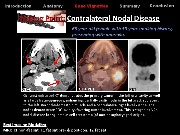
Introduction Anatomy Case Vignettes Summary Conclusion Tipping Point: Contralateral Nodal Disease 65 year old female with 50 year smoking history, presenting with anorexia. CT+C CT + PET Contrast-enhanced CT demonstrates the primary tumor in the left oral cavity as well as a large heterogeneous, enhancing, partially cystic node in the left neck subjacent to the left sternocleidomastoid muscle and a contralateral right level 2 node. The nodes demonstrate FDG-avidity, favoring tumor involvement. This is staged as N 2 c nodal disease for squamous cell carcinoma (of non-nasopharyngeal origin). Best Imaging Modality: MRI: T 1 non-fat sat, T 1 fat sat pre- & post-con, T 2 fat sat
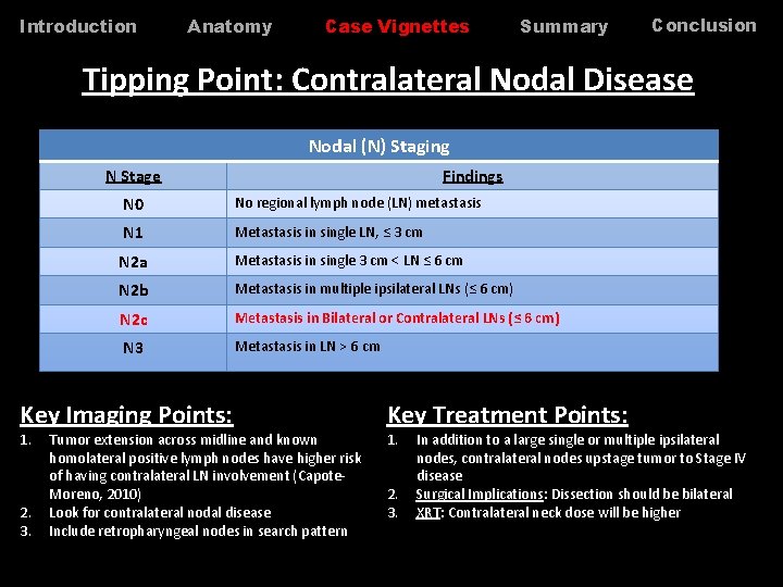
Introduction Anatomy Case Vignettes Summary Conclusion Tipping Point: Contralateral Nodal Disease Nodal (N) Staging N Stage N 0 No regional lymph node (LN) metastasis N 1 Metastasis in single LN, ≤ 3 cm N 2 a Metastasis in single 3 cm < LN ≤ 6 cm N 2 b Metastasis in multiple ipsilateral LNs (≤ 6 cm) N 2 c Metastasis in Bilateral or Contralateral LNs (≤ 6 cm) N 3 Metastasis in LN > 6 cm Key Imaging Points: 1. 2. 3. Findings Tumor extension across midline and known homolateral positive lymph nodes have higher risk of having contralateral LN involvement (Capote. Moreno, 2010) Look for contralateral nodal disease Include retropharyngeal nodes in search pattern Key Treatment Points: 1. 2. 3. In addition to a large single or multiple ipsilateral nodes, contralateral nodes upstage tumor to Stage IV disease Surgical Implications: Dissection should be bilateral XRT: Contralateral neck dose will be higher
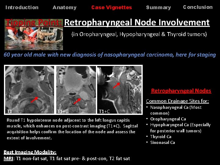
Introduction Anatomy Case Vignettes Summary Conclusion Tipping Point: Retropharyngeal Node Involvement (in Oropharyngeal, Hypopharyngeal & Thyroid tumors) 60 year old male with new diagnosis of nasopharyngeal carcinoma, here for staging Retropharyngeal Nodes Common Drainage Sites for: T 1+C Round T 1 hypointense node adjacent to the left longus capitis muscle, which enhances on post-contrast imaging (T 1+C). Sagittal acquisition helps confirm the location of the node and assess the extent of involvement. Best Imaging Modality: MRI: T 1 non-fat sat, T 1 fat sat pre- & post-con, T 2 fat sat • Nasopharyngeal Ca (Most common) • Oropharyngeal Ca • Hypopharyngeal Ca (Especially for posterior wall tumors) • Thyroid Ca • Sinonasal Ca
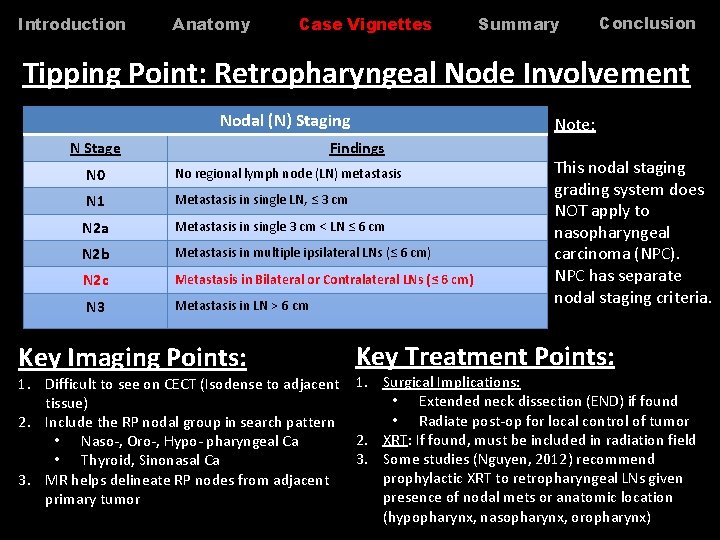
Introduction Anatomy Case Vignettes Summary Conclusion Tipping Point: Retropharyngeal Node Involvement Nodal (N) Staging N Stage Note: Findings N 0 No regional lymph node (LN) metastasis N 1 Metastasis in single LN, ≤ 3 cm N 2 a Metastasis in single 3 cm < LN ≤ 6 cm N 2 b Metastasis in multiple ipsilateral LNs (≤ 6 cm) N 2 c Metastasis in Bilateral or Contralateral LNs (≤ 6 cm) N 3 Metastasis in LN > 6 cm Key Imaging Points: This nodal staging grading system does NOT apply to nasopharyngeal carcinoma (NPC). NPC has separate nodal staging criteria. Key Treatment Points: 1. Difficult to see on CECT (Isodense to adjacent 1. Surgical Implications: • Extended neck dissection (END) if found tissue) • Radiate post-op for local control of tumor 2. Include the RP nodal group in search pattern 2. XRT: If found, must be included in radiation field • Naso-, Oro-, Hypo- pharyngeal Ca 3. Some studies (Nguyen, 2012) recommend • Thyroid, Sinonasal Ca prophylactic XRT to retropharyngeal LNs given 3. MR helps delineate RP nodes from adjacent presence of nodal mets or anatomic location primary tumor (hypopharynx, nasopharynx, oropharynx)
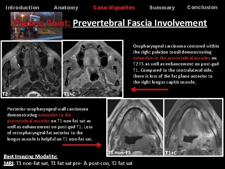
Introduction Anatomy Case Vignettes Summary Conclusion Tipping Point: Prevertebral Fascia Involvement Oropharyngeal carcinoma centered within the right palatine tonsil demonstrating extension to the prevertebral muscles on T 2 FS as well as enhancement on post-gad T 1. Compared to the contralateral side, there is loss of the fat plane anterior to the right longus capitis muscle. T 2 T 1+C Posterior oropharyngeal wall carcinoma demonstrating extension to the prevertebral muscles on T 1 non fat sat as well as enhancement on post-gad T 1. Loss of retropharyngeal fat anterior to the longus muscle is helpful on T 1 non-fat sat. T 1 non-FS Best Imaging Modality: MRI: T 1 non-fat sat, T 1 fat sat pre- & post-con, T 2 fat sat T 1+C
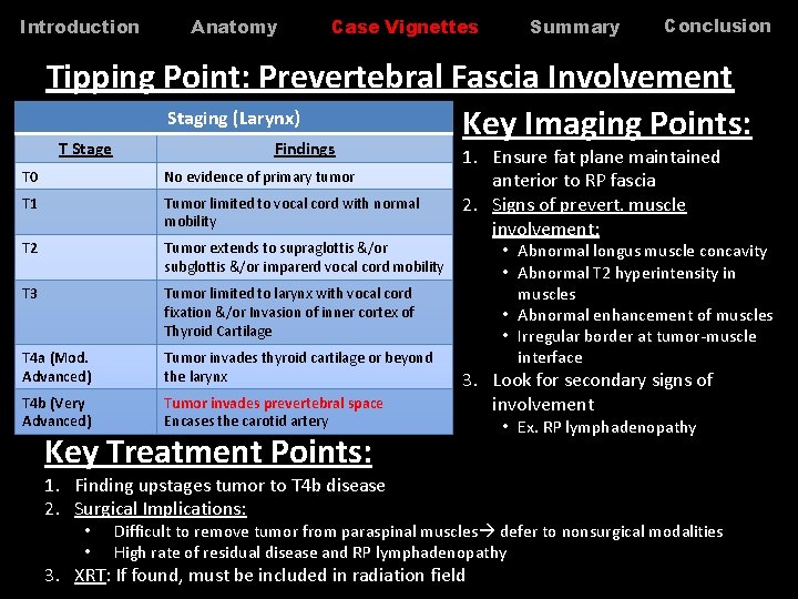
Introduction Anatomy Case Vignettes Summary Conclusion Tipping Point: Prevertebral Fascia Involvement Staging (Larynx) Key Imaging Points: T Stage Findings T 0 No evidence of primary tumor T 1 Tumor limited to vocal cord with normal mobility T 2 Tumor extends to supraglottis &/or subglottis &/or imparerd vocal cord mobility T 3 Tumor limited to larynx with vocal cord fixation &/or Invasion of inner cortex of Thyroid Cartilage T 4 a (Mod. Advanced) Tumor invades thyroid cartilage or beyond the larynx T 4 b (Very Advanced) Tumor invades prevertebral space Encases the carotid artery 1. Ensure fat plane maintained anterior to RP fascia 2. Signs of prevert. muscle involvement: 3. Look for secondary signs of involvement Key Treatment Points: 1. Finding upstages tumor to T 4 b disease 2. Surgical Implications: • • • Abnormal longus muscle concavity • Abnormal T 2 hyperintensity in muscles • Abnormal enhancement of muscles • Irregular border at tumor-muscle interface • Ex. RP lymphadenopathy Difficult to remove tumor from paraspinal muscles defer to nonsurgical modalities High rate of residual disease and RP lymphadenopathy 3. XRT: If found, must be included in radiation field
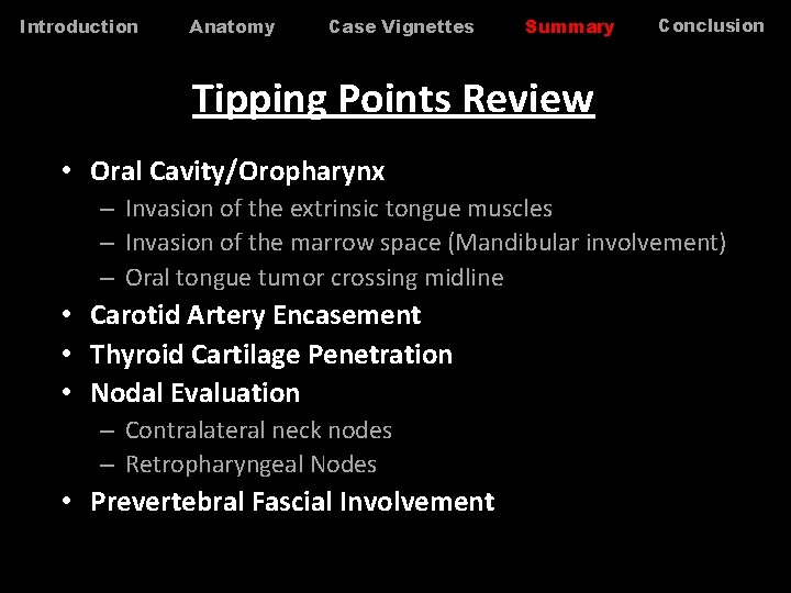
Introduction Anatomy Case Vignettes Summary Conclusion Tipping Points Review • Oral Cavity/Oropharynx – Invasion of the extrinsic tongue muscles – Invasion of the marrow space (Mandibular involvement) – Oral tongue tumor crossing midline • Carotid Artery Encasement • Thyroid Cartilage Penetration • Nodal Evaluation – Contralateral neck nodes – Retropharyngeal Nodes • Prevertebral Fascial Involvement
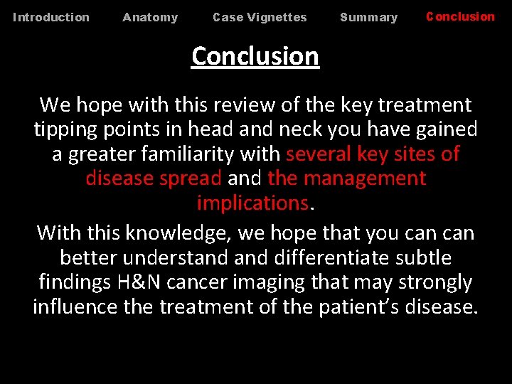
Introduction Anatomy Case Vignettes Summary Conclusion We hope with this review of the key treatment tipping points in head and neck you have gained a greater familiarity with several key sites of disease spread and the management implications. With this knowledge, we hope that you can better understand differentiate subtle findings H&N cancer imaging that may strongly influence the treatment of the patient’s disease.
Introduction Anatomy Case Vignettes Summary Conclusion References • • AJCC Cancer Staging Manual Capote-Moreno A, et al. Prognostic Factors Influencing Contralateral Neck Lymph Nodes Metastases in Oral and Oropharyngeal Cancers. J Oral Maxillofac Surg. 2010 Feb; 68 (2) 268 -75. Godeny M. Prognostic factors in advanced pharyngeal and oral cavity cancer; significance of multimodality imaging in terms of 7 th edition of TNM. Cancer Imaging. 2014, 14: 15. Landry D, Glastonbury CM. Squamous cell carcinoma of the upper aerodigestive tract: A review. Radiol Clin North Am. 2015 Jan; 53(1): 81 -97. Nguyen N, et al. Effectiveness of prophylactic retropharyngeal lymph node irradiation in patients with locally advanced head and neck cancer. BMC Cancer. 2012, 12: 253. Razek AA, Huang BY. Soft tissue tumors of the head and neck. Radiographics. 2011 Nov-Dec; 31 (7): 1923 -54. Yousem D, et al. Resectability issues with head and neck cancer. AJNR Am J Neuroradiol. 2006. Nov-Dec; 27(10): 2024 -36.