DXA in Osteoporosis Key Points In Indications Interpretation


- Slides: 51
DXA in Osteoporosis Key Points In Indications, Interpretation and Diagnosis DR MANGESH PANAT Associate Professor and Unit Head Dept. Of Orthopaedics MGM MEDICAL COLLEGE AURANGABAD
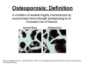
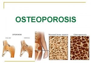
Diagnosis Of osteoporosis Clinical Assessment Diagnostic Criteria Bone Densitometry
Medical History • Symptoms • Osteoporosis symptoms: none • Fracture symptoms: variable • Pain and deformity (hip, spine, forearm, etc. ) • Frequently no symptoms (vertebral fractures)- 1. Asymptomatic fracture-- H/o LBP few months back. 2. If LBP+ Point tenderness Do X ray to Diagnose fracture • Risk factors for osteoporosis • Risk factors for fracture
Clinical Risk Factors for Low Bone Mass • Loss of height • Low body weight • Advanced age • Late age at menarche • Menopausal • Time since menopause • Smoking • Dietary calcium • Alcohol intake • Medications • Inflammatory diseases • Prior fragility fracture Riggs BL. NEJM. 1986; 314: 1676. Ribot C. Clin Endocrinol. 1992; 36: 225.
Risk Factors for Osteoporotic Fracture Are Not The Same as Risk Factors for Low BMD • • • Low BMD Advancing age Prior fragility fracture Family history of osteoporosis or fragility fracture in a first degree relative (genetics) Current smoker Low body weight Falls Sarcopenia Dementia Marshall D et al. BMJ. 1996; 312: 1254. Hui SL et al. J Clin Invest. 1988; 81: 1804. NOF Clinician’s Guide. 2008. Lang, et. al. Bone. 2008; 42: 798 -805
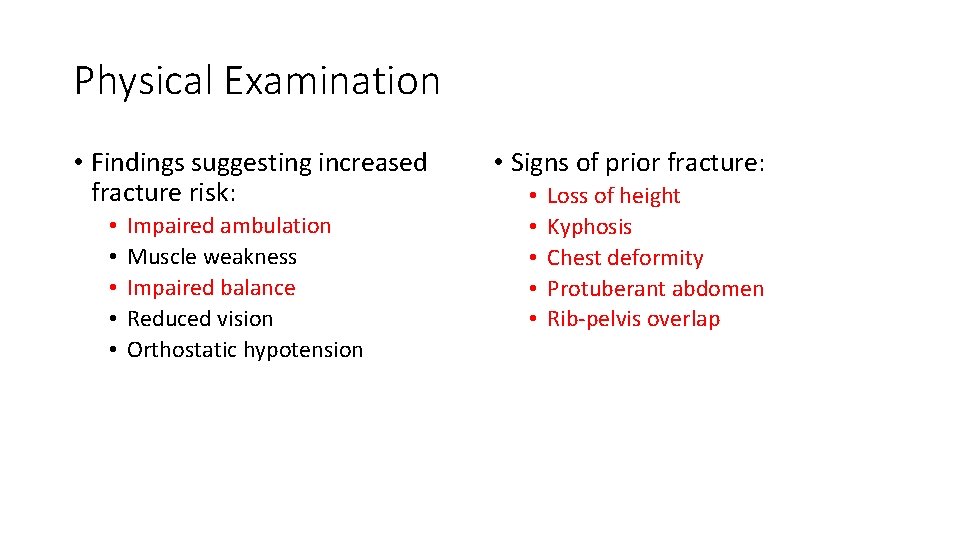
Physical Examination • Findings suggesting increased fracture risk: • • • Impaired ambulation Muscle weakness Impaired balance Reduced vision Orthostatic hypotension • Signs of prior fracture: • • • Loss of height Kyphosis Chest deformity Protuberant abdomen Rib-pelvis overlap
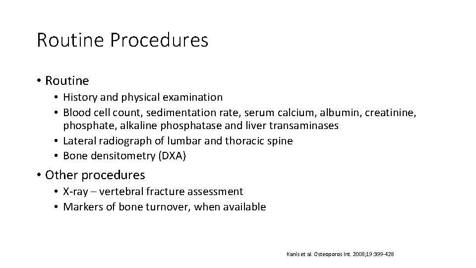
Routine Procedures • Routine • History and physical examination • Blood cell count, sedimentation rate, serum calcium, albumin, creatinine, phosphate, alkaline phosphatase and liver transaminases • Lateral radiograph of lumbar and thoracic spine • Bone densitometry (DXA) • Other procedures • X-ray – vertebral fracture assessment • Markers of bone turnover, when available Kanis et al. Osteoporos Int. 2008; 19: 399 -428
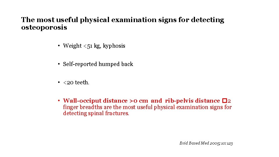
The most useful physical examination signs for detecting osteoporosis • Weight <51 kg, kyphosis • Self-reported humped back • <20 teeth. • Wall-occiput distance >0 cm and rib-pelvis distance � 2 finger breadths are the most useful physical examination signs for detecting spinal fractures. Evid Based Med 2005; 10: 123
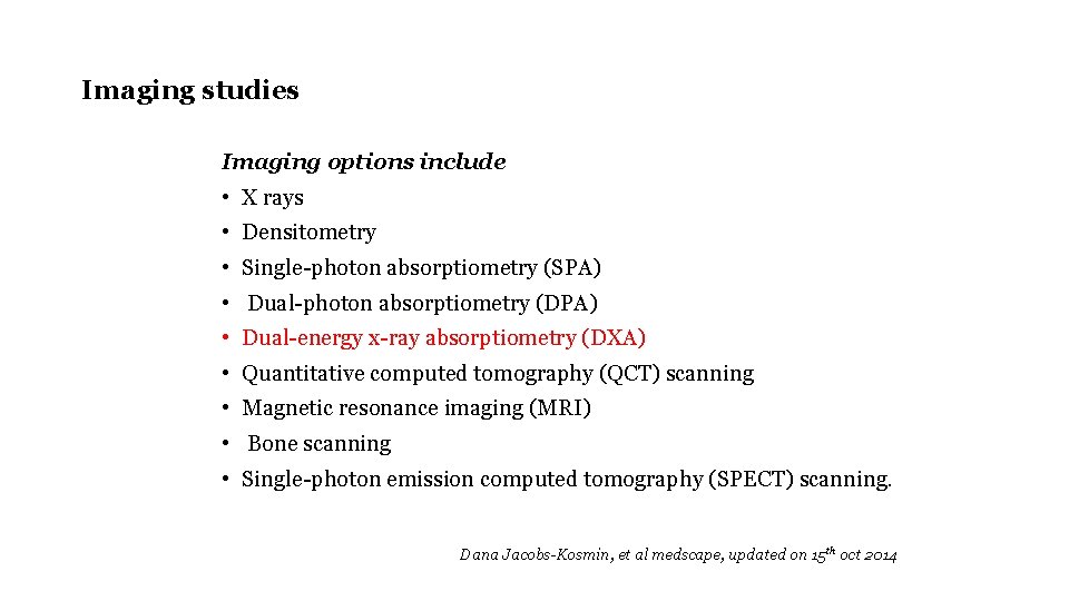
Imaging studies Imaging options include • X rays • Densitometry • Single-photon absorptiometry (SPA) • Dual-photon absorptiometry (DPA) • Dual-energy x-ray absorptiometry (DXA) • Quantitative computed tomography (QCT) scanning • Magnetic resonance imaging (MRI) • Bone scanning • Single-photon emission computed tomography (SPECT) scanning. Dana Jacobs-Kosmin, et al medscape, updated on 15 th oct 2014
Principles of DXA interpretation and Reporting 2/27/2021 10
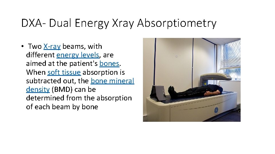
DXA- Dual Energy Xray Absorptiometry • Two X-ray beams, with different energy levels, are aimed at the patient's bones. When soft tissue absorption is subtracted out, the bone mineral density (BMD) can be determined from the absorption of each beam by bone
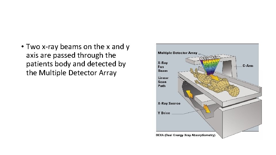
• Two x-ray beams on the x and y axis are passed through the patients body and detected by the Multiple Detector Array
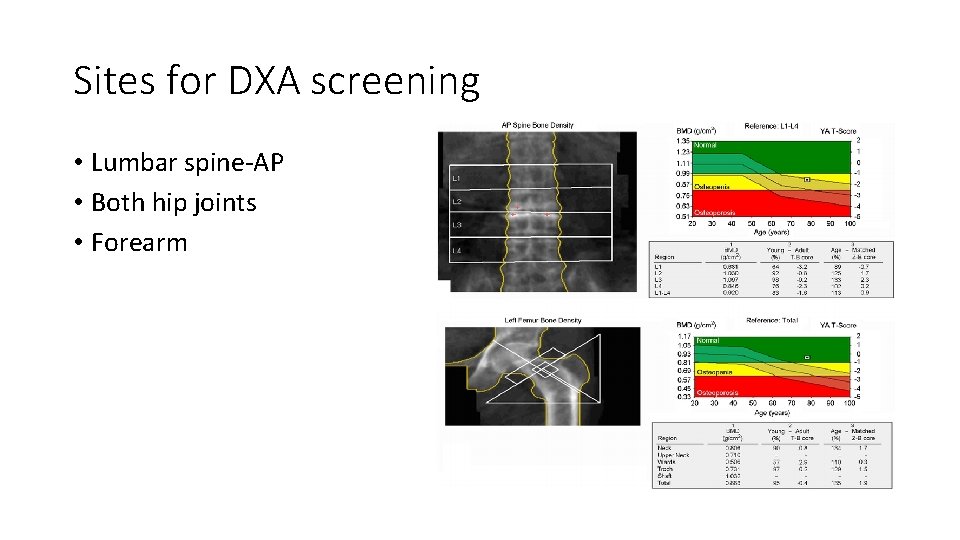
Sites for DXA screening • Lumbar spine-AP • Both hip joints • Forearm
Indications for DXA screening • Females more than 65 yrs. • Females with risk factors • Males more than 70 yrs • Monitoring of treatment
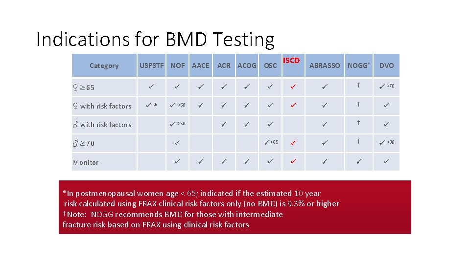
Indications for BMD Testing Category ♀ 65 ♀ with risk factors ♂ with risk factors USPSTF NOF AACE ACR ACOG OSC ISCD ABRASSO NOGG† DVO † >70 * >50 † >50 ♂ 70 Monitor >65 † >80 *In postmenopausal women age < 65; indicated if the estimated 10 year risk calculated using FRAX clinical risk factors only (no BMD) is 9. 3% or higher †Note: NOGG recommends BMD for those with intermediate fracture risk based on FRAX using clinical risk factors
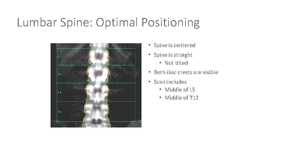
Lumbar Spine: Optimal Positioning • Spine is centered • Spine is straight • Not tilted • Both iliac crests are visible • Scan includes • Middle of L 5 • Middle of T 12
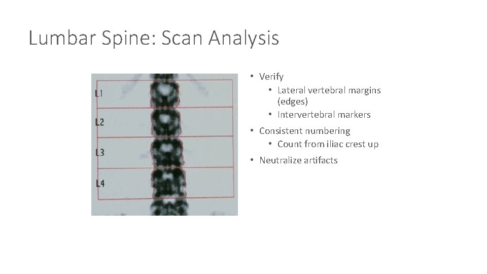
Lumbar Spine: Scan Analysis • Verify • Lateral vertebral margins (edges) • Intervertebral markers • Consistent numbering • Count from iliac crest up • Neutralize artifacts
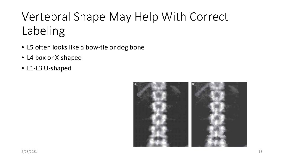
Vertebral Shape May Help With Correct Labeling • L 5 often looks like a bow-tie or dog bone • L 4 box or X-shaped • L 1 -L 3 U-shaped 2/27/2021 18
Baseline DXA Report Minimum Requirements — Results • Sites • PA L 1 -L 4 spine and total hip/femoral neck • Scan forearm if a site is invalid, cannot be imaged, or if there is a history of hyperparathyroidism • Results • BMD in g/cm 2 for each site • The T-score and/or Z-score where appropriate The Writing Group for the ISCD Position Development Conference. J Clin Densitom. 2004; 7: 45.
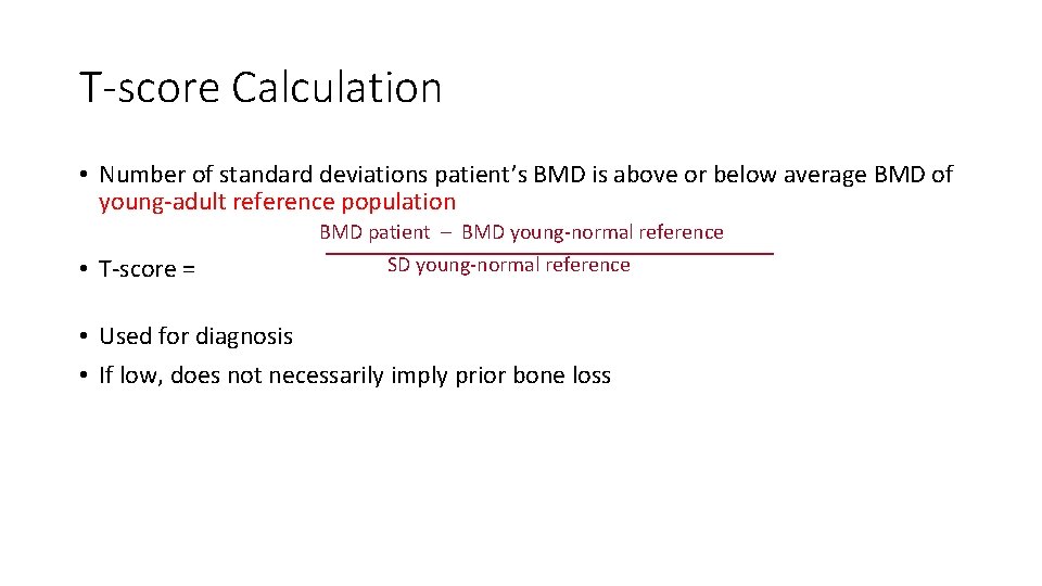
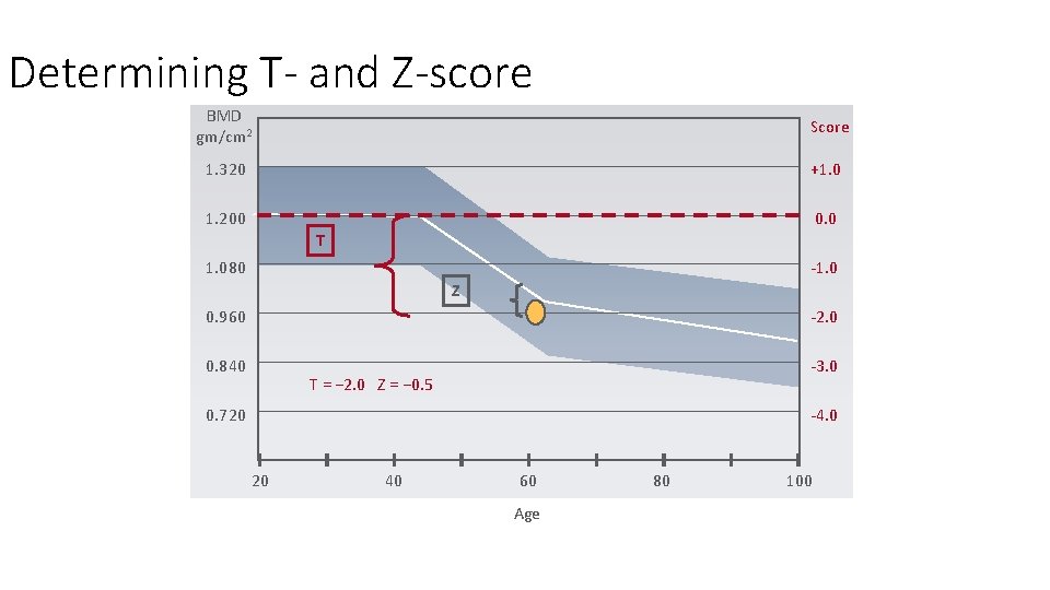
T-score Calculation • Number of standard deviations patient’s BMD is above or below average BMD of young-adult reference population BMD patient – BMD young-normal reference • T-score = SD young-normal reference • Used for diagnosis • If low, does not necessarily imply prior bone loss
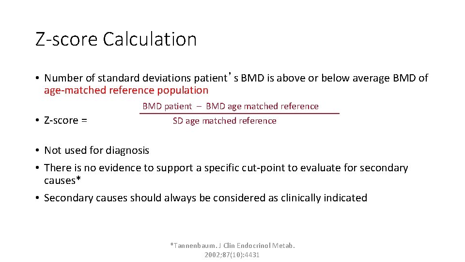
Z-score Calculation • Number of standard deviations patient’s BMD is above or below average BMD of age-matched reference population BMD patient – BMD age matched reference • Z-score = SD age matched reference • Not used for diagnosis • There is no evidence to support a specific cut-point to evaluate for secondary causes* • Secondary causes should always be considered as clinically indicated *Tannenbaum. J Clin Endocrinol Metab. 2002; 87(10): 4431
Determining T- and Z-score BMD gm/cm 2 Score 1. 320 +1. 0 1. 200 0. 0 T 1. 080 -1. 0 Z 0. 960 -2. 0 0. 840 -3. 0 T = − 2. 0 Z = − 0. 5 0. 720 -4. 0 20 40 60 Age 80 100
WHO Classification for Postmenopausal Osteoporosis The T-score compares an individual’s BMD with the mean value for young normals and expresses the difference as a standard deviation score T-score (SD) Normal Equal to -1. 0 or higher Low Bone Mass (Osteopenia) Between -1. 0 and -2. 5 Osteoporosis Equal to -2. 5 or lower Severe Osteoporosis Equal to -2. 5 or lower with fracture World Health Organization. Technical Report Series 843; WHO, Geneva. 1994. Kanis JA et al. J Bone Miner Res. 1994; 9: 1137.
Why Use T-score Instead of Z-score for Diagnosis? • Bone strength related to BMD • Risk of fracture related to BMD • Using Z-score would suggest that osteoporosis does not increase with age and many “normal” patients (Z-score � -1) have osteoporotic fractures • Hence using the T-Score enables comparison with young adult
Baseline DXA Report Minimum Requirements — Results • Diagnosis based on lowest T-score of spine, total hip or femoral neck; if hip or spine not interpretable, use the 33% site of the forearm • Report only one diagnosis • Don’t say: “osteoporosis at spine, osteopenia at hip” • Use T-scores for diagnosis in postmenopausal women and men age 50+ The Writing Group for the ISCD Position Development Conference. J Clin Densitom. 2004; 7: 45.
DXA(Dual Energy X ray Absorptiometry) Key Points • Use L 1 -L 4 for BMD measurement • Presence of fragility fracture is Diagnostic of OP. BMD in such cases is done to assess the treatment effect during follow up. • Repeat DXA should be done on same machine or at least machine made by same manufacturer. • Don’t repeat BMD frequently( repeat only after 1. 5 -2 years) • Look for the position of patient on baseline and follow up visit. • Be suspicious of sudden rise in BMD between baseline and follow up values. 2/27/2021 26
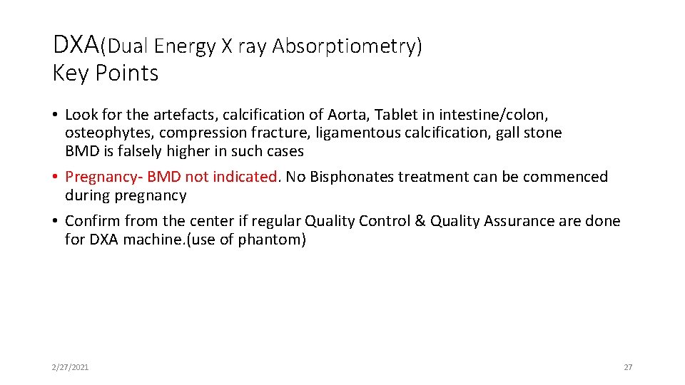
DXA(Dual Energy X ray Absorptiometry) Key Points • Look for the artefacts, calcification of Aorta, Tablet in intestine/colon, osteophytes, compression fracture, ligamentous calcification, gall stone BMD is falsely higher in such cases • Pregnancy- BMD not indicated. No Bisphonates treatment can be commenced during pregnancy • Confirm from the center if regular Quality Control & Quality Assurance are done for DXA machine. (use of phantom) 2/27/2021 27
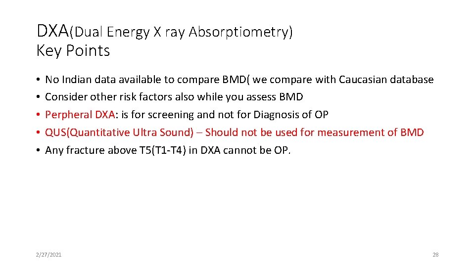
DXA(Dual Energy X ray Absorptiometry) Key Points • • • No Indian data available to compare BMD( we compare with Caucasian database Consider other risk factors also while you assess BMD Perpheral DXA: is for screening and not for Diagnosis of OP QUS(Quantitative Ultra Sound) – Should not be used for measurement of BMD Any fracture above T 5(T 1 -T 4) in DXA cannot be OP. 2/27/2021 28
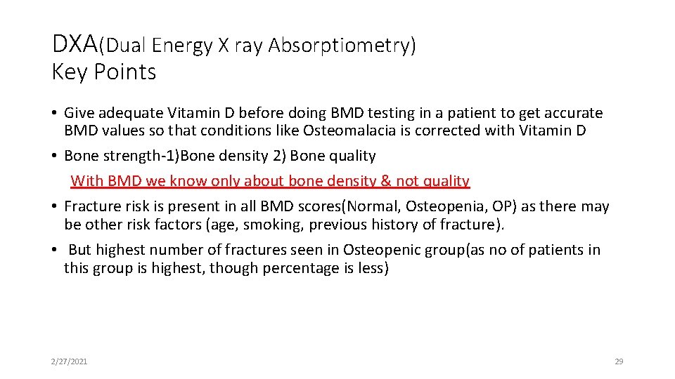
DXA(Dual Energy X ray Absorptiometry) Key Points • Give adequate Vitamin D before doing BMD testing in a patient to get accurate BMD values so that conditions like Osteomalacia is corrected with Vitamin D • Bone strength-1)Bone density 2) Bone quality With BMD we know only about bone density & not quality • Fracture risk is present in all BMD scores(Normal, Osteopenia, OP) as there may be other risk factors (age, smoking, previous history of fracture). • But highest number of fractures seen in Osteopenic group(as no of patients in this group is highest, though percentage is less) 2/27/2021 29
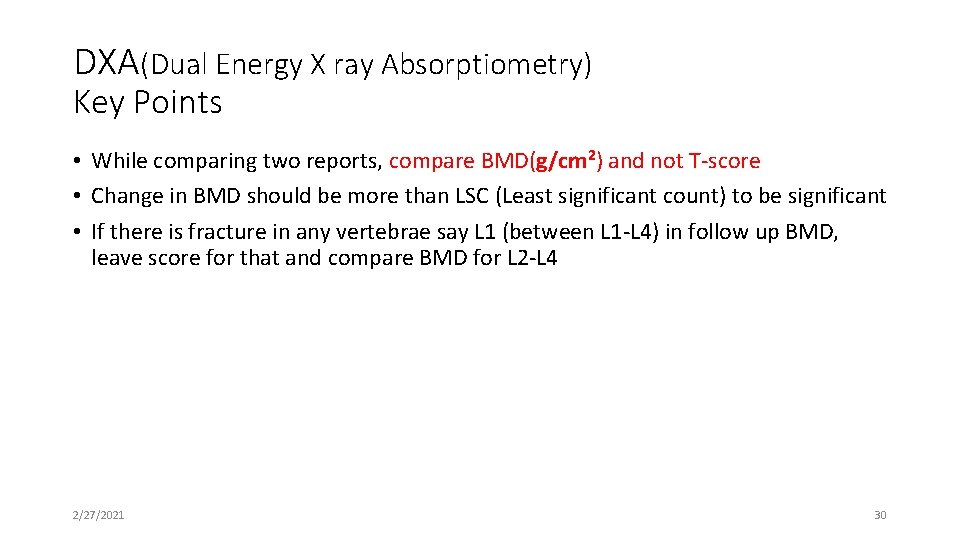
DXA(Dual Energy X ray Absorptiometry) Key Points • While comparing two reports, compare BMD(g/cm 2) and not T-score • Change in BMD should be more than LSC (Least significant count) to be significant • If there is fracture in any vertebrae say L 1 (between L 1 -L 4) in follow up BMD, leave score for that and compare BMD for L 2 -L 4 2/27/2021 30
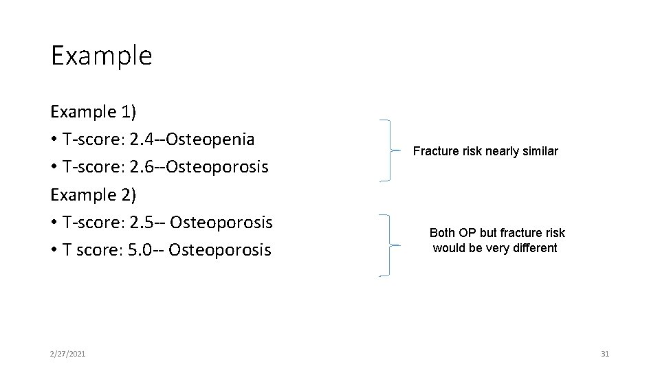
Example 1) • T-score: 2. 4 --Osteopenia • T-score: 2. 6 --Osteoporosis Example 2) • T-score: 2. 5 -- Osteoporosis • T score: 5. 0 -- Osteoporosis 2/27/2021 Fracture risk nearly similar Both OP but fracture risk would be very different 31
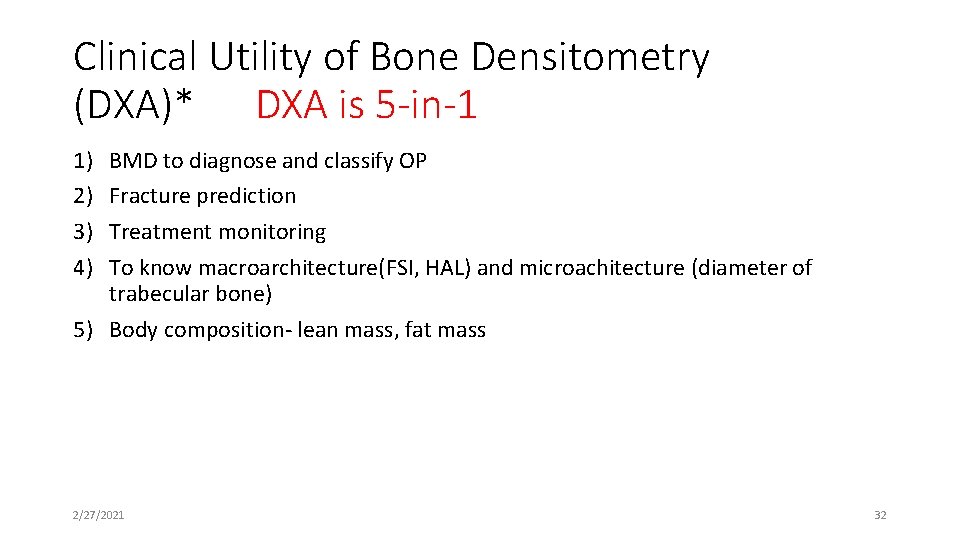
Clinical Utility of Bone Densitometry (DXA)* DXA is 5 -in-1 1) 2) 3) 4) BMD to diagnose and classify OP Fracture prediction Treatment monitoring To know macroarchitecture(FSI, HAL) and microachitecture (diameter of trabecular bone) 5) Body composition- lean mass, fat mass 2/27/2021 32
Limitations of 1994 WHO Classification • Not intended as treatment guidelines • Definitions do not necessarily apply to other populations (e. g. , men, non. Caucasians, premenopausal women) • Does not recognize that a presumptive diagnosis of osteoporosis can be made by a low-trauma (fragility) fracture regardless of the patient’s BMD • Does not differentiate between osteoporosis and other causes of low BMD
ISCD Official Position Diagnosis in Premenopausal Women (age 20 and older) • WHO classification should not be applied to healthy premenopausal women • For women prior to menopause, Z-scores, rather than T-scores, are preferred. This is particularly important in children. • A Z-score of -2. 0 or lower is defined as “below the expected range for age” and a Z-score above -2. 0 is “within the expected range for age. ” • The diagnosis of osteoporosis in premenopausal women should not be made on the basis of densitometric criteria alone
ISCD Official Position Diagnosis in Perimenopausal Women • The WHO diagnostic criteria may be applied to women in the menopausal transition • BMD measurement is indicated for women during the menopausal transition who have clinical risk factors for fracture such as low body weight, prior fracture or high-risk medications Simonelli, C. , et. al. , J Clin Densitom; 2008; 11: 109 -122
ISCD Official Position Diagnosis in Children and Adolescents Males and Females Ages 5 -19 • Should NOT be made on the basis of densitometric criteria alone • The diagnosis of osteoporosis requires the presence of both a clinically significant fracture history and low bone mineral content or bone mineral density • Clinically significant fracture history is defined as: • Long bone fracture of the lower extremities, and/or • Vertebral compression fracture, and/or • Two or more long-bone fractures of the upper extremities • Low BMC or BMD is defined as a Z-score ≤ -2. 0 adjusted for age, gender and body size as appropriate Rauch F, et. al. , J Clin Densitom; 2008; 11: 22 -28
ISCD Official Position Diagnosis in Men • Age 50 and older • T-scores are preferred • The WHO densitometric classification is applicable • In men younger than age 50 • Z-scores, not T-scores are preferred • A Z-score of -2 or lower is defined as “below the expected range for age” and a Z-score above -2 is “within the expected range for age” • Osteoporosis cannot be diagnosed on the basis of BMD alone
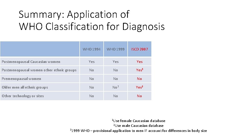
Summary: Application of WHO Classification for Diagnosis WHO 1994 WHO 1999 ISCD 2007 Postmenopausal Caucasian women Yes Yes Postmenopausal women other ethnic groups No No Yes¹ Premenopausal women No No No Older men all ethnic groups No No 3 Yes² Other technology or sites No No No 1 Use female Caucasian database 2 Use male Caucasian database 31999 WHO – provisional application to men IF account for differences in body size
Vertebral Fractures • Important to detect because they • Predict future fractures • Are associated with increased mortality and morbidity (reduced quality of life) • Often missed in the course of routine medical care • Can be prevented with appropriate therapy • Traditionally require X-ray for diagnosis • X-ray is usually not obtained • Possible solution: VFA can diagnose fracture at the time of DXA BMD measurement
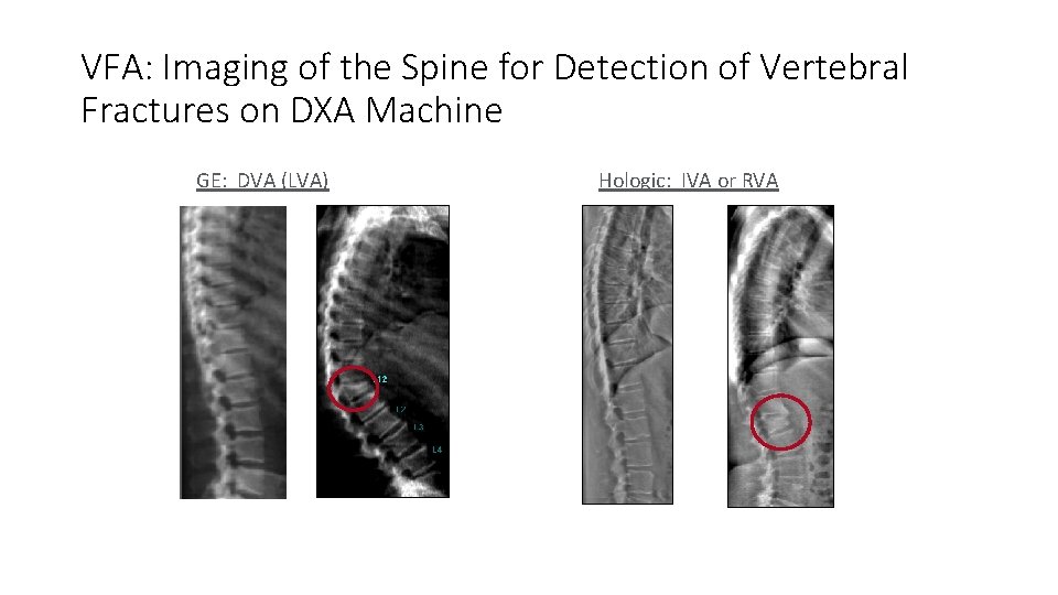
What Is VFA? • Vertebral fracture assessment – diagnosing vertebral fractures using a DXA machine • A visual technique – different than the quantitative methodology of DXA for measurement of BMD Lateral imaging of Plain X ray spine/ VFA are important to diagnose fracture
VFA: Imaging of the Spine for Detection of Vertebral Fractures on DXA Machine GE: DVA (LVA) Hologic: IVA or RVA
ISCD Official Position Indications for VFA • Consider VFA when the results may influence clinical management • Osteopenia plus any of the following: Female Male • Age of patient >70 > 80 • Historical height loss ≥ 4 cm (1. 6 in) ≥ 6 cm (2. 4 in) • Prospective height loss ≥ 2 cm (0. 8 in) ≥ 3 cm (1. 2 in) • Self-reported vertebral fracture not previously documented • 2 or more: female 60 -69 or male 70 -79, non-vertebral fracture, chronic systemic disease, androgen deprivation therapy, height loss 2 -4 cm female or 3 -6 cm male • Chronic steroids ≥ 5 mg ≥ 3 months • DXA diagnosis of osteoporosis if finding ≥ 1 vertebral fracture would alter clinical management Schousboe, et. al. , J Clin Densitom 11: 92 -108, 2008
Relative Contraindications for Central DXA • Pregnancy • Recent contrast study (generally less than 72 hours) • Spine measurement only • Recent nuclear medicine scan • Extensive orthopedic instrumentation • Body weight greater than table limit • Solution: Measure the forearm
Fracture Risk Assessment
Bone Density and Fracture Risk • Bone mineral density is important determinant of bone strength • Bone strength is important predictor of fracture risk • But there is more…
Multiple Fracture Risk Factors Exist Which Ones to Use? • • • Age Prior fracture Low body weight Weight loss Inactivity Glucocorticoids Hyperparathyroidism Diabetes type 1 Anorexia Gastrectomy • • • Gender (female) Current smoking Low sunlight exposure Family Hx of fracture Surgical menopause Low calcium intake Hyperthyroidism Diabetes type 2 Rheumatoid arthritis Espallargues, M. et al. Osteoporos Int. 2001; 12: 811 -822
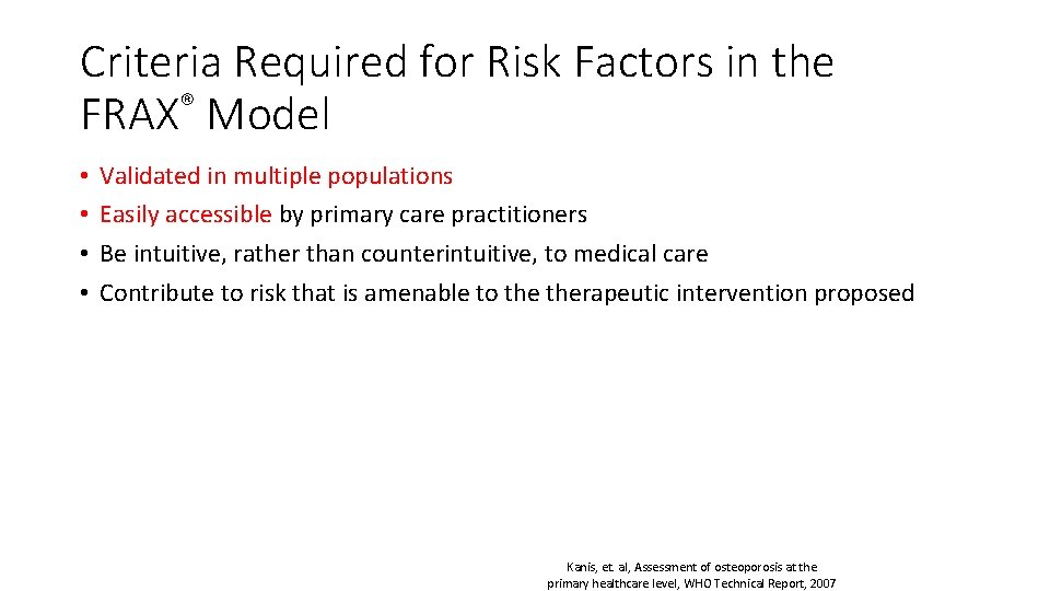
Criteria Required for Risk Factors in the FRAX® Model • • Validated in multiple populations Easily accessible by primary care practitioners Be intuitive, rather than counterintuitive, to medical care Contribute to risk that is amenable to therapeutic intervention proposed Kanis, et. al, Assessment of osteoporosis at the primary healthcare level, WHO Technical Report, 2007
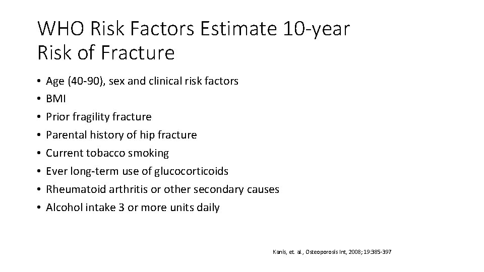
WHO Risk Factors Estimate 10 -year Risk of Fracture • • Age (40 -90), sex and clinical risk factors BMI Prior fragility fracture Parental history of hip fracture Current tobacco smoking Ever long-term use of glucocorticoids Rheumatoid arthritis or other secondary causes Alcohol intake 3 or more units daily Kanis, et. al. , Osteoporosis Int, 2008; 19: 385 -397
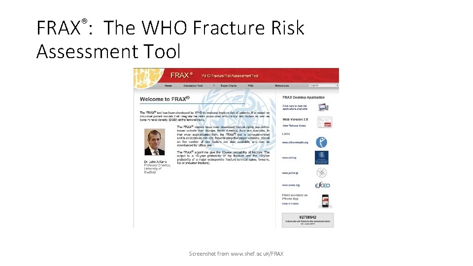
FRAX®: The WHO Fracture Risk Assessment Tool Screenshot from www. shef. ac. uk/FRAX
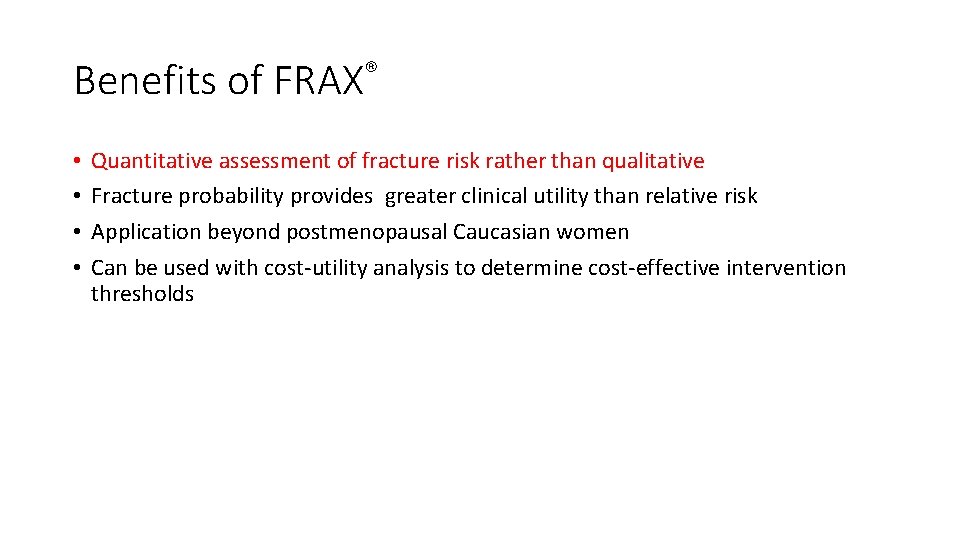
Benefits of FRAX® • • Quantitative assessment of fracture risk rather than qualitative Fracture probability provides greater clinical utility than relative risk Application beyond postmenopausal Caucasian women Can be used with cost-utility analysis to determine cost-effective intervention thresholds
THANK YOU