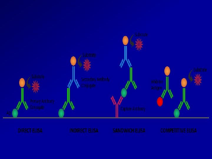
Direct Examination 1 Antigen Detection immunofluorescence ELISA etc


 The absorption spectrum of the purified physical particle in the ultraviolet range should")
- Slides: 16
Direct Examination 1. Antigen Detection immunofluorescence, ELISA etc. 2. Electron Microscopy morphology of virus particles immune electron microscopy 3. Light Microscopy histological appearance inclusion bodies 4. Viral Genome Detection hybridization with specific nucleic acid probes polymerase chain reaction (PCR)
Serology Detection of rising titres of antibody between acute and convalescent stages of infection, or the detection of Ig. M in primary infection.
Molecular Methods based on the detection of viral genome are also commonly known as molecular methods. It is often said that molecular methods is the future direction of viral diagnosis. However in practice, although the use of these methods is indeed increasing, the role played by molecular methods in a routine diagnostic virus laboratory is still small compared to conventional methods. It is certain though that the role of molecular methods will increase rapidly in the near future.
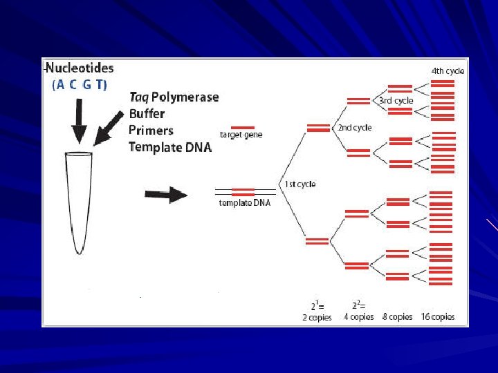
Polymerase Chain Reaction PCR allows the in vitro amplification of specific target DNA sequences by a factor of 106 and is thus an extremely sensitive technique. It is based on an enzymatic reaction involving the use of synthetic oligonucleotides flanking the target nucleic sequence of interest. These oligonucleotides act as primers for thermostable Taq polymerase. Repeated cycles (usually 25 to 40) of denaturation of the template DNA (at 94 o. C), annealing of primers to their complementary sequences (50 o. C), and primer extension (72 o. C) result in the exponential production of the specific target fragment. Detection and identification of the PCR product is usually carried out by agarose gel electrophoresis, hybridization with a specific oligonucleotide probe, restriction enzyme analysis, or DNA sequencing.
Schematic of PCR Each cycle doubles the copy number of the target
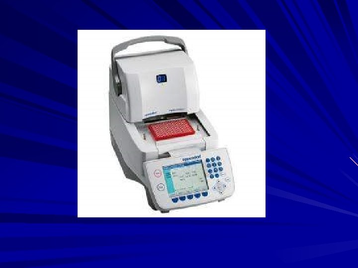
Viral genome detection: PCR
Detection of Virus-Infected Cells 1. Development of cytopathic effects 2. Appearance of a virus-encoded protein, such as the hemagglutinin of influenza virus. 3. Adsorption (hemadsorption) of erythrocytes to infected cells, due to the presence of virus-encoded hemagglutinin (parainfluenza, influenza) in cellular membranes 4. Detection of virus-specific nucleic acid 5. Inclusion Body Formation
Quantitation of Viruses A- Physical Methods 1 -polymerase chain reaction 2 -serologic tests such as radioimmunoassays (RIA) and enzymelinked immunosorbent assays (ELISA) 3 -Hemagglutination assays 4 - Virus particles can be counted directly in the electron microscope by comparison with a standard suspension of latex particles of similar small size
B-Biologic Methods 1 -End point biologic assays depend on the measurement of animal death, animal infection, or cytopathic effects in tissue culture at a series of dilutions of the virus being tested. The titer is expressed as the 50% infectious dose (ID 50), which is the reciprocal of the dilution of virus that produces the effect in 50% of the cells or animals inoculated. 2 -Plaque assay: The most widely used assay for infectious virus a single plaque can arise from a single infectious virus particle, termed a plaque-forming unit (PFU).
3 - Pock formation: viruses, eg, herpes and vaccinia, form pocks when inoculated onto the chorioallantoic membrane of an embryonated egg. Such viruses can be quantitated by relating the number of pocks counted to the viral dilution inoculated.
Identification of a Particle as a Virus When a characteristic physical particle has been obtained, it should fulfill the following criteria before it is identified as a virus particle: (1) The particle can be obtained only from infected cells or tissues. (2) Particles obtained from various sources are identical regardless of the cellular origin in which the virus is grown. (3) The degree of infective activity of the preparation varies directly with the number of particles present. (4) Destruction of the physical particle by chemical or physical means is associated with a loss of viral activity. (5) Certain properties of the particles and infectivity must be shown to be identical, e. g. , their sedimentation behavior in the ultracentrifuge and their p. H stability curves.
(6) The absorption spectrum of the purified physical particle in the ultraviolet range should coincide with the ultraviolet inactivation spectrum of the virus. (7) Antisera prepared against the infectious virus should react with the characteristic particle and vice versa. Direct observation of an unknown virus can be accomplished by electron microscopic examination of aggregate formation in a mixture of antisera and crude viral suspension. (8) The particles should be able to induce the characteristic disease in vivo. (9) Passage of the particles in tissue culture should result in the production of progeny with biologic and antigenic properties of the virus.