
MICROBIOLOGY WITH DISEASES BY TAXONOMY THIRD EDITION Chapter
MICROBIOLOGY WITH DISEASES BY TAXONOMY, THIRD EDITION Chapter Microscopy, Staining, and Classification 4 Lecture prepared by Mindy Miller-Kittrell, University of Tennessee, Knoxville Copyright © 2011 Pearson Education Inc.

Microscopy and Staining Animation: Microscopy and Staining: Overview Copyright © 2011 Pearson Education Inc.
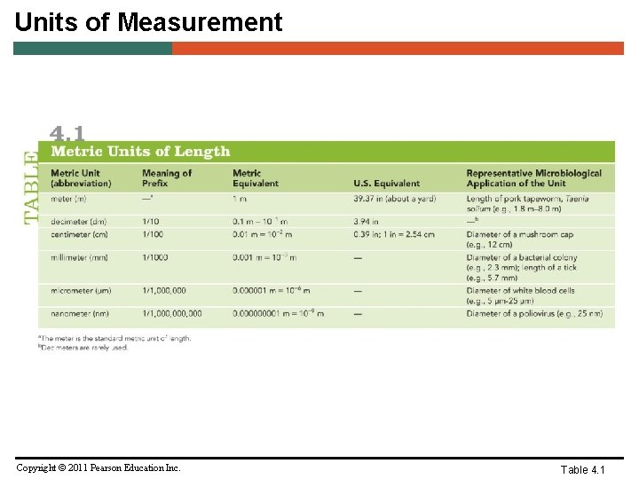
Units of Measurement Copyright © 2011 Pearson Education Inc. Table 4. 1
Microscopy • General Principles of Microscopy – Wavelength of radiation – Magnification – Resolution – Contrast Copyright © 2011 Pearson Education Inc.
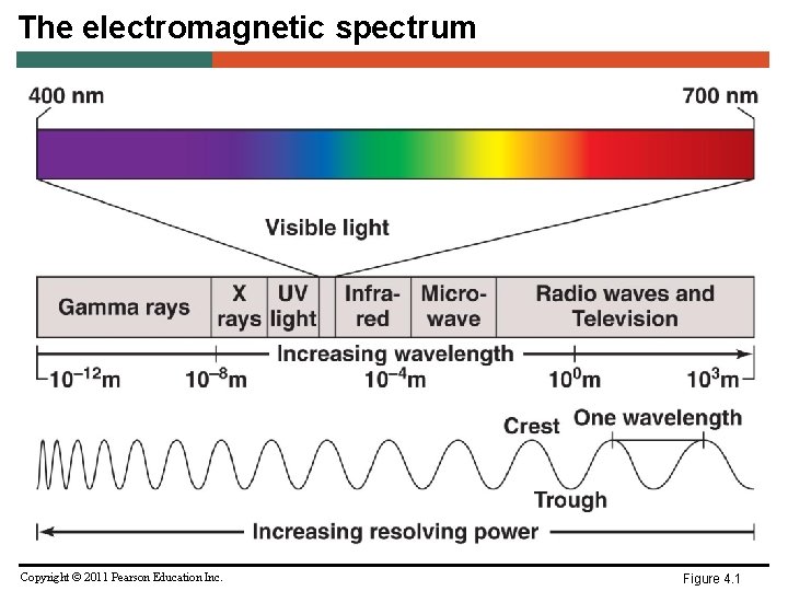
The electromagnetic spectrum Copyright © 2011 Pearson Education Inc. Figure 4. 1

Light refraction and image magnification Copyright © 2011 Pearson Education Inc. Figure 4. 2
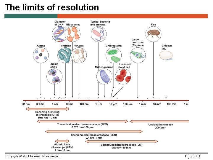
The limits of resolution Copyright © 2011 Pearson Education Inc. Figure 4. 3
Microscopy • General Principles of Microscopy – Contrast – Differences in intensity between two objects, or between an object and background – Important in determining resolution – Staining increases contrast – Use of light that is in phase increases contrast Copyright © 2011 Pearson Education Inc.
Microscopy • Light Microscopy – Bright-field microscopes – Simple – Contain a single magnifying lens – Similar to magnifying glass – Leeuwenhoek used simple microscope to observe microorganisms Copyright © 2011 Pearson Education Inc.
Microscopy • Light Microscopy – Bright-field microscopes – Compound – Series of lenses for magnification – Light passes through specimen into objective lens – Oil immersion lens increases resolution – Have one or two ocular lenses – Total magnification = magnification of objective lens X magnification of ocular lens – Most have condenser lens (direct light through specimen) Copyright © 2011 Pearson Education Inc.
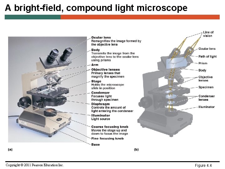
A bright-field, compound light microscope Copyright © 2011 Pearson Education Inc. Figure 4. 4
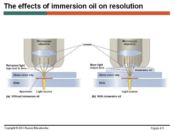
The effects of immersion oil on resolution Copyright © 2011 Pearson Education Inc. Figure 4. 5
Microscopy • Light Microscopy – Dark-field microscopes – Best for observing pale objects – Only light rays scattered by specimen enter objective lens – Specimen appears light against dark background – Increases contrast and enables observation of more details Copyright © 2011 Pearson Education Inc.
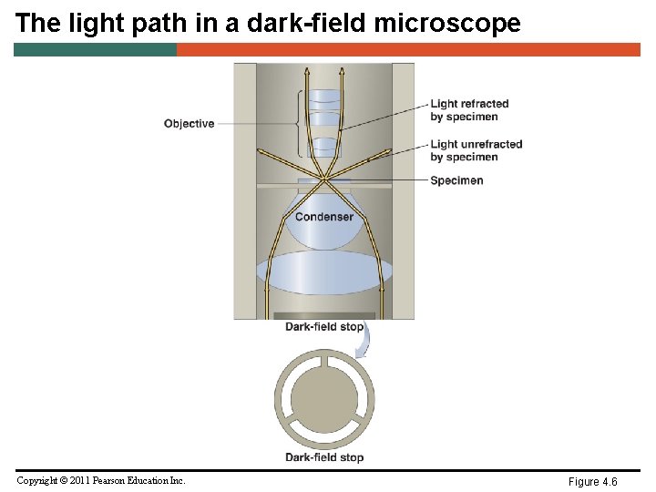
The light path in a dark-field microscope Copyright © 2011 Pearson Education Inc. Figure 4. 6
Microscopy • Light Microscopy – Phase microscopes – Used to examine living organisms or specimens that would be damaged/altered by attaching them to slides or staining – Light rays in phase produce brighter image, while light rays out of phase produce darker image – Contrast is created because light waves are out of phase – Two types – Phase-contrast microscope – Differential interference contrast microscope Copyright © 2011 Pearson Education Inc.
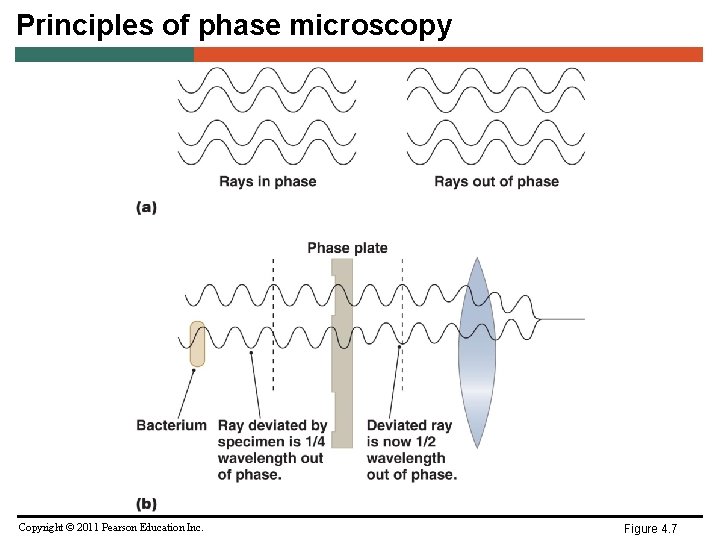
Principles of phase microscopy Copyright © 2011 Pearson Education Inc. Figure 4. 7
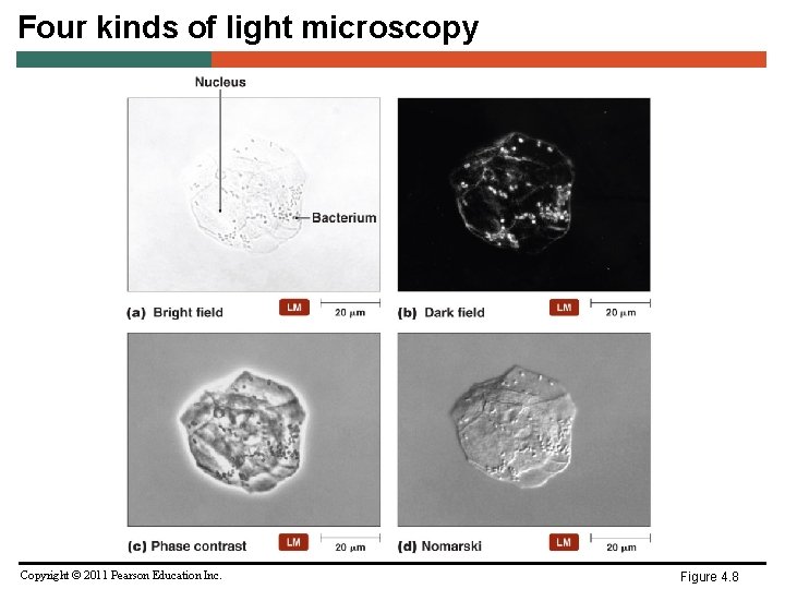
Four kinds of light microscopy Copyright © 2011 Pearson Education Inc. Figure 4. 8
Microscopy • Light Microscopy – Fluorescent microscopes – Direct UV light source at specimen – Specimen radiates energy back as a longer, visible wavelength – UV light increases resolution and contrast – Some cells are naturally fluorescent; others must be stained – Used in immunofluorescence to identify pathogens and to make visible a variety of proteins Copyright © 2011 Pearson Education Inc.
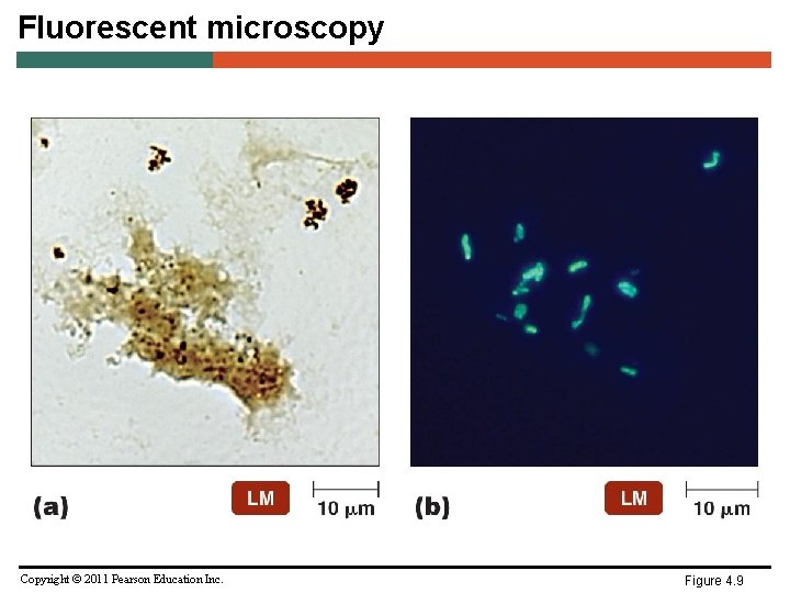
Fluorescent microscopy Copyright © 2011 Pearson Education Inc. Figure 4. 9
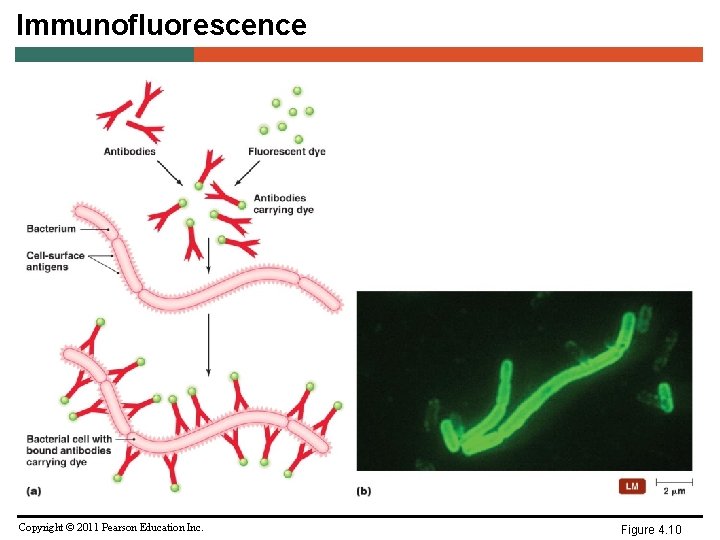
Immunofluorescence Copyright © 2011 Pearson Education Inc. Figure 4. 10
Microscopy • Light Microscopy – Confocal microscopes – Use fluorescent dyes – Use UV lasers to illuminate fluorescent chemicals in a single plane – Resolution increased because emitted light passes through pinhole aperture – Computer constructs 3 -D image from digitized images Copyright © 2011 Pearson Education Inc.
Microscopy • Electron Microscopy – Light microscopes cannot resolve structures closer than 200 nm – Electron microscopes have greater resolving power and magnification – Magnifies objects 10, 000 X to 100, 000 X – Detailed views of bacteria, viruses, internal cellular structures, molecules, and large atoms – Two types – Transmission electron microscopes – Scanning electron microscopes Copyright © 2011 Pearson Education Inc.
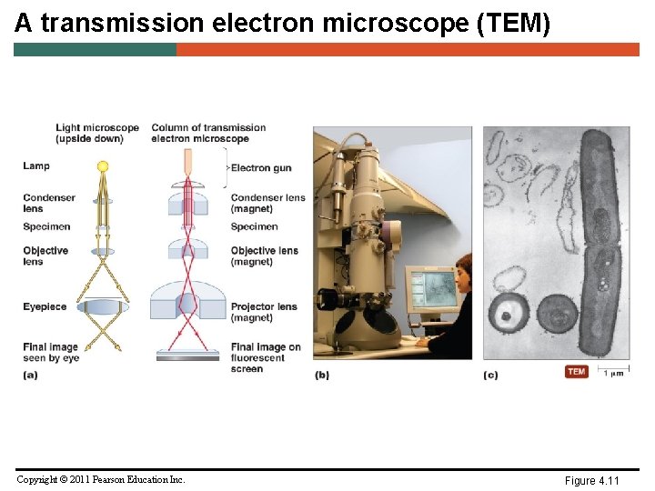
A transmission electron microscope (TEM) Copyright © 2011 Pearson Education Inc. Figure 4. 11
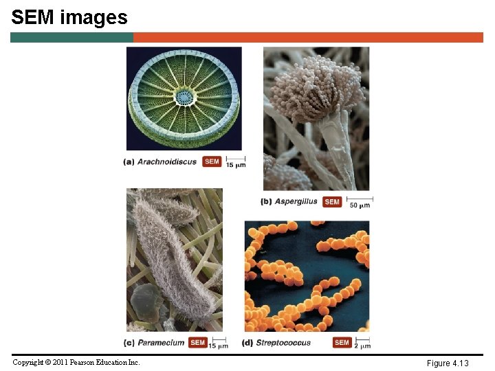
SEM images Copyright © 2011 Pearson Education Inc. Figure 4. 13
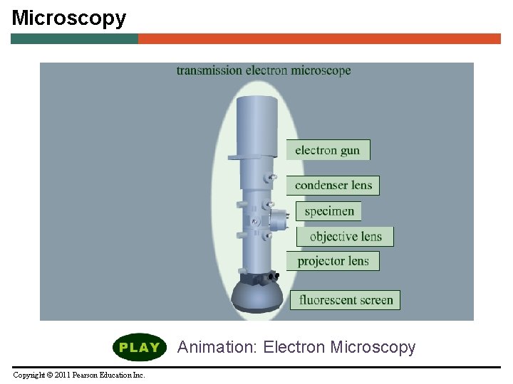
Microscopy Animation: Electron Microscopy Copyright © 2011 Pearson Education Inc.
Microscopy • Probe Microscopy – Magnifies more than 100, 000 times – Two types – Scanning tunneling microscopes – Atomic force microscopes Copyright © 2011 Pearson Education Inc.
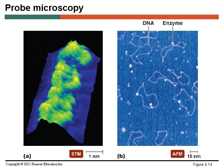
Probe microscopy Copyright © 2011 Pearson Education Inc. Figure 4. 14
Staining • Increases contrast and resolution by coloring specimens with stains/dyes • Smear of microorganisms (thin film) made prior to staining • Microbiological stains contain chromophore • Acidic dyes stain alkaline structures; more commonly, basic dyes stain acidic structures Copyright © 2011 Pearson Education Inc.
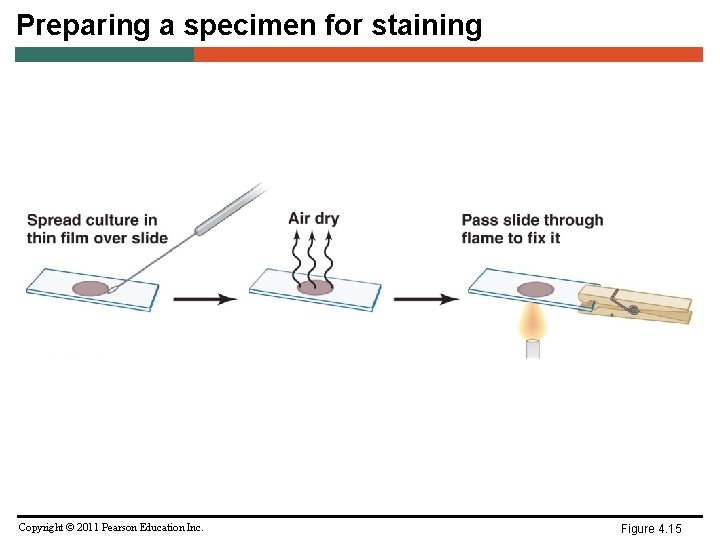
Preparing a specimen for staining Copyright © 2011 Pearson Education Inc. Figure 4. 15
Staining • Simple stains • Differential stains – Gram stain – Acid-fast stain – Endospore stain • Special stains – Negative (capsule) stain – Flagellar stain Copyright © 2011 Pearson Education Inc.
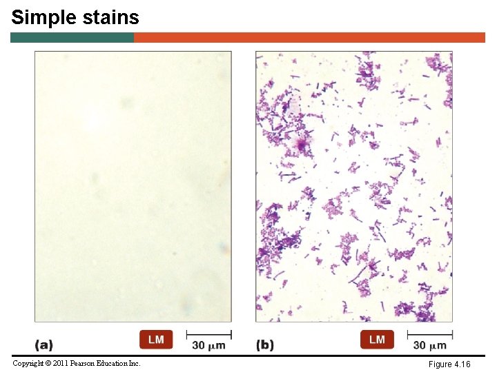
Simple stains Copyright © 2011 Pearson Education Inc. Figure 4. 16
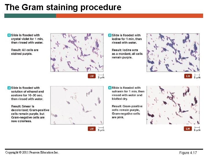
The Gram staining procedure Copyright © 2011 Pearson Education Inc. Figure 4. 17
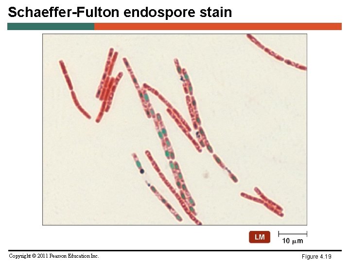
Schaeffer-Fulton endospore stain Copyright © 2011 Pearson Education Inc. Figure 4. 19
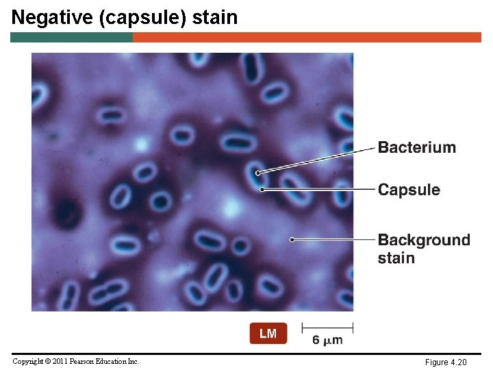
Negative (capsule) stain Copyright © 2011 Pearson Education Inc. Figure 4. 20
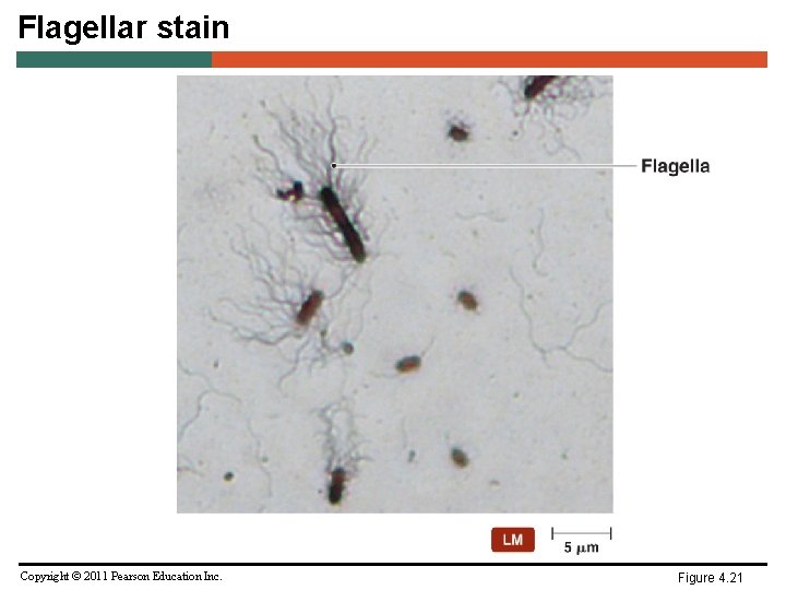
Flagellar stain Copyright © 2011 Pearson Education Inc. Figure 4. 21
Staining • Staining for Electron Microscopy – Chemicals containing heavy metals used for transmission electron microscopy – Stains may bind molecules in specimens or the background Copyright © 2011 Pearson Education Inc.
Classification of Microbes • Taxonomy - The science of classifying, naming and identifying organisms. • Classification- Grouping of related organisms – Hierarchal in nature – Establishes criteria in identification of microbes. – Aids understanding of evolution of organisms. – Organism sorted on basis on degrees of similarities. • Identification – – Methods to determine the taxon in which a Copyright © 2011 Pearson Education Inc.
Systems of Classification • Nomenclature – rules for naming organisms – Carolus Linnaeus (1758) Father of Taxonomy – Binomial Nomenclature – Each living organism has two names, genus & species – Latin or Latinized names – Genus always a noun, capitalized and written first – Specific epithet usually adjective, all lowercase and written last – Two part name must be underlined or italicized Copyright © 2011 Pearson Education Inc.
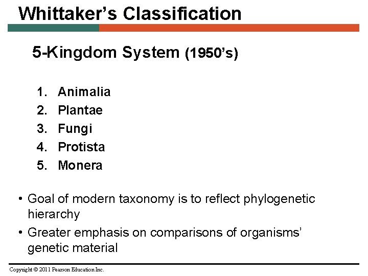
Whittaker’s Classification 5 -Kingdom System (1950’s) 1. 2. 3. 4. 5. Animalia Plantae Fungi Protista Monera • Goal of modern taxonomy is to reflect phylogenetic hierarchy • Greater emphasis on comparisons of organisms’ genetic material Copyright © 2011 Pearson Education Inc.
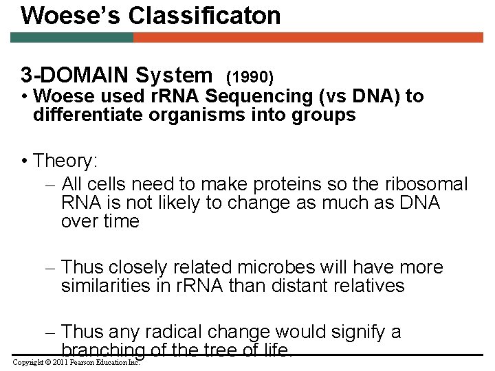
Woese’s Classificaton 3 -DOMAIN System (1990) • Woese used r. RNA Sequencing (vs DNA) to differentiate organisms into groups • Theory: – All cells need to make proteins so the ribosomal RNA is not likely to change as much as DNA over time – Thus closely related microbes will have more similarities in r. RNA than distant relatives – Thus any radical change would signify a branching of the tree of life. Copyright © 2011 Pearson Education Inc.
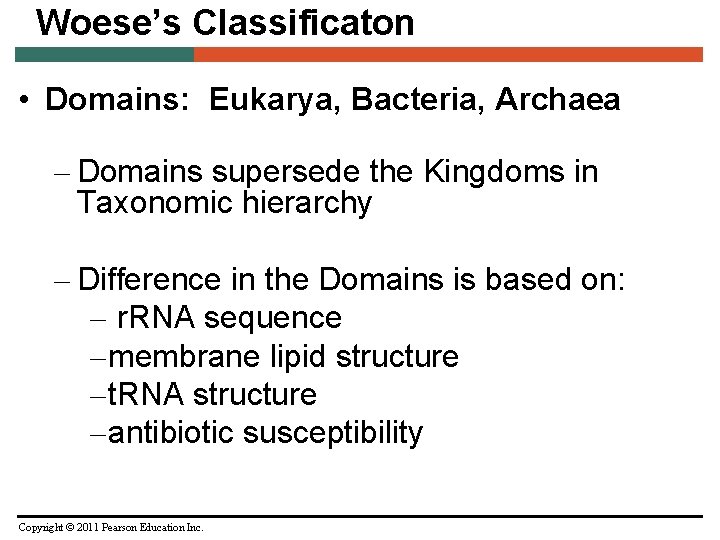
Woese’s Classificaton • Domains: Eukarya, Bacteria, Archaea – Domains supersede the Kingdoms in Taxonomic hierarchy – Difference in the Domains is based on: – r. RNA sequence – membrane lipid structure – t. RNA structure – antibiotic susceptibility Copyright © 2011 Pearson Education Inc.
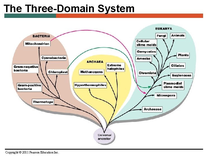
The Three-Domain System Copyright © 2011 Pearson Education Inc.
Eukarya Domain Kingdoms: • Animalia - multicellular; no cell walls; obtain nutrients and energy by ingestion (chemoheterotroph) • Plantae - multicellular; cellulose in cell walls; obtain energy by photosynthesis (photoautotroph) • Fungi - unicellular or multicellular; chitin in cell walls; obtain energy by absorption (chemoheterotroph); develop from spores or hyphal fragments • Protista - a catchall for eukaryotic organisms that do not fit other kingdoms; Unicellular; no cell walls Copyright © 2011 Pearson Education Inc.
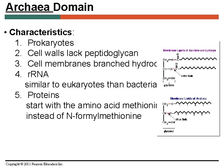
Archaea Domain • Characteristics: 1. Prokaryotes 2. Cell walls lack peptidoglycan 3. Cell membranes branched hydrocarbons 4. r. RNA similar to eukaryotes than bacteria 5. Proteins start with the amino acid methionine instead of N-formylmethionine Copyright © 2011 Pearson Education Inc.
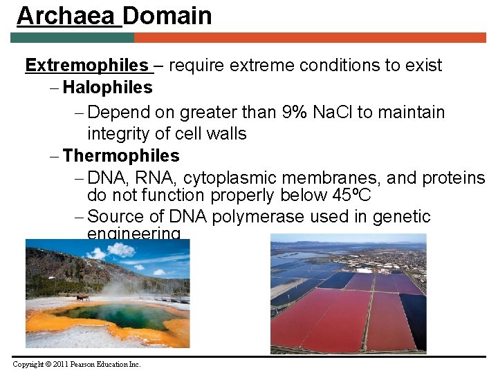
Archaea Domain Extremophiles – require extreme conditions to exist – Halophiles – Depend on greater than 9% Na. Cl to maintain integrity of cell walls – Thermophiles – DNA, RNA, cytoplasmic membranes, and proteins do not function properly below 45ºC – Source of DNA polymerase used in genetic engineering Copyright © 2011 Pearson Education Inc.
Archaea Domain Methanogens – Largest group of archaea – Convert carbon dioxide, hydrogen gas, and organic acids to methane gas – Convert organic wastes in pond, lake, and ocean sediments to methane – Some live in colons of animals – One of primary sources of environmental methane – Have produced ~10 trillion tons of methane that is buried in mud on ocean floor Copyright © 2011 Pearson Education Inc.
Archaea Domain • Phyla: 1. Crenarchaeota 2. Euryarchaeota 3. Korarchaeota -No organisms have been seen 4. Nanoarchaeota (? ) Known only from RNA samples in the envronment Often live in extreme environments Often carry out unusual metabolic processes NONE OF THEM IS KNOWN TO CAUSE DISEASE Copyright © 2011 Pearson Education Inc.
Classification and Identification of Microorganisms • Linnaeus, Whittaker, and Taxonomic Categories – Linnaeus – System classified organisms based on characteristics in common – Grouped organisms that can successfully interbreed into categories called species – Used binomial nomenclature in his system Copyright © 2011 Pearson Education Inc.
Classification and Identification of Microorganisms • Taxonomic and Identifying Characteristics – Physical characteristics – Biochemical tests – Serological tests – Phage typing – Analysis of nucleic acids Copyright © 2011 Pearson Education Inc.
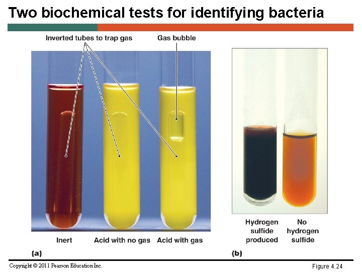
Two biochemical tests for identifying bacteria Copyright © 2011 Pearson Education Inc. Figure 4. 24
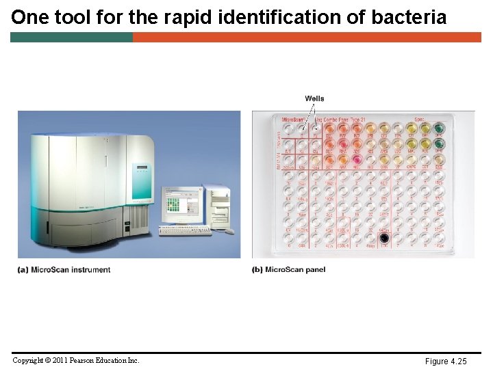
One tool for the rapid identification of bacteria Copyright © 2011 Pearson Education Inc. Figure 4. 25
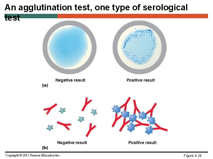
An agglutination test, one type of serological test Copyright © 2011 Pearson Education Inc. Figure 4. 26
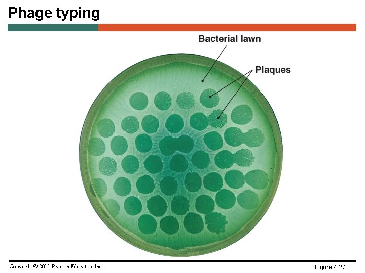
Phage typing Copyright © 2011 Pearson Education Inc. Figure 4. 27
Classification and Identification of Microorganisms • Taxonomic Keys – Dichotomous keys – Series of paired statements where only one of two “either/or” choices applies to any particular organism – Key directs user to another pair of statements, or provides name of organism Copyright © 2011 Pearson Education Inc.
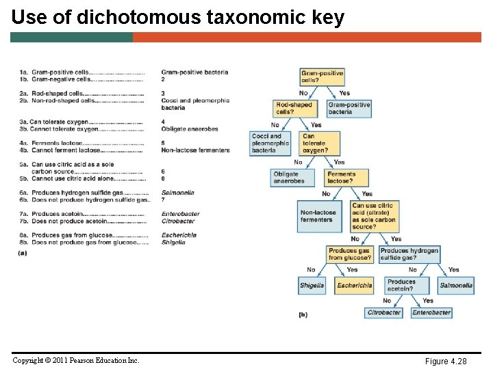
Use of dichotomous taxonomic key Copyright © 2011 Pearson Education Inc. Figure 4. 28
- Slides: 55