Cysts of jaws and oral soft tissues including


 cyst Eruption cyst")
 cyst n Nasolabial (nasoalveolar) cyst n Median cysts")
 n Aneurysmal")
 Non-odontogenic c. (10%) Radicular")


 and eruption cyst n n n n Most frequently involve impacted/late-erupting teeth")

 n n n AD Multiple naevoid BCC")


 cyst Commonest of the non-odontogenic")


- Slides: 29
Cysts of jaws and oral soft tissues, including developmental cysts. Markéta Hermanová
Definition of a cyst n A pathological cavity, lined wholly or in part by epithelium, having fluid or semifluid contents n Broader definition: a pathological cavity, having fluid or semifluid contents, which has not been created by the accumulation of pus
Classification of cysts of the jaws Odontogenic cysts - developmenal - inflammatory n Non-odontogenic cysts n Non-epitheliazed primary bone cysts n Cysts of the soft tissues n
Odontogenic cysts n n - - Developmental Odontogenic keratocysts Dentigerous (follicular) cyst Eruption cyst Lateral periodontal cysts Gingival cyst Glandular odontogenic cyst Inflammatory Radicular cyst (a) apical (b) lateral (c) residual periapical Paradental cyst
Non-odontogenic cysts Nasopalatine duct (incisive canal) cyst n Nasolabial (nasoalveolar) cyst n Median cysts n Palatal cyst of the newborn (Epstein perls; Bohn´s nodules) n
Non-epitheliazed primary bone cysts Solitary bone cyst (simple, traumatic, haemorrhagic bone cyst) n Aneurysmal bone cyst n Stafne´s idiopathic bone cavity n
Incidence of cyst of the jaws Odontogenic cysts (90 %) Non-odontogenic c. (10%) Radicular cysts % Dentigerous cyst % Keratocyst % Paradental cyst Gingival cyst 60 -75 10 -15 5 -10 3 -5 % <1 % Lateral periodontal c. <1 % Nasopalatine cyst 5 -10 % Other non-odontogenic and primary bone cysts <1 %
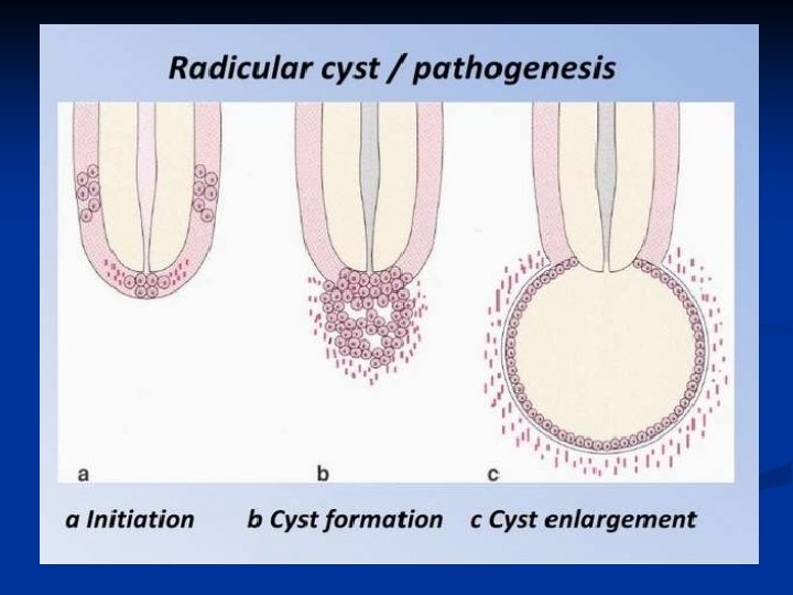
Origins of odontogenic cysts n n - - - Derived from epithelial residues of the tooth-forming organ The main cyst types derived from each residue are: Dental lamina rests/gland of Serres (a) odontogenic keratocysts (b) some lateral periodontal and gingival cysts Reduced enamel epithelium (a) dentigerous cysts (b) paradental cysts Rests of Malassez (a) radicular cysts
Radicular cysts Apical, residual periapical, or lateral subtypes n Apical most common n Associated with non-vital tooth n Apical radiolucency indistinguishable from a periapical granuloma n May be symptomless n Enlargement of cyst leads to bone resorption n
Radicular cyst apikální laterální reziduální
Radicular cyst
=Calcifying odontogenic tumour
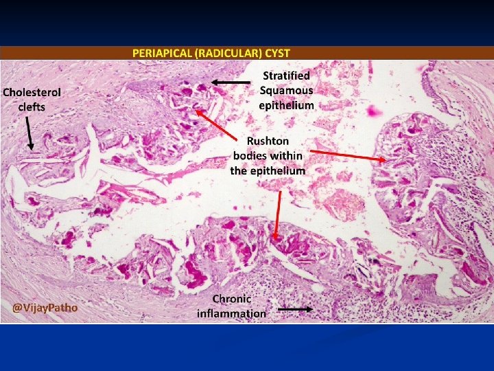
Radicular cyst-histology Arise from proliferation of the rests of Malassez within chronic periapical granulomas n Lined by non-keratinizing squamous epithelium n Supported by a chronically inflammed capsule n Capsule may contain collections of cholesterols n Hypertonic content: breakdown products, n
Expansion of radicular cyst Hydrostatic pressure of the cyst fluid increased due to hypertonic content n Water drawn into the cyst cavity along this osmotic gradient n Cyst expansion n Bone resorption n
Dentigerous (follicular) and eruption cyst n n n n Most frequently involve impacted/late-erupting teeth Develop between reduced enamel epithelium and crown Surround part or all of the involved crown Cysts attached to amelocemental junction Lined by thin, non-keratinizing squamous epithelium; often shows mucous cell metaplasia Non-inflamed capsule; may contain odontogenic epithelial rests Eruption cyst = extraalveolar dentigerous cyst
Odontogenic keratocysts n n n Bimodal age distribution – 2 nd-3 rd decades and 5 th decade Few symptoms; cause little expansion; may reach large size Unilocular/multilocular radiolucency; may mimic dentigerous cyst More common in mandibula than in maxilla Tendency to recur May be multiple; associated with nevoid basal cell carcinoma syndrome
Naevoid basal cell carcinoma syndrome (Gorlin syndrome) n n n AD Multiple naevoid BCC + multiple odontogenic keratocysts + skeletal abnormalities (rib abnormalitites, vertebral deformities, polydactyly, cleft lip/palate) + calcified falx cerebri + brain tumours Mutation in tumor supperssor gene PTCH (9 q) Mutations of PTCH affect the normal function of Hedgehog signalling pathway controls transcription of the genes involved in the developlment, patterning, and growth of numerous tissues and organs
Odontogenic keratocysts n n n - Thin, easily torn wall Lined by an even layer of parakeratinized squamous epithelium Palisaded basal cell layer Satellite cysts in capsule Tendency to recur due to difficulty of surgical removal thin, easily ruptured wall Projection into cancellous spaces easily torn Satellite cysts in capsule Cyst enlargement involves Focal areas of active growth of the cyst wall Extension of proliferating areas along cancellous spaces Production of bone resorbing factors
Gingival cyst - in neonates, arise from remnants of the dental lamina, disappear spontaneously n Developmental lateral periodontal cyst - Canine and premolar region of the mandibula, vital teeth - Non-keratinizing or cuboidal n Glandular odontogenic cyst - Anterior part of mandibula, potentially aggresive - Lining by cuboidal, columnar and mucous cells n Paradental cyst - Alongside partly eruptive 3 rd molar involved by pericoronitits - Histologically resemble radicular cysts-inflammatory n
Odontogenic keratocyst
Non-odontogenic cysts n n - Nasopalatine duct (incisive canal) cyst Commonest of the non-odontogenic cysts Derived from nasopalatine duct residues; midline anterior palate Lining: stratified squamous epithelium, pseudostratified ciliated columnar epithelium, mucous cells, columnar or cuboidal epithelium Nasolabial cyst In soft tissue of the upper lip; also bilateral Lining: pseudostratified columnar epithelium, stratified squamous epithelium, mucous cell, ciliated cells Derived from remnants of the lower part of the embryonic nasolacrimal duct Palatal cyst of the newborn (Epstein perls; Bohn´s nodules) 1 -3 mm papules, midline near the junction of soft and hard palate Keratin filled, lined by stratified squamous epithelium Median cyst Palate, mandibula Displaced nasopalatine duct cyst? ? ? In mandibula odontogenic? ? ?
Non-epitheliazed primary bone cysts n n n - Solitary bone cyst Mainly molar region mandible; second decade Empty cavity, no epithelial lining; loose fibrous tissue covering the bone Pathogenesis: haemodynamic distrubance in medullary bone (trauma, haemorrhage) Aneurysmal bone cyst Primary or secondary; uni- or multilocular Blood filled spaces separated by cellular fibrous tissue Pathogenesis: haemodynamic distrubance in medullary bone Stafne´s idiopathic bone cavity Developmental anomaly of the mandible Usually contains ectopic salivary tissue in continuity to submandibular salivary gland
Aneurysmal bone cyst
Cysts of the soft tissues n - n n n - Salivary mucoceles Mucous extravasation cyst (lower lip, cheek, floor of the mouth; mucin-filled cystic cavity lined by inflammed granulation tissue, mucophages; ranula – clinical term, swelling of the floor of the mouth; usually mucous extravasation cyst) Mucous retention cyst (no in lower lip; cystic dilatation of the duct) Dermoid and epidermoid cysts Dermoid cysts: Developmental lesions; lined by orthokeratinized stratified squamoud epithelium, with skin appendages in the wall Epidermoid cysts: usually aquired, traumatic implantation of epithelium, cystic change and expansion Lymphoepithelial cyst Also classified as branchial cyst Lined by stratified squamous epithelium with well-organized lymphoid tissue in the wall Thyreoglossal cyst developmental, from the embryonic thareoglossal duct, localised in the midline of the tongue
Mucocele
Thyreoglossal cyst
Thank you for your attention …