Cranium RTEC 233 Fall 2011 Week 1 1
- Slides: 52
Cranium RTEC 233 Fall 2011 Week 1 1
Human skull is made of how many bones? 2
What are the two distinct groups they are divided into? 3
What is the purpose of the : facial bones? Cranial bones? 4
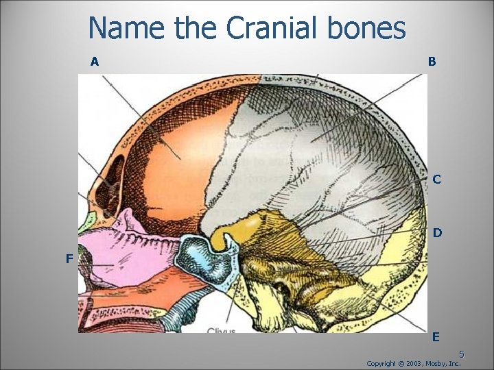
Name the Cranial bones A B C D F E 5 Copyright © 2003, Mosby, Inc.
Cranial bones are further divided into: 6
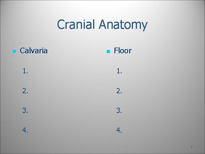
Cranial Anatomy n Calvaria n Floor 1. 2. 3. 4. 7
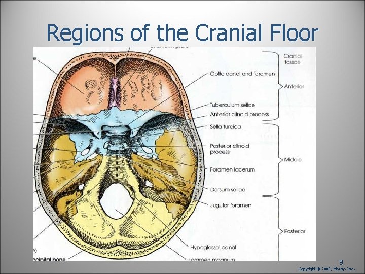
Name the 3 regions of the cranial floor 1. 2. 3. 8
Regions of the Cranial Floor 9 Copyright © 2003, Mosby, Inc .
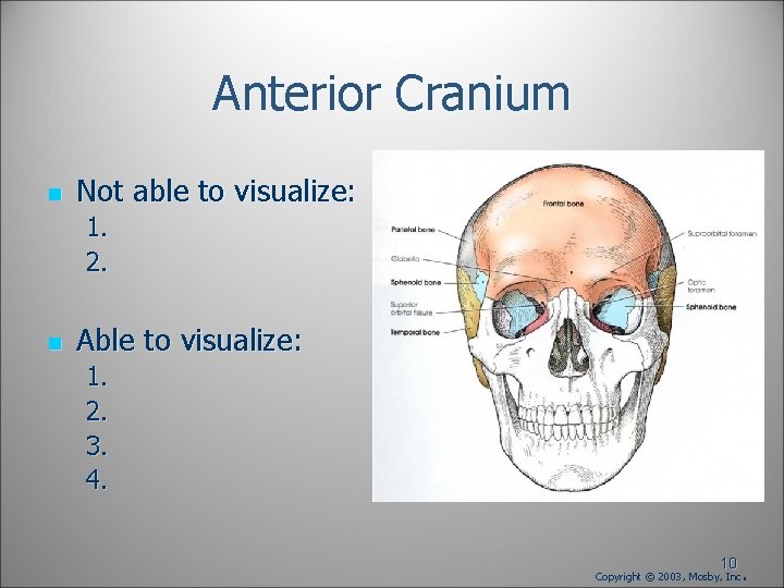
Anterior Cranium n Not able to visualize: 1. 2. n Able to visualize: 1. 2. 3. 4. 10 Copyright © 2003, Mosby, Inc .
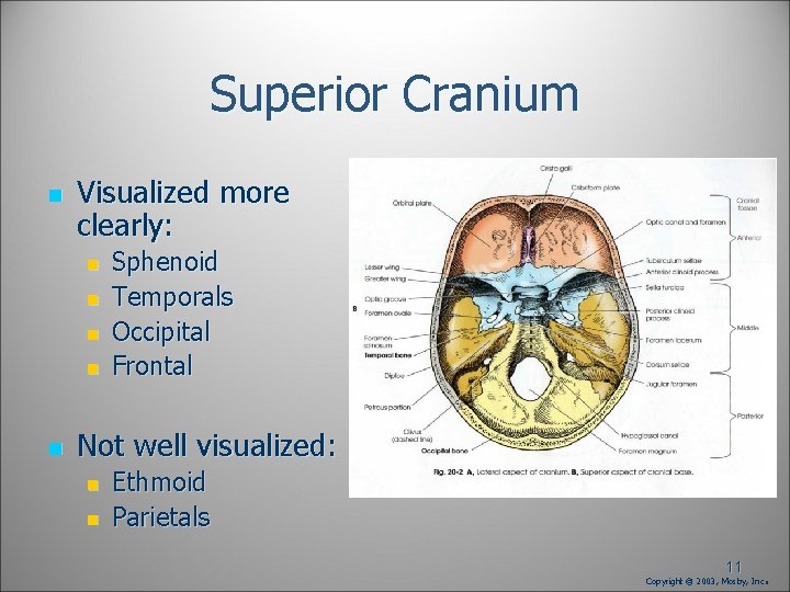
Superior Cranium n Visualized more clearly: n n n Sphenoid Temporals Occipital Frontal Not well visualized: n n Ethmoid Parietals 11 Copyright © 2003, Mosby, Inc .
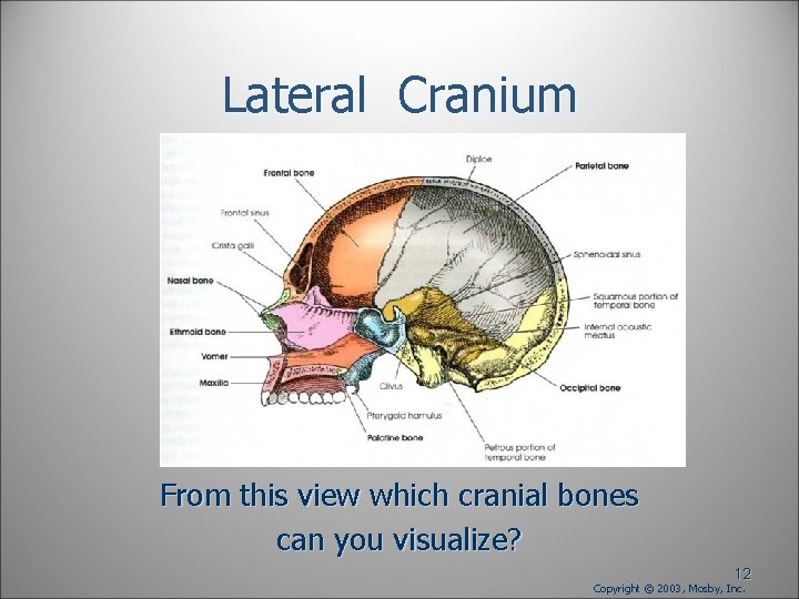
Lateral Cranium From this view which cranial bones can you visualize? 12 Copyright © 2003, Mosby, Inc.
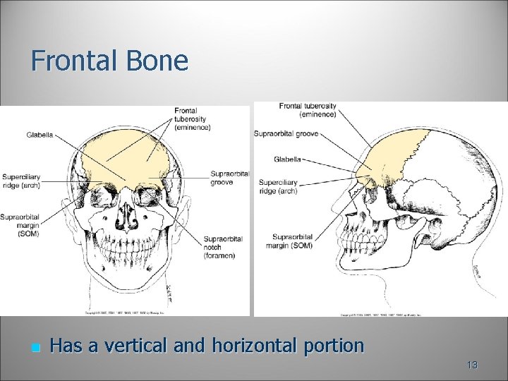
Frontal Bone n Has a vertical and horizontal portion 13
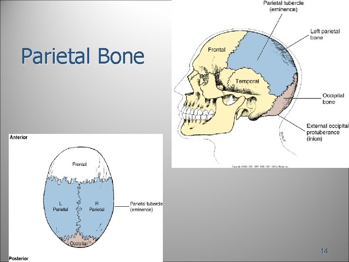
Parietal Bone 14
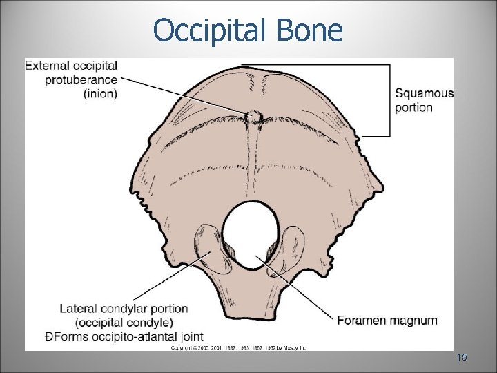
Occipital Bone 15
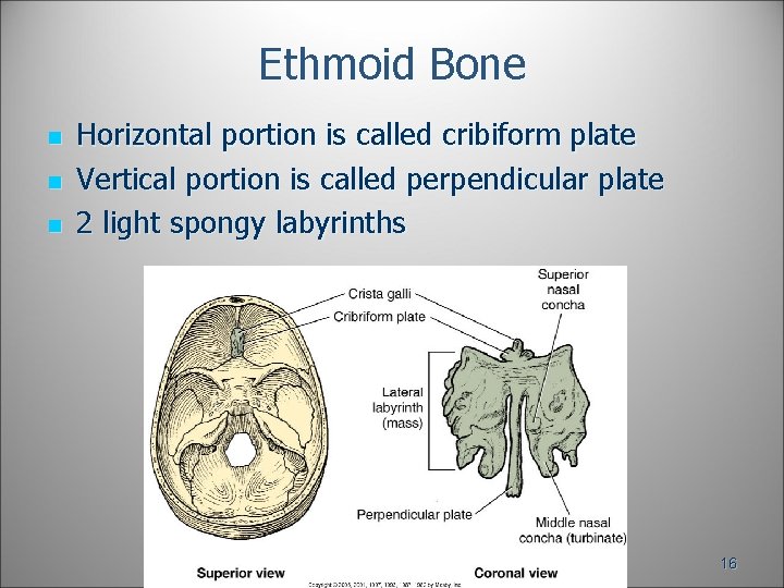
Ethmoid Bone n n n Horizontal portion is called cribiform plate Vertical portion is called perpendicular plate 2 light spongy labyrinths 16
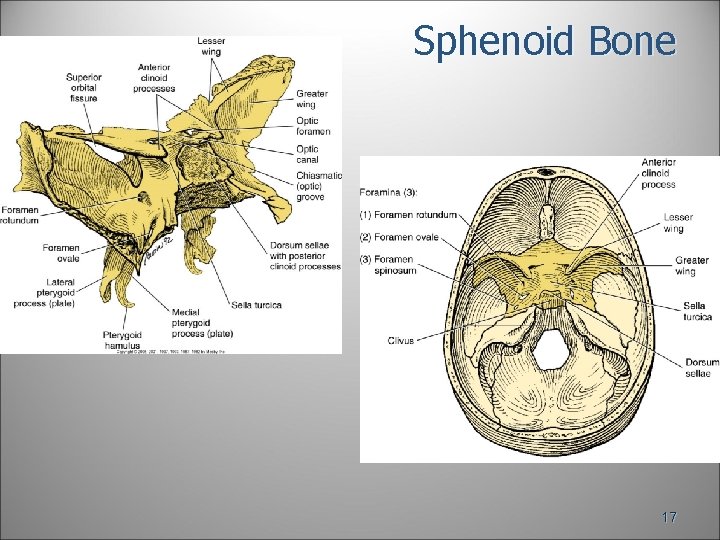
Sphenoid Bone 17
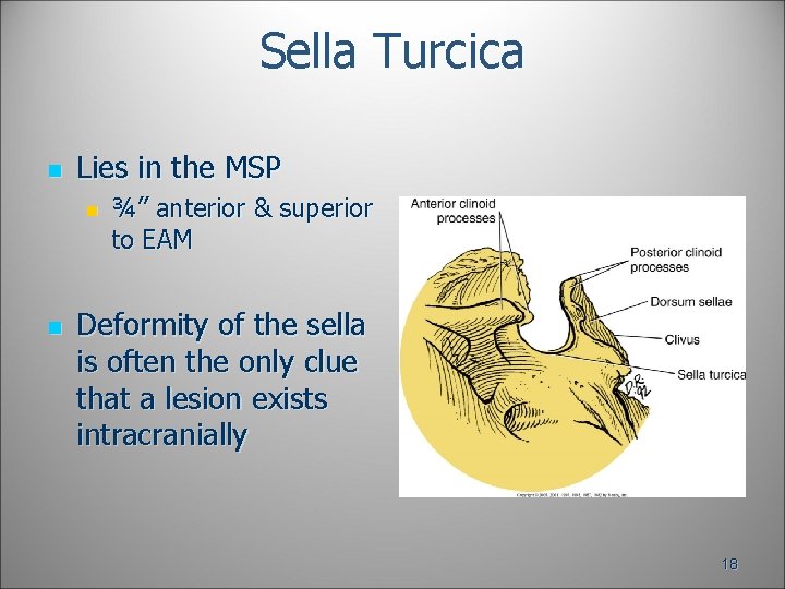
Sella Turcica n Lies in the MSP n n ¾” anterior & superior to EAM Deformity of the sella is often the only clue that a lesion exists intracranially 18
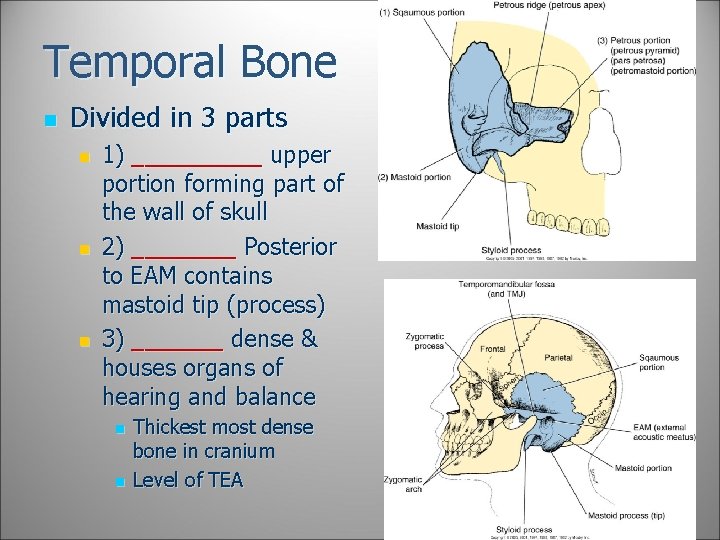
Temporal Bone n Divided in 3 parts n n n 1) _____ upper portion forming part of the wall of skull 2) ____ Posterior to EAM contains mastoid tip (process) 3) _______ dense & houses organs of hearing and balance n n Thickest most dense bone in cranium Level of TEA 19
Name the four sutures: 20
Name the junctions of the sutures: 21
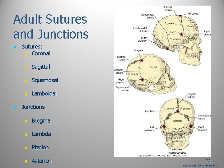
Adult Sutures and Junctions n n Sutures: n Coronal n Sagittal n Squamosal n Lamboidal Junctions n Bregma n Lambda n Pterion n Asterion 22 Copyright © 2003, Mosby, Inc.
Name the fontanels and where are they found? 23
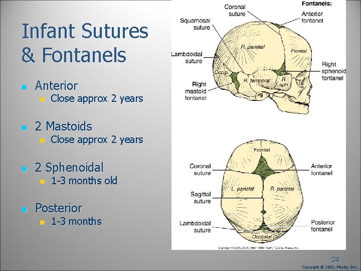
Infant Sutures & Fontanels n Anterior n n 2 Mastoids n n Close approx 2 years 2 Sphenoidal n n Close approx 2 years 1 -3 months old Posterior n 1 -3 months 24 Copyright © 2003, Mosby, Inc.
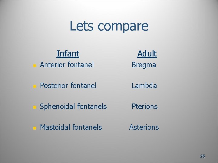
Lets compare Infant Adult n Anterior fontanel Bregma n Posterior fontanel Lambda n Sphenoidal fontanels Pterions n Mastoidal fontanels Asterions 25
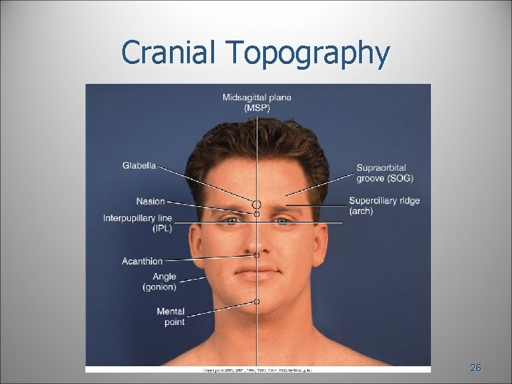
Cranial Topography 26
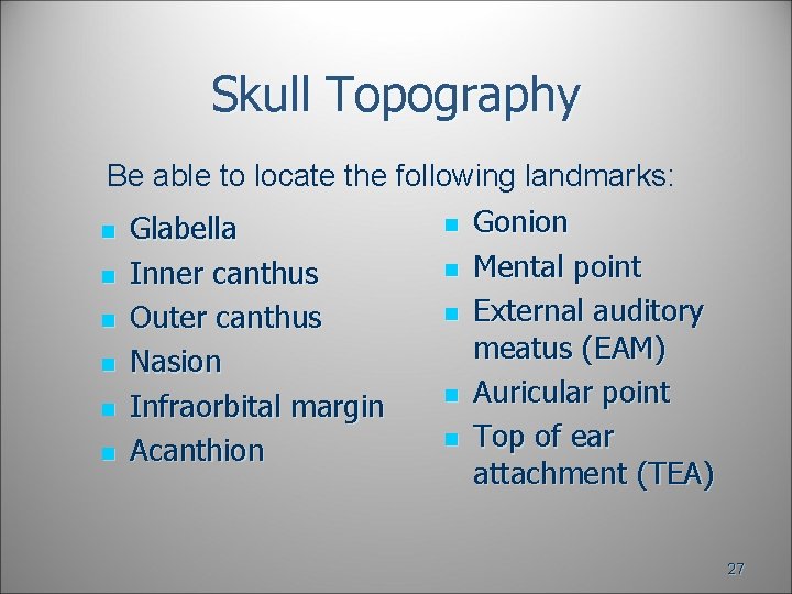
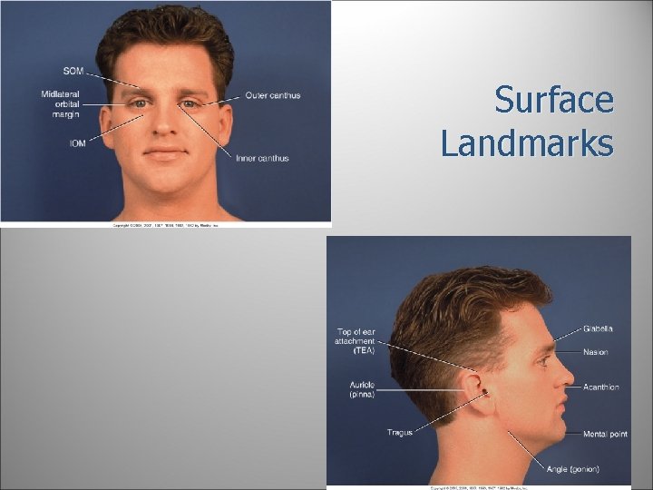
Skull Topography Be able to locate the following landmarks: n n n Glabella Inner canthus Outer canthus Nasion Infraorbital margin Acanthion n n Gonion Mental point External auditory meatus (EAM) Auricular point Top of ear attachment (TEA) 27
Surface Landmarks 28
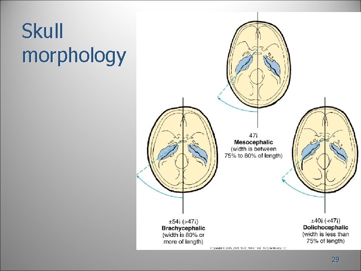
Skull morphology 29
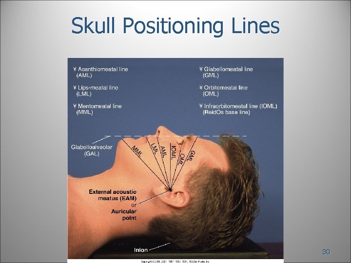
Skull Positioning Lines 30
Skull Positioning Lines n Interpupillary line (IPL) a) n Acanthiomeatal line (AML) b) n Mentomeatal line (MML) c) 31
Skull Positioning Lines n Orbitomeatal line (OML) a) n Infraorbitomeatal line (IOML) b) n Glabellomeatal line (GML) c) 32
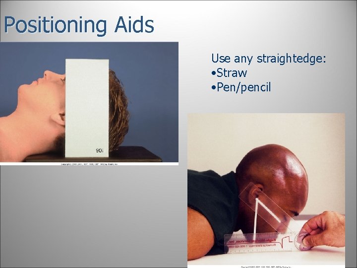
Positioning Aids Use any straightedge: • Straw • Pen/pencil 33
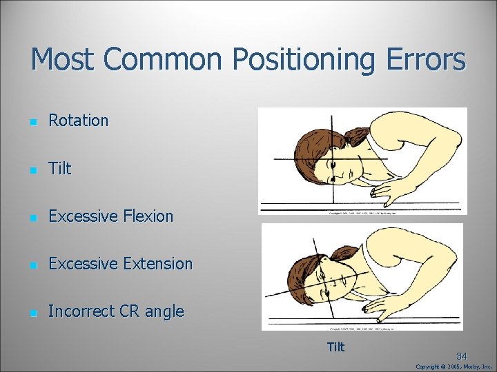
Most Common Positioning Errors n Rotation n Tilt n Excessive Flexion n Excessive Extension n Incorrect CR angle Rotation Tilt 34 Copyright © 2005, Mosby, Inc.
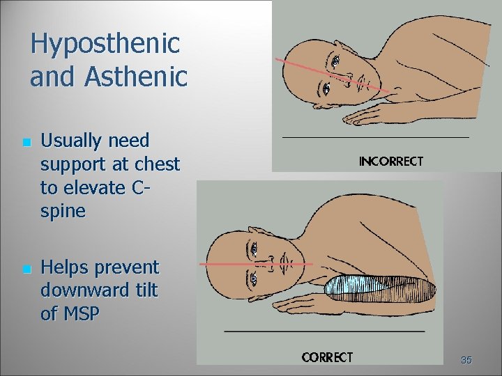
Hyposthenic and Asthenic n n Usually need support at chest to elevate Cspine Helps prevent downward tilt of MSP 35
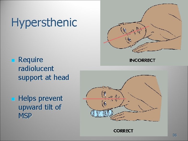
Hypersthenic n n Require radiolucent support at head Helps prevent upward tilt of MSP 36
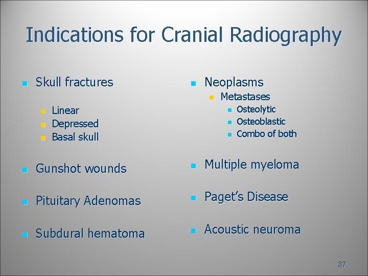
Indications for Cranial Radiography n Skull fractures n Neoplasms n n Linear Depressed Basal skull Metastases n n n Osteolytic Osteoblastic Combo of both n Gunshot wounds n Multiple myeloma n Pituitary Adenomas n Paget’s Disease n Subdural hematoma n Acoustic neuroma 37
Disinfect the Table or Bucky!! 38
Cleanliness n n Hair and skin of face are naturally oily; illness often increases oiliness Cranial procedures require direct contact of patient’s face with VBS n Clean device after each patient n Wash your hands!!! 39
Radiation Protection n n Collimate to anatomy of interest Shield gonads/abdomen of pediatric patients and those of reproductive age Shield thyroid and thymus of pediatric patient when doing so will not interfere with demonstration of anatomy of interest Good communication and positioning skills reduce chance of need for repeat radiographs 40
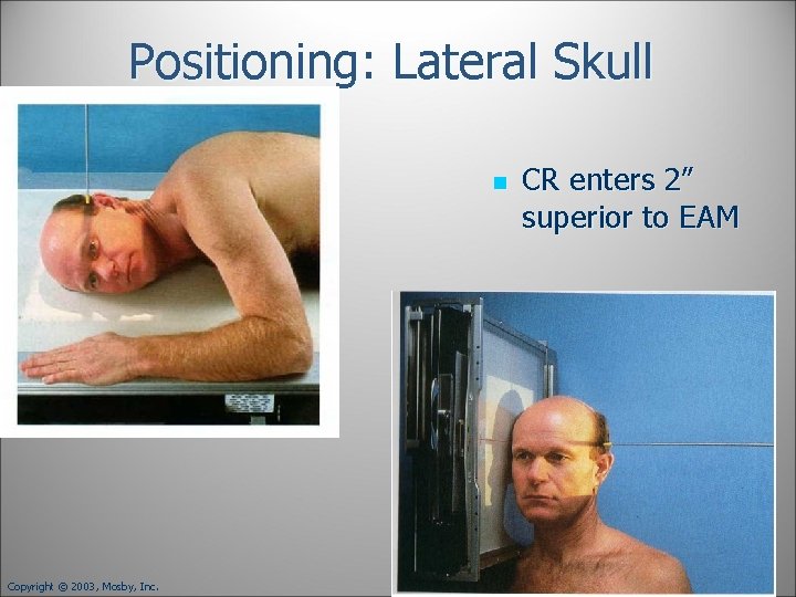
Positioning: Lateral Skull n Copyright © 2003, Mosby, Inc. CR enters 2” superior to EAM 41
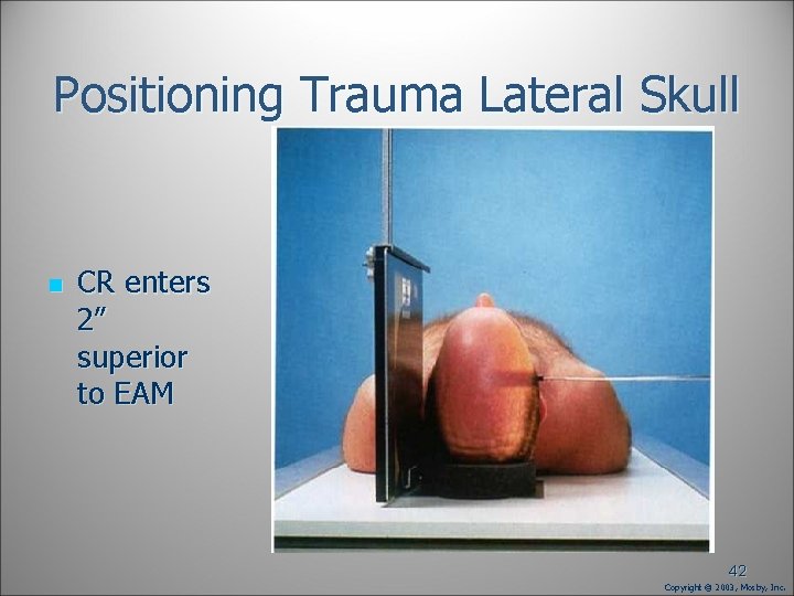
Positioning Trauma Lateral Skull n CR enters 2” superior to EAM 42 Copyright © 2003, Mosby, Inc.
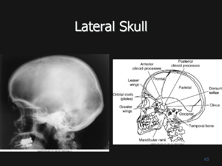
Lateral Skull 43
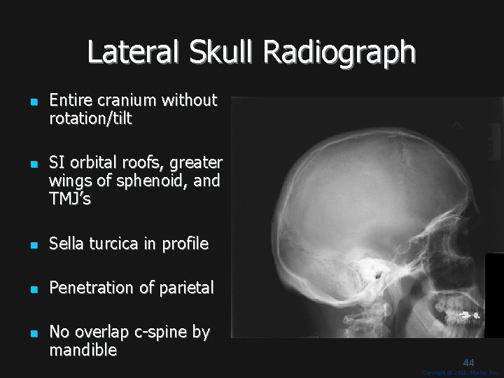
Lateral Skull Radiograph n n Entire cranium without rotation/tilt SI orbital roofs, greater wings of sphenoid, and TMJ’s n Sella turcica in profile n Penetration of parietal n No overlap c-spine by mandible 44 Copyright © 2003, Mosby, Inc.
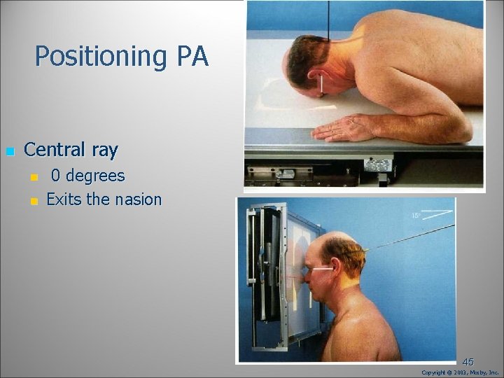
Positioning PA n Central ray n n 0 degrees Exits the nasion 45 Copyright © 2003, Mosby, Inc.
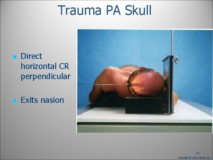
Trauma PA Skull n n Direct horizontal CR perpendicular Exits nasion 46 Copyright © 2003, Mosby, Inc.
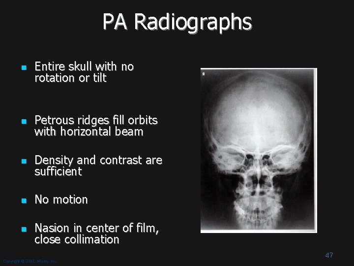
PA Radiographs n Entire skull with no rotation or tilt n Petrous ridges fill orbits with horizontal beam n Density and contrast are sufficient n No motion n Nasion in center of film, close collimation Copyright © 2003, Mosby, Inc. 47
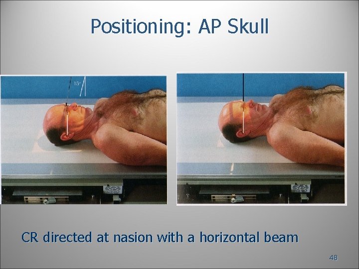
Positioning: AP Skull CR directed at nasion with a horizontal beam 48
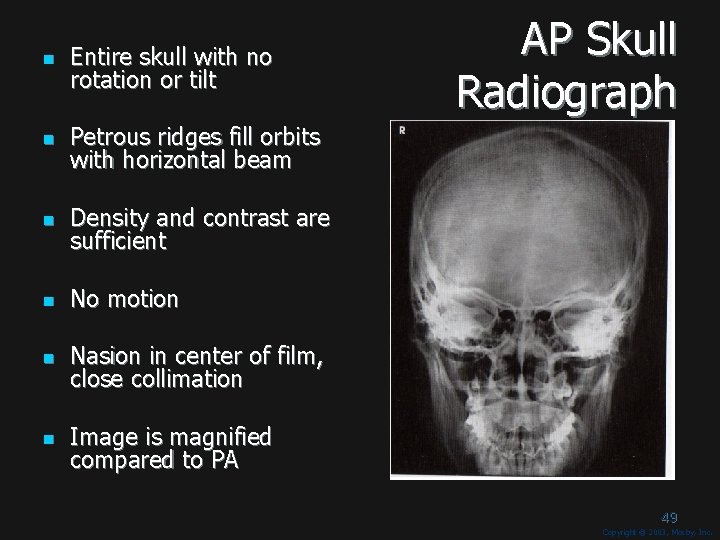
n Entire skull with no rotation or tilt n Petrous ridges fill orbits with horizontal beam n Density and contrast are sufficient n No motion n Nasion in center of film, close collimation n Image is magnified compared to PA AP Skull Radiograph 49 Copyright © 2003, Mosby, Inc.
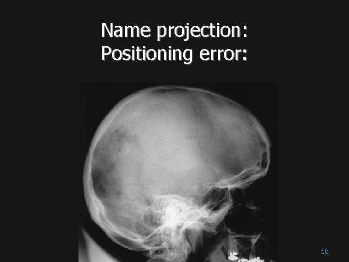
Name projection: Positioning error: 50
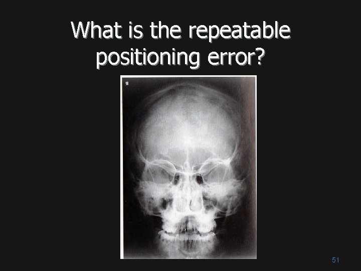
What is the repeatable positioning error? 51
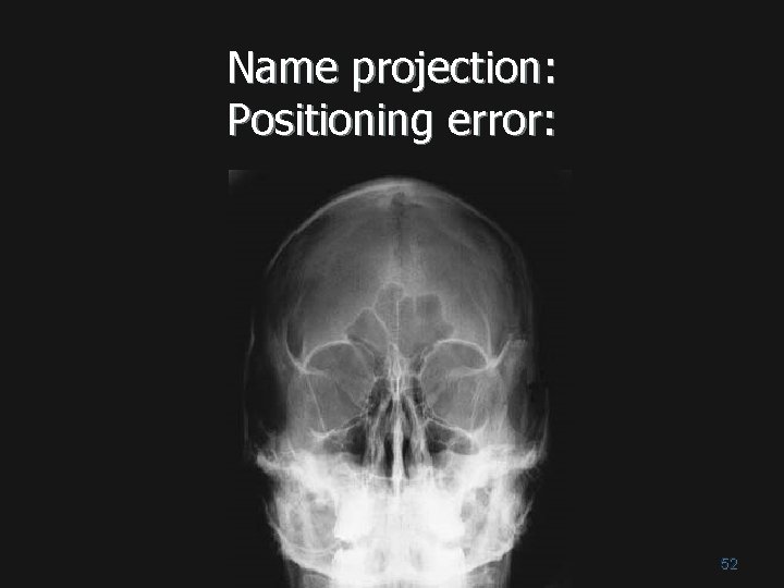
Name projection: Positioning error: 52