COR TRIATUM SINISTER IN ADULT Dr Smruti Hindaria


 cor triatum, or cor triatum sinister, is a")


- Slides: 19
COR TRIATUM SINISTER IN ADULT Dr Smruti Hindaria Assistant Professor, Department of CVTS
CASE • 32 years old lady • c/o breathlessness and palpitations on exertion since childhood, aggravated since 3 months • 2 D ECHO - All 4 PV draining into a confluence, right lower PV is narrowed & entering into the confluence, confluence draining into RA at SVC-RA junction, RA-RV dilated, severe PAH +, membrane seen in LA • Room air Sp. O 2 88% • o/e - central cyanosis+ • Patient started on sildenafil 1 month prior to planned surgery
CARDIAC CT • Right upper & middle lobe veins; and the right lower lobe vein are draining into a smallchamber seen posterior and to the right of the left atrium measuring 35 x 28 mm in size. • This chamber is separated from the left atrium by a thin septum except a small communication inferolaterally, measuring 8 mm in diameter. • Confluence of the right upper lobe and middle lobe veins is also continuous with a smallcaliber horizontal draining channel extending to the left in subcarinal location.
• Thehorizontal draining channel measures 13 x 4 mm in diameter at its right end and 16 x 14 mm in diameter at its left end at its confluence with left-sided pulmonary veins. The left upper and lower lobe pulmonary veins are seen to drain into this common channel whichthen extends superiorly to drain into the left innominate vein. • The vertical vein measures 29 mm in diameter in left parahilar region and 26 mm at its opening into the innominate vein.
CARDIAC CT
CARDIAC CT
SURGICAL PLAN • Total intracardiac repair of Cor triatum on Cardiopulmonary Bypass
INTRAOPERATIVE FINDINGS • Dilated RV, MPA, PA pressure 60/30 • Large 5 cm diameter vertical vein arising from left venous chamber draining into innominate vein • Left venous chamber communicating via narrow horizontal channel to cor chamber • Cor triatum with right pulmonary veins draining into it, 6 mm perforation in cor septum • 5 mm OS ASD
SURGICAL CORRECTION 1. Excision of cor septum 2. Common posterior venous channel anastamosis to LA roof extending upto LA appendage 3. Ligation and closure of vertical vein 4. Patch closure of IAS
POST OPERATIVE CARE • Patient electively ventilated for 24 hours i/v/o severe preoperative PAH • milrinone infusion continued for 24 hours post-op • Extubated on POD 1 • Post extubation Sp. O 2 99% on 4 lit O 2 • Drainage was minimal • Oral pulmonary vasodilators started immediately after referral to Pulmonary medicine department (tab endoblo T)
• Patient was discharged on POD 5 on warf Ecosprin Metoprolol XL Cardace Lasilactone Endobloc-T
• On follow-up 2 D ECHO after 1 month, pulmonary flow was seen entering LA, normal PA pressure, good BVF • Patient remains well on 6 month follow up. Only low dose cardace continued.
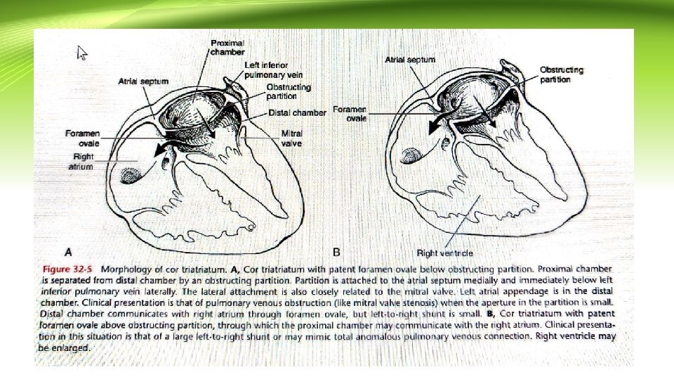
COR TRIATUM • Classic (or typical) cor triatum, or cor triatum sinister, is a rare congenital cardiac anomaly in which the pulmonary veins typically enter a “proximal” left atrial chamber separated from the “distal” left atrial chamber by a partition in which there are one or more restrictive ostia
• Infants with classic cor triatum, with a small aperture between the proximal and distal chambers, usually present with evidence of low cardiac output, including pallor, tachypnea, poor peripheral pulses, and growth failure. • When there is associated left-to-right shunting due to connection of the proximal chamber to the right atrium or because of associated PAPVC, evidence of pulmonary overcirculation and venous obstruction may be present in the chest radiograph, and right ventricular enlargement is prominent.
• In children and young adults, the classic presentation is with signs and symptoms of pulmonary venous hypertension.
• In most patients, the aperture is severely restrictive, and approximately 75% of persons born with classic cor triatum die in infancy. • However, when the proximal chamber communicates with the right atrium through a fossa ovalis atrial septal defect, the prognosis is better because the proximal chamber decompresses into the right atrium. These patients present with signs of a large left-to-right shunt and generally survive longer than those with an obstructive aperture. Rarely, cor triatum may be discovered in an adult.
Conclusions • Rare case - On review of literature, only 8 cases of Cor triatum in adults have been reported so far in individual case series. Our patient survived to adulthood, also had uneventful pregnancy before presentation. • Complex anomaly - not straightforward anatomy • Repeated preop discussions and planning to be prepared for on table possibilities • Excellent preoperative images - Radiology dept • Management of Pulmonary hypertension - Pulmo med dept