Chapter 15 Sense Organs SENSE OF SMELL l


 l Olfactory pathway: when the level of odorproducing chemicals")
 stimuli;")
 l Neural pathway for taste ¡ Taste sensation begins")

 l Middle ear (Figure 15")
 l Inner ear (Figure 15")

 l Cochlea and cochlear duct")
 l Sense of hearing ¡")
 ¡Pathway of sound waves l.")
 ¡Neural pathway of hearing l.")
 l Vestibule and semicircular canals")
 l Sense of balance ¡")
 ¡ Dynamic equilibrium: needed to")
 ¡Accessory")
 l. Lacrimal apparatus: structures that secrete tears and drain")

 ¡Muscles of the eye l. Extrinsic eye muscles: skeletal")
 ¡Layers of the eyeball: three coats of tissues make")

 l. Vascular layer: middle coat • Contains many blood")
 l. Inner layer: incomplete innermost coat of the eyeball")
 • Three layers of neurons make up the sensory")
 ¡Cavities and humors l. Cavities: eyeball has a large")
 l. Humors • Aqueous humor: fills both chambers of")
 l The process of seeing ¡Formation of retinal image")
 ¡Formation of retinal image (cont. ) l. Constriction of")
 ¡Formation of retinal image (cont. ) l. Convergence of")
 ¡The role of photopigments: light-sensitive pigmented compounds undergo structural")
 l. Cones: three types of cones in the retina,")
 ¡Neural pathway of vision (Figure 15 -30) l. Fibers")
- Slides: 56
Chapter 15: Sense Organs
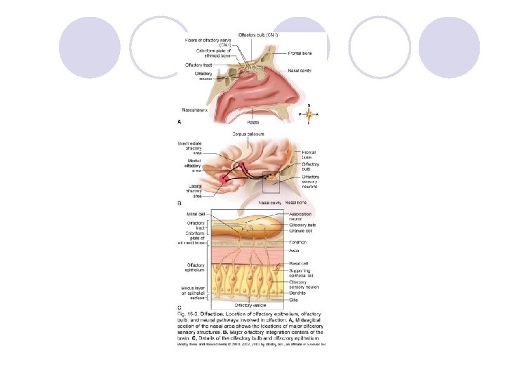
SENSE OF SMELL l Olfactory receptors ¡Olfactory sense organs consist of epithelial support cells and olfactory sensory neurons (Figure 15 -6) l. Olfactory cilia: line the upper surface of the nasal cavity l. Olfactory cells: chemoreceptors; gas molecules or chemicals dissolved in the mucus covering the nasal epithelium stimulate the olfactory cells l. Olfactory epithelium: located in most superior portion of the nasal cavity l. Olfactory receptors: extremely sensitive and easily fatigued; rapidly adapt with continual stimuli
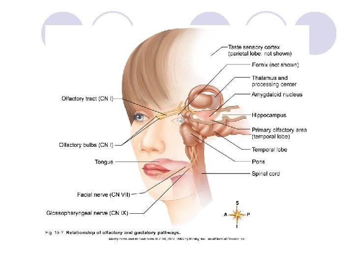
SENSE OF SMELL (cont. ) l Olfactory pathway: when the level of odorproducing chemicals reaches a threshold level, the following occur (Figure 15 -7): ¡Receptor potential and then action potential are generated and passed to the olfactory nerves in the olfactory bulb ¡The impulse then passes through the olfactory tract and into the thalamic and olfactory centers of the brain for interpretation, integration, and memory storage
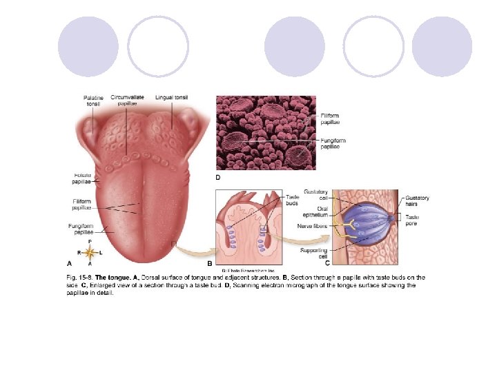
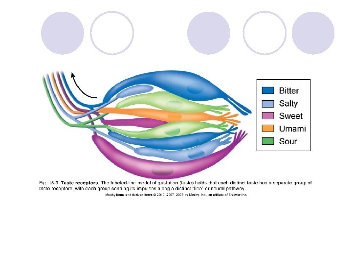
SENSE OF TASTE l Taste buds: sense organs that respond to gustatory (taste) stimuli; associated with papillae ¡ Chemoreceptors stimulated by chemicals dissolved in the saliva ¡ Gustatory cells: sensory cells in taste buds ¡ Sense of taste depends on the creation of a receptor potential in gustatory cells because of taste-producing chemicals in the saliva ¡ Taste buds are similar structurally; functionally, each taste bud responds most effectively to one of five primary taste sensations: sour, sweet, bitter, umami, and salty (and perhaps others, such as metallic) (Figures 15 -8 and 15 -9) ¡ Adaptation and sensitivity thresholds differ for each primary taste sensation
SENSE OF TASTE (cont. ) l Neural pathway for taste ¡ Taste sensation begins with a receptor potential in the gustatory cells of a taste bud; an action potential is generated and activated, which then transmits the sensory input to the brain ¡ Nerve impulses from the anterior two thirds of the tongue travel over the facial nerve; those from the posterior third of the tongue travel over the glossopharyngeal nerve ¡ Nerve impulses are carried to the medulla oblongata, relayed into the thalamus, and then relayed into the gustatory area of the cerebral cortex in the parietal lobe of the brain
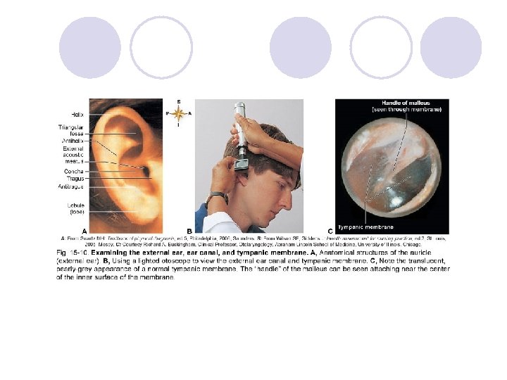
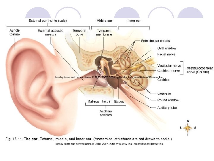
SENSES OF HEARING AND BALANCE: THE EAR l External ear: two divisions (Figures 15 -10 and 15 -11) ¡Auricle, or pinna: the visible portion of the ear ¡External acoustic meatus: tube leading from the auricle into the temporal bone and ending at the tympanic membrane
SENSES OF HEARING AND BALANCE: THE EAR (cont. ) l Middle ear (Figure 15 -11) ¡Tiny, epithelium-lined cavity hollowed out of the temporal bone ¡Contains three auditory ossicles l. Malleus (hammer): attached to the inner surface of the tympanic membrane l. Incus (anvil): attached to the malleus and stapes l. Stapes (stirrup): attached to the incus ¡Openings into the middle ear cavity l. Opening from the external acoustic meatus covered with tympanic membrane l. Oval window: opening into inner ear; stapes fits here l. Round window: opening into inner ear; covered by a membrane l. Opening into the auditory (eustachian) tube
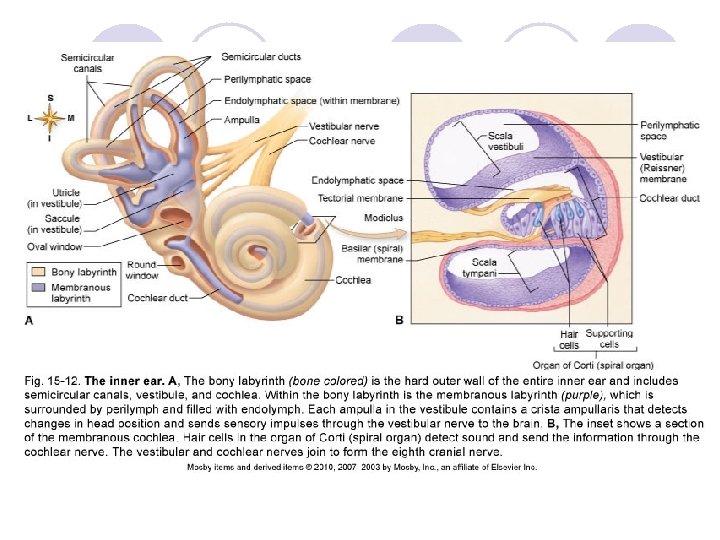
SENSES OF HEARING AND BALANCE: THE EAR (cont. ) l Inner ear (Figure 15 -12) ¡Structure of the inner ear l Bony labyrinth: composed of the vestibule, cochlea, and semicircular canals l Membranous labyrinth: composed of utricle and saccule inside the vestibule, cochlear duct inside the cochlea, and membranous semicircular ducts inside the bony semicircular canals l Vestibule and semicircular canal organs are involved with balance l Cochlea: involved with hearing l Endolymph: clear, potassium-rich fluid filling the membranous labyrinth l Perilymph: similar to cerebrospinal fluid; surrounds the membranous labyrinth, filling the space between the membranous tunnel and its contents and the bony walls that surround it
SENSES OF HEARING AND BALANCE: THE EAR (cont. ) l Cochlea and cochlear duct ¡Cochlea: bony labyrinth ¡Cochlear duct l Lies inside the cochlea; only part of the internal ear concerned with hearing; contains endolymph l Shaped like a triangular tube l Divides the cochlea into the scala vestibuli, the upper section, and the scala tympani, the lower section; both sections filled with perilymph l Vestibular membrane: the roof of the cochlear duct l Basilar (spiral) membrane: floor of the cochlear duct l Organ of Corti: rests on the basilar membrane; consists of supporting cells and hair cells; also called spiral organ l Axons of the neurons that begin around the organ of Corti and extend in the cochlear nerve to the brain to produce the sensation of hearing
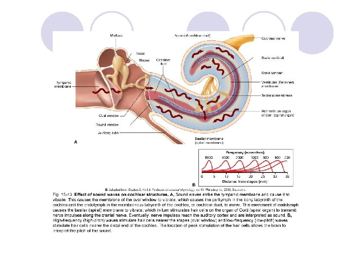
SENSES OF HEARING AND BALANCE: THE EAR (cont. ) l Sense of hearing ¡ Sound is created by vibrations ¡ Ability to hear sound waves depends on volume, pitch, and other acoustic properties ¡ Sound waves must be of sufficient amplitude to move the tympanic membrane and have a frequency capable of stimulating the hair cells in the organ of Corti (spiral organ) (Figure 15 -13) ¡ Basilar membrane width and thickness varies throughout its length l High-frequency sound waves vibrate the narrow portion near the oval window l Low frequencies vibrate the wider, thicker portion near the apex of the cochlea l Each frequency stimulates different hair cells and facilitates perception of different pitches l Perception of loudness is determined by movement amplitude; the greater the movement, the louder the perceived sound l Hearing results from stimulation of the auditory area of the cerebral cortex
SENSES OF HEARING AND BALANCE: THE EAR (cont. ) ¡Pathway of sound waves l. Enter external auditory canal l. Strike tympanic membrane, causing vibrations l. Tympanic vibrations move the malleus, which moves the incus and then the stapes l. The stapes moves against the oval window, which begins the fluid conduction of sound waves l. The perilymph in the scala vestibuli of the cochlea begins a “ripple” that is transmitted through the vestibular membrane to the endolymph inside the duct, to the basilar membrane, and then to the organ of Corti l. From the basilar membrane, the ripple is transmitted through the perilymph in the scala tympani and then expends itself against the round window
SENSES OF HEARING AND BALANCE: THE EAR (cont. ) ¡Neural pathway of hearing l. A movement of hair cells against the tectorial membrane stimulates the dendrites that terminate around the base of the hair cells and initiates impulse conduction by the cochlear nerve to the brainstem l. Impulses pass through “relay stations” in the nuclei in the medulla, pons, midbrain, and thalamus before reaching the auditory area of the temporal lobe
SENSES OF HEARING AND BALANCE: THE EAR (cont. ) l Vestibule and semicircular canals (Figure 1512) ¡Vestibule: the central section of the bony labyrinth; the utricle and saccule are the membranous structures within the vestibule ¡Three semicircular canals l. Each canal is at a right angle to the other l. Membranous semicircular ducts within the canals; each contains endolymph and connects with the utricle l. Each canal enlarges into an ampulla near junction with utricle
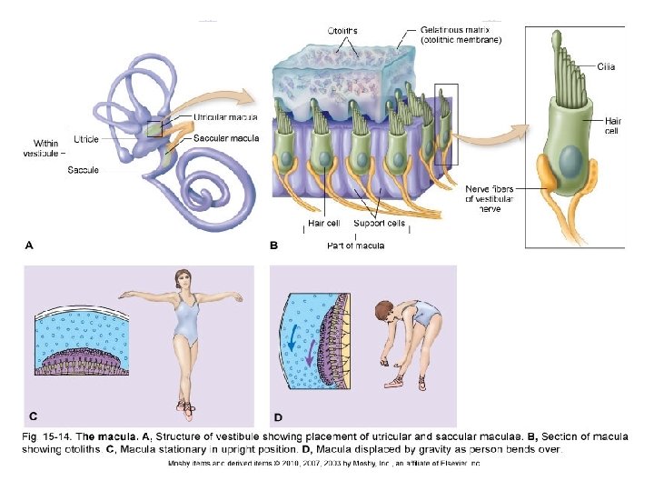
SENSES OF HEARING AND BALANCE: THE EAR (cont. ) l Sense of balance ¡ Static equilibrium: ability to sense head position relative to gravity or acceleration/deceleration (Figure 15 -14) l Movements of the maculae, located in both the utricle and saccule, provide information related to head position or acceleration l Otoliths are located within the matrix of the macula l Changing head position produces a change of pressure on the otolith-weighted matrix, stimulating the hair cells that stimulate the receptors of the vestibular nerve l Vestibular nerve fibers conduct impulses to the brain and sense head position and a change in the pull of gravity l Righting reflexes: muscular responses to restore the body and its parts to their normal position when displaced; caused by stimuli of the macula and impulses from proprioceptors and from the eyes
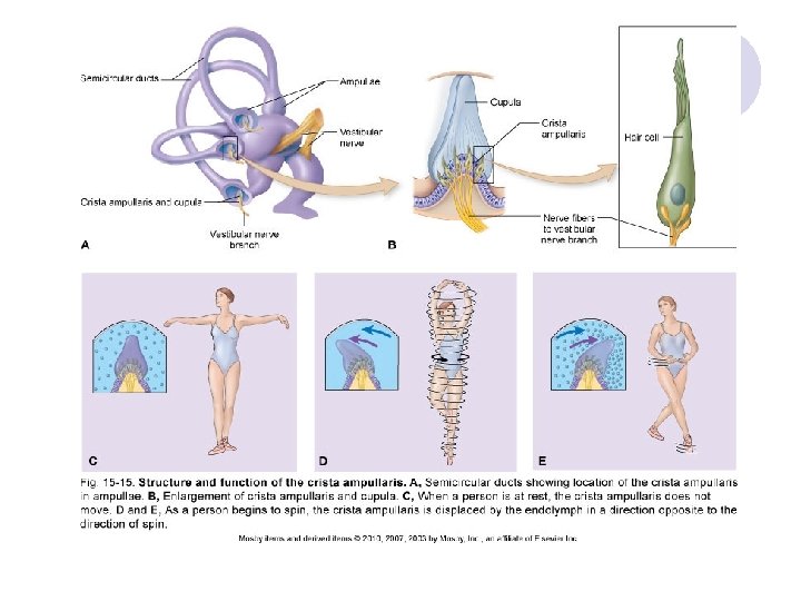
SENSES OF HEARING AND BALANCE: THE EAR (cont. ) ¡ Dynamic equilibrium: needed to maintain balance when the head or body is rotated or suddenly moved; able to detect changes in direction and rate at which movement occurs (Figure 15 -15) l Depends on the functioning of the cristae ampullaris, located in the ampulla of each semicircular duct l Cupula: gelatinous cap where the hair cells of cristae are embedded • Does not respond to gravity • Moves with the flow of endolymph in the semicircular ducts l Semicircular ducts are arranged at nearly right angles to each other to detect movement in all directions l Hair cells bend as cupula moves, producing a receptor potential followed by an action potential • Action potential passes through the vestibular portion of the eighth cranial nerve to the medulla oblongata • Sent next to other areas of the brain and spinal cord for interpretation, integration, and response
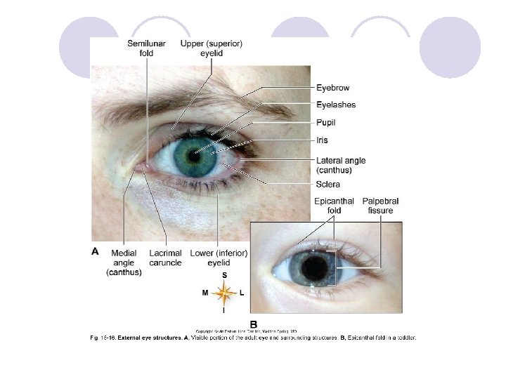
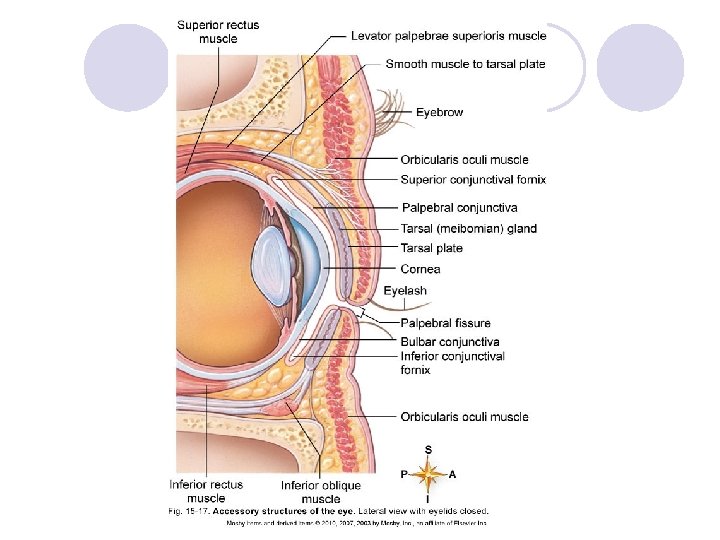
VISION: THE EYE l Structure of the eye (Figures 15 -16 to 1525) ¡Accessory structures (Figures 15 -16 to 15 -20) l. Eyebrows and eyelashes give some protection against foreign objects entering the eye; cosmetic purposes l. Eyelids consist of voluntary muscle and skin • Lined with conjunctiva, a mucous membrane • Palpebral fissure: opening between the eyelids • Angle or canthus: where the upper and lower eyelids join
VISION: THE EYE (cont. ) l. Lacrimal apparatus: structures that secrete tears and drain them from the surface of the eyeball (Figure 15 -19) • Lacrimal glands: size and shape of a small almond Located at the upper, outer margin of each orbit Approximately 12 small ducts lead from each gland Drain tears onto the conjunctiva • Lacrimal canals: small channels that empty into lacrimal sacs • Lacrimal sacs: located in a groove in the lacrimal bone • Nasolacrimal ducts: small tubes that extend from the lacrimal sac into the inferior meatus of the nose
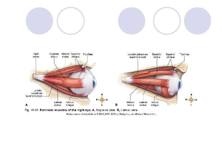
VISION: THE EYE (cont. ) ¡Muscles of the eye l. Extrinsic eye muscles: skeletal muscles that attach to the outside of the eyeball and bones of the orbit • Named according to their position on the eyeball • Include the superior, inferior, medial, and lateral rectus muscles and superior and inferior oblique muscles l. Intrinsic eye muscles: smooth muscles located within the eye • Iris: regulates size of pupil • Ciliary muscle: controls shape of lens
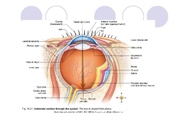
VISION: THE EYE (cont. ) ¡Layers of the eyeball: three coats of tissues make up the eyeball (Figure 15 -21) l. Fibrous layer: outer coat • Sclera: tough, white, fibrous tissue • Cornea: the transparent anterior portion that lies over the iris; no blood vessels found in the cornea or in the lens • Scleral venous sinus (canal of Schlemm): ring-shaped venous sinus found deep within the anterior portion of the sclera at its junction with the cornea
VISION: THE EYE (cont. ) l. Vascular layer: middle coat • Contains many blood vessels and a large amount of pigment • Choroid: pigmented membrane lining more than two thirds of the posterior fibrous outer coat • Anterior portion has three different structures Ciliary body: thickening of choroid; fits between anterior margin of retina and posterior margin of iris; Suspensory ligament: attached to the ciliary processes and blends with the elastic capsule of the lens to hold it in place Iris: colored part of the eye; consists of circular and radial smooth muscle fibers that form a doughnutshaped structure; attaches to the ciliary body
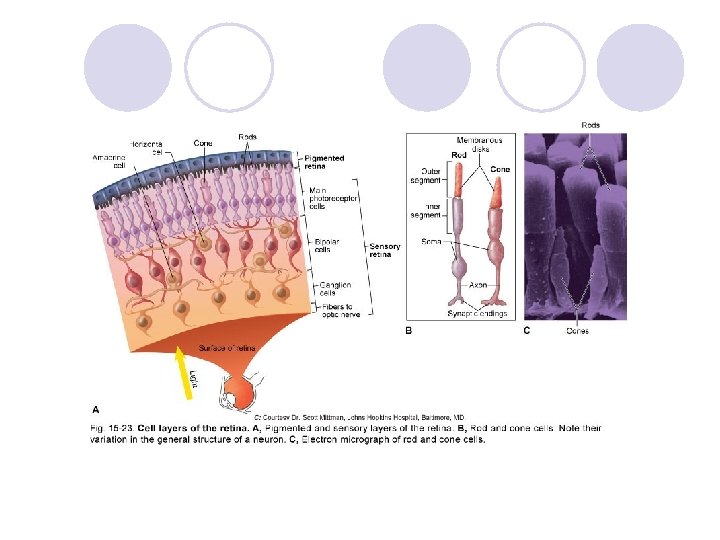
VISION: THE EYE (cont. ) l. Inner layer: incomplete innermost coat of the eyeball • Retina: composed of an outer layer of pigmented epithelium (pigmented retina) and an inner layer of nervous tissue (sensory retina) • Three layers of neurons make up the sensory retina 1. Photoreceptor cells: visual receptors, sensitive to light rays Rods: absent from the fovea and macula; increased in density toward the periphery of the retina Cones: less numerous than rods; most densely concentrated in the fovea centralis in the macula lutea 2. Bipolar cells
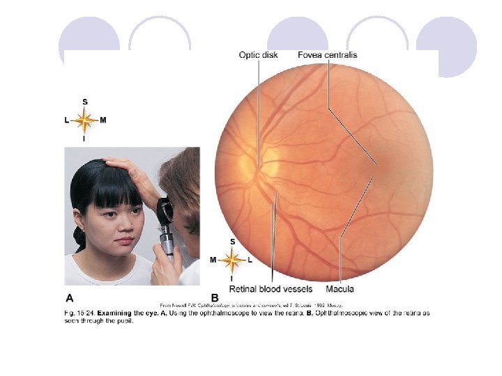
VISION: THE EYE (cont. ) • Three layers of neurons make up the sensory retina (cont. ) 3. Ganglionic cells: all axons of these neurons extend back to the optic disk; part of the sclera, which contains perforations through which the fibers emerge from the eyeball as the optic nerve Horizontal and amacrine cells allow lateral connections within the sensory retina • Optic nerve: second cranial nerve extends from the eyeball to the brain • Retinal blood vessels: critical to normal visual function (Figure 15 -24)
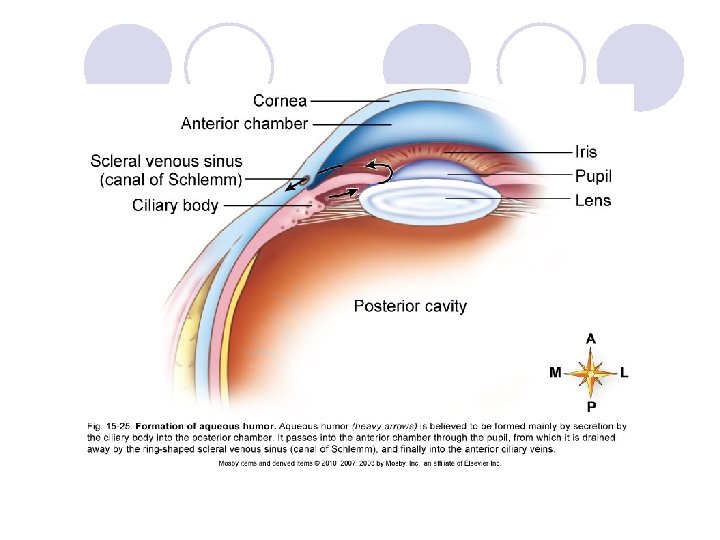
VISION: THE EYE (cont. ) ¡Cavities and humors l. Cavities: eyeball has a large interior space divided into two cavities • Anterior cavity lies in front of the lens; has two subdivisions Anterior chamber: space anterior to the iris and posterior to the cornea Posterior chamber: small space posterior to the iris and anterior to the lens • Posterior cavity is larger than the anterior cavity; occupies all the space posterior to the lens, suspensory ligament, and ciliary body
VISION: THE EYE (cont. ) l. Humors • Aqueous humor: fills both chambers of the anterior cavity; clear, watery fluid that often leaks out when the eye is injured; formed from blood in capillaries located in the ciliary body (Figure 15 -25) • Vitreous humor: fills the posterior cavity; semisolid material; helps maintain sufficient intraocular pressure, with aqueous humor, to give the eyeball its shape
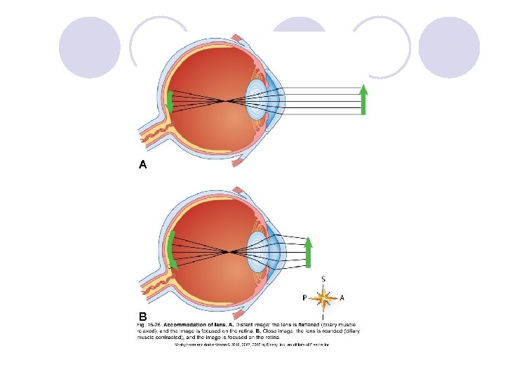
VISION: THE EYE (cont. ) l The process of seeing ¡Formation of retinal image l. Refraction of light rays: deflection, or bending, of light rays produced when they pass obliquely from one transparent medium to another of different optical density; cornea, aqueous humor, lens, and vitreous humor are the refracting media of the eye l. Accommodation of lens: increase in curvature of the lens to achieve the greater refraction needed for near vision (Figure 15 -26)
VISION: THE EYE (cont. ) ¡Formation of retinal image (cont. ) l. Constriction of pupil: muscles of iris are important to formation of a clear retinal image • Pupil constriction prevents divergent rays from object from entering eye through periphery of the cornea and lens • Near reflex: constriction of pupil that occurs with accommodation of the lens in near vision • Photopupil reflex: pupil constricts in bright light
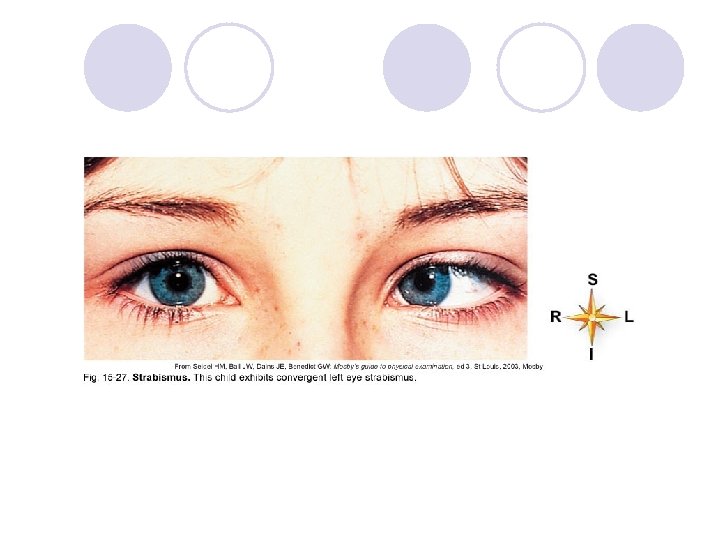
VISION: THE EYE (cont. ) ¡Formation of retinal image (cont. ) l. Convergence of eyes: movement of the two eyeballs inward so that their visual axes come together at the object viewed • The closer the object, the greater the degree of convergence necessary to maintain single vision • For convergence to occur, a functional balance between antagonistic extrinsic muscles must exist • Strabismus is abnormal convergence (Figure 15 -27)
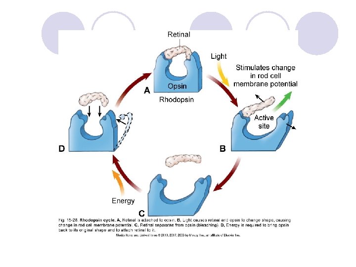
VISION: THE EYE (cont. ) ¡The role of photopigments: light-sensitive pigmented compounds undergo structural changes that result in generation of nerve impulses interpreted by the brain as sight l. Rods: photopigment in rods is rhodopsin • Highly light sensitive • Breaks down into opsin and retinal • Separation of opsin and retinal in the presence of light causes an action potential in rod cells • Energy is needed to re-form rhodopsin (Figure 15 -28)
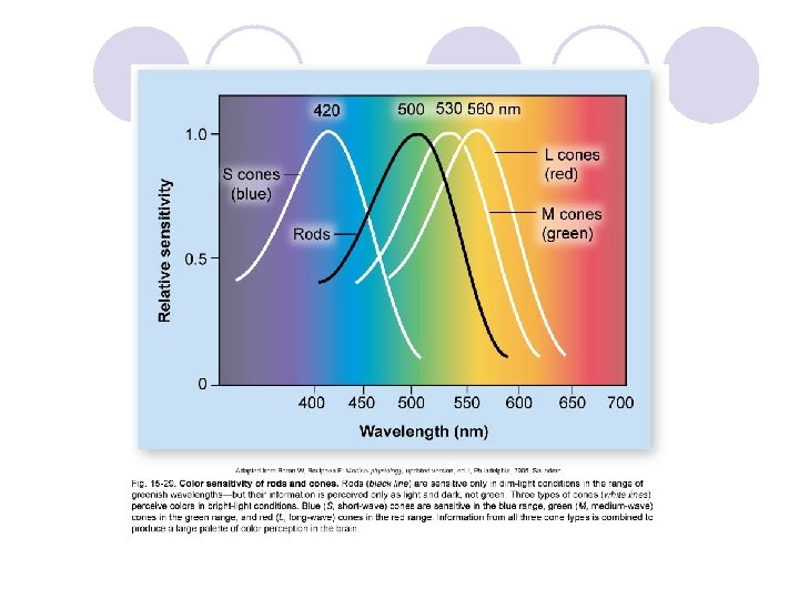
VISION: THE EYE (cont. ) l. Cones: three types of cones in the retina, each with a different variation of the rhodopsin photopigment: blue, green, and red • Perception of a large variety colors results from the combination of signals from different cone types • Cone pigments are less light sensitive than rhodopsin and need brighter light to break down (Figure 15 -29) l. Ganglion cells: relay information from rods and cones (by way of bipolar cells) but also relay nonimage light information to the body’s biological clock; photopigment is melanopsin
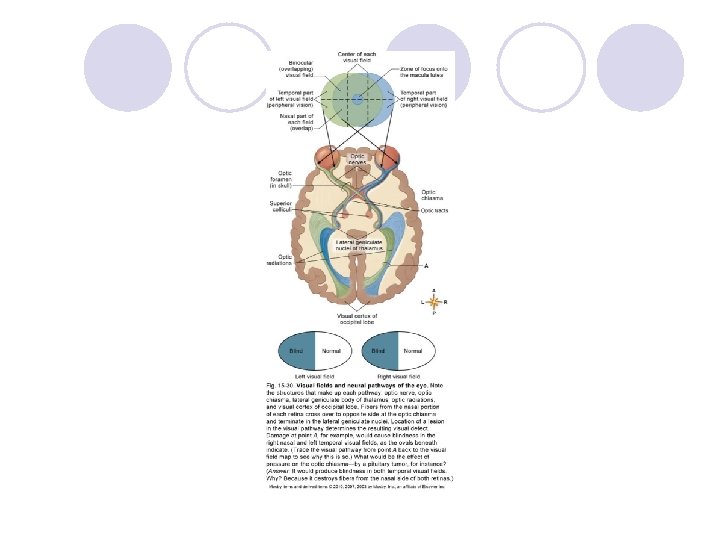
VISION: THE EYE (cont. ) ¡Neural pathway of vision (Figure 15 -30) l. Fibers that conduct impulses from the rods and cones reach the visual cortex in the occipital lobes by way of the optic nerves, optic chiasma, optic tracts, and optic radiations l. Optic nerve contains fibers from only one retina, but optic chiasma contains fibers from the nasal portion of both retinas; these anatomical facts explain peculiar visual abnormalities that sometimes occur