Central nervous system Dr Heyam Awad FRCPATH 2017


")
? ? ? • It is the pressure")


- Slides: 39
Central nervous system Dr Heyam Awad FRCPATH 2017
LECTURE 2: disturbed fluid balance and increased intracranial pressure Topics to be covered: - Increased intracranial pressure. - Brain edema - Hydrocephalus - Herniation - Cerebral ischemia
ILOs • Understand causes and symptoms of increased intracranial pressure. • Define cerebral edema and know its types and causes. • Define hydrocephalus and know its types and causes. • Define herniation and know its types and complications • Understand autoregulationo f blood flow in the brain • List causes of hypoxia and ischemia • Understand outcomes of global brain ischemia • Apply the above knowledge in clinical cases.
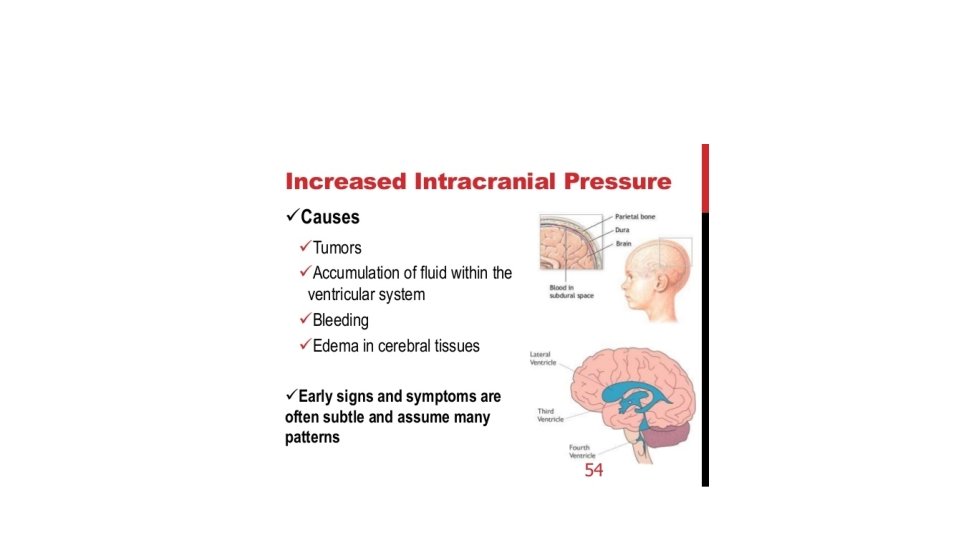
The cranium. . • The brain is enclosed within the skull, which is a rigid box that protects it. • In adults, skull bones cannot expand • So if the material within the cranium increases. . Pressure will increase= increased intracranial pressure
What's inside the cranium? • ROUGHLY: 80% brain tissue ( including fluid; around 75%) : 10% blood : 10% CSF (cerebrospinal fluid) IF any of these components increases, the intracranial pressure increases.
OK, so what is intracranial pressure (ICP)? ? ? • It is the pressure inside the skull and is measured in millimeters of mercury • at rest, it is normally 7– 15 mm. Hg for a supine adult. • The upper limit of ICP is 20– 25 mm Hg • If pressure in the cranium is higher than this upper limit= increased intracranial pressure (= intracranial hypertension.
Causes of increased intracranial pressure • mass effect : brain tumor, hematoma, or abscess. • generalized brain swelling : ischemic-anoxia states, hypertension • increase in venous pressure : heart failure • obstruction to CSF flow and/or absorption or increased CSF production: hydrocephalus. • Idiopathic or unknown
clinical presentation according to severity:
Brain edema= cerebral edema • = accumulation of excess fluid within the brain parenchyma. • Two types: vasogenic and cytotoxic edema. . Usually coexist
Vasogenic edema • Due to disruption of blood brain barrier. • So: shift of fluids from vessels to brain tissue. • Lymphatic vessels are rare in the brain. . So there is little or no resorption of excess edema fluid. • Can be generalised ( due to hypoxia) or localised ( due to inflammation or tumors)
Cytotoxic edema • Due to neuronal or glial cell membrane injury. • Causes: toxins or hypoxia. • Here fluid moves from cells to interstitial tissue.
morphology • With edema, the brain becomes swollen. . And its weight increases. • The normal adult human brain weighs on average about 1. 2– 1. 4 kg , or about 2% of total body weight, although there is substantial individual variation. • Edema causes flat gyri and narrow sulci
Brain edema
hydrocephalus • Increased CSF within ventricles. • Caused by overproduction or decreased resorption of CSF. • Overproduction: rare, due to choroid plexus tumors. • Decreased resorption. . Can be localised or generalised.
hydrocephalus • Localised: noncommunicating hydrocephalus. • Generalised: communicating hydrocephalus.
• In infancy, before closure of the cranial sutures , the head enlarges. • After closure of the cranial sutures: increased intracranial pressure occurs. Of course there is no increase in head circumference
hydrocephalus
hydrocephalus
herniation • Increased volume of tissue inside the skull. . Increased intracranial pressure which causes focal expansion of the brain tissue. • Because the cranial vault is subdivided by rigid dural folds (falx and tentorium)…. The expanded brain tissue is displaced in relation to these folds. • Expansion: herniation
herniation • Subfalcine = cingulate • Transtentorial = uncinate • Tonsillar.
herniation
Cingulate herniation -cingulate gyrus displaced under edge of falx -Can cause compression of anterior cerebral artery
Transtentorial herniation • Medial aspect of temporal lobe compressed against the free margin of the tentorium. • Third cranial nerve compressed. . Dilated pupil, impaired ocular movement on the side of the lesion • Posterior cerebral artery can be affected. . Ischemic injury to tissues supplied by it including visual cortex.
Tonsillar herniation • Displaced cerebellar tonsils through foramen magnum • Brain stem compression… respiratory and cardiac centres in medulla compromised. • LIFE THREATENING
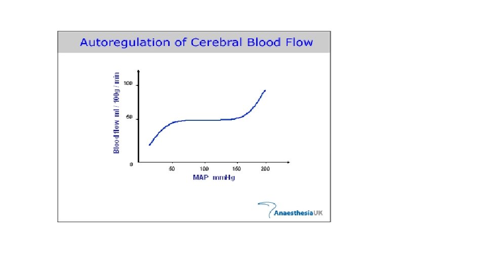
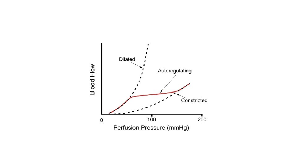
Hypoxia and ischemia • Brain is highly oxygen dependent. • Brain 2% of body weight but receives 15% of cardiac output • 20% of total body oxygen consumption. • Autoregulation of vascular resistance allows stability of cerebral blood flow over a wide range of blood pressures and intracranial pressure. • If blood pressure very low (systolic less than 50)… hypoxia
Brain hypoxia • Functional hypoxia. • ischemic hypoxia
Functional hypoxia • Low partial pressure of oxygen: high altitude • Impaired oxygen carrying capacity: anaemia and CO poisoning • Decreased oxygen use by tissues: cyanide poisoning
Functional hypoxia
Ischemic hypoxia • Hypo-perfusion due to hypotension or vascular obstruction • Ischemia can be global or focal • Focal ischemia causes infarctions and this will be discussed in the next lecture.
Global cerebral ischemia Occurs due to severe hypotension, systolic below 50 mm Hg: • Cardiac arrest • Shock • Severe hypotension • Outcome depends on severity and duration of insult
Global ischemia • Neurons more susceptible to hypoxic injury than glial cells. • Most susceptible neurons: pyramidal cells of hippocampus and neocortex + Purkinje cells of the cerebellum
ischemia • If mild: transient confessional state • severe : neural death, if survive: severely impaired neurologically • Severest forms result in brain death.
Morphology of reversible global ischemia • Swelling • Wide gyri • Narrow sulci • Poor grey white matter demarcation
Irreversible global ischemia can cause brain death • Diffuse cortical injury with flat EEG ( isoelectric EEG) • Brain stem damage: No reflexes and no respiration • If on mechanical support: autolysis of brain= respirator brain /
Suggested reading about brain death…for those who are interested • https: //www. ncbi. nlm. nih. gov/pmc/articles/PMC 2772257/ • Also a pdf is downloaded in my webpage…. This is an interesting read I encourage you to have a look!!