Neoplasia lecture 4 Dr Heyam Awad FRCPath Molecular


. . Governor:")


 MYC becomes under")


. •")
 • Noncoding, micro (22 nucleotides) that are negative regulators of")


 that")


- Slides: 37
Neoplasia lecture 4 Dr Heyam Awad FRCPath
Molecular basis of cancer • Neoplasms are caused by nonlethal, genetic damage, which causes uncontrolled cellular proliferation. • Nonlethal: so cells can still multiply! • Genetic damage: mutations or non-mutational damages (epigenetic changes) • uncontrolled proliferation… not all genetic damages produce tumors, they only do so if they result in a crazy cell that can multiply continuously in an uncontrolled, uninhibited fashion!
Tumor clonality • Because tumor cells originate from one single genetically damaged crazy cell…. They are clonal • What does a clone mean? • Note: tumors start as a clone, but with time they acquire several mutations in some of the cells. . They become heterogeneous. This is because some cells develop mutations that make them acquire characteristics like: ability to invade, to metastasize. . etc
Genetic damages in neoplasms • These need to be damages causing changes in cell proliferation So: four types of regulatory genes are mainly affected: • 1. growth promoting proto-oncogenes • 2. growth inhibiting tumor suppressor genes • 3. genes that regulate apoptosis • 4. genes involved in DNA repair.
oncogenes • Normally: our cells have proto-oncogenes. These cause cell proliferation in a regulated manner • If the proto-oncogenes are mutated or overexpressed: they are called oncogenes • Proto-oncogenes encode for proteins: proto-oncoproteins, or oncoproteins • These oncoproteins include: transcription factors, growth regulating proteins, proteins involved in cell survival.
oncogenes • Oncogenes cause overexpression of proteins involved in cell growth. • If one allele is mutated or overexpressed: there will be increase in the growth proteins • So oncogenes are dominant. • Important oncogenes : RAS and ABL
How oncogenes overexpressed? ? • 1. point mutation resulting in activation • 2. amplification : increased number of copies of the oncogenes • 3. translocations • 4. Epigenetic modification • Details will follow. Don’t worry
Tumor suppressor genes • They normally inhibit cell growth • If mutated or lost: loss of growth inhibition : so tumor • Both alleles need to be lost or mutated for the tumors to develop…. Because if only one allele is lost , the other can compensate! • So they are recessive genes • in some cases loss of one allele is enough for transformation… haploinsufficiency ( see next slide for definition)
• Haploinsufficiency: A situation in which the total level of a gene product (a particular protein) produced by the cell is about half of the normal level and that is not sufficient to permit the cell to function normally. • Another way to define haploinsufficiency is as a condition that arises when the normal phenotype requires the protein product of both alleles, and reduction of 50% of gene function results in an abnormal phenotype
Tumor suppressor genes • Most important examples: • 1. RB gene(retinoblastoma gene). . Governor: controls growth and puts a brake in cellular proliferation • 2. TP 53 gene … guardian of the genome… sense genetic damage. So if there is damage it causes cessation of proliferation or if the damage cannot be repaired it causes apoptosis.
TP 53 • Mutations in TP 53 do not directly transform cells. They permit and accelerate acquisition of mutations in oncogenes or tumor suppressor genes. • Same rule applies for mutations in genes that are responsible for DNA repair • Ability of a cell to accumulate mutations = mutator phenotype
note • Genes that regulate apoptosis or DNA repair may be dominant or recessive.
Note • TP 53 is present on chromosome 17 p ( note the p in both and note that 5+3=1+7 • If you don’t like the comment above. . Ignore it !!!! • By the way , RB is present on chromosome 13 q
Genetic lesions in cancer • Mutations or nonmutational • Mutations: 1. subtle: point mutations, insertions, deletions • : or 2. large, karyotypic change: translocations, deletions, gene amplification, aneuploidy • Non mutational: Micro. RNAs and epigenetic modifications
Genetic lesions in cancer
Balanced translocations • Translocations can cause cancer if they increase expression of a proto -oncogene. • This can happen by two mechanisms: • 1. removing the proto-oncogene from its normal, regulated locus to a new position where it becomes under influence of a highly active promoter. • 2. translocation forms a new fusion gene that encodes a novel protein.
translocations • Occur mainly in haematogenous neoplasms ; why ? ? • Because lymphoid cells make DNA breaks during antibody or T cell receptor recombination. ( loads of cutting and rearrangements of the genes… so there is more chance that a gene that was cut will be “pasted “ in a new locus!
Tumor type translocation Oncogene affected mechanism notes BURKITT lymphoma t(8; 14) MYC becomes under stimulation of heavy chain gene elements 90% of Burkitt cases have the mutation overexpression Follicular B cell lymphoma t(14, 18) BCL 2 (antiapoptotic) Overexpression of BCL 2 by immunoglobulin gene elements overexpression Chronic t(9; 22) myelogenous leukemia (CML) BCR-ABL rearrangement New fusion gene ( Philadelphia chromosome) 90% of cases. More details on next slide! Ewing sarcoma EWS – Fli 1 fusion Fusion gene EWS is a transcription factor Fusion product Prostate carcinoma ETS Fusion gene Lung cancer ALK Fusion gene causing activation of ALK kinase t(11; 22) Only 4% of lung tumors have this fusion…these respond to ALK kinase inhibitors
Philadelphia chromosome
deletions • More in non-hematopoietic solid tumors • Second most common karyotypic abnormality. • Result in loss of tumor suppressor gene • 2 copies of the tumor suppressor gene need to be lost, usually one by point mutation and another by deletion • Deletions result in loss of heterozygosity (LOH). • Deletions in 13 q 14. site of RB gene • Deletions of 17 p… TP 53.
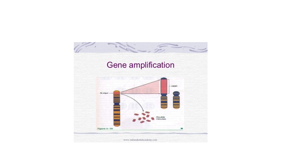
Gene amplifications • Proto-oncogenes can be amplified and overexpressed. . Converted to oncogenes. • Two patterns : homogenously stained region : Double minutes Exampl: 1. NMYC amplification in neuroblastoma , 25 -30% of neuroblastoma cases have the amplification and it is associated with poor prognosis 2. ERBB 2= HER 2/NEU amplification in 20% of breast cancer. Antibodies against the protein receptor effective in treatment
HER 2 in breast cancer. Can be detected by immunostain please chech lecture 4 activity for details
aneuploidy • = number of chromosomes not multiple of the haploid state (23). • Result from errors of the mitotic checkpoint • Aneupolidy. . Cause or result of neopalsia?
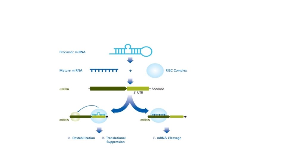
micro. RNAs (mi. RNAs) • Noncoding, micro (22 nucleotides) that are negative regulators of the genes. • They inhibit gene expression post-transcriptionally = repress translation or cleave m. RNA.
mi. RNAs
• mi. RNAs that target oncogenes…. If reduced, then inhibition lost…. Overexpression of oncogenes. . ras in lung carcinoma and myc in some leukemias • mi. RNAs that target tumor suppressor genes… if increased… downregulation of tumor suppressor genes.
mi. RNA • Cause cancer by increasing oncogene expression or decreasing tumor suppressor gene expression.
epigenetics • the study of changes in organisms caused by modification of gene expression rather than alteration of the genetic code itself • Epigenetics are reversible, heritable changes in gene expression that occur without mutation.
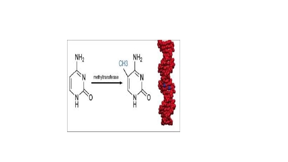
Epigenetic modifications • Reversible , heritable changes in gene expression without mutation. • Two types: Histone modifications and DNA methylation.
Epigenetic mutations • functionally relevant changes to the genome that do not involve a change in the nucleotide sequence. Examples of mechanisms that produce such changes are DNA methylation and histone modification, each of which alters how genes are expressed without altering the underlying DNA sequence.
Epigenetics and cancer • Gene expression is silenced by DNA methylation= more methyl groups more silencing. In cancer cells: • 1. Global DNA hypo methylation : increases expression of genes. also causes chromosomal instability • 2. Selective promoter hyper methylation of tumor suppressor genes: silenced
NOTE • Some epigenetic modifications can result from mutations of DNA sequences (gens) that regulate epigenetic modifications!!!!!
• Because epigenetic modifications regulate gene expression. . They might affect the response of one gene in different cells. • E. g NOTCH 1 gene in oncogenic in T cell leukemia but is tumor suppressor in squamous cell carcinoma • As if NOTCH is Spiderman : you have the red Spiderman and the black one. . They act differently in different situations according to the encvironment they are in that is according to the epigenetics!!!