Cell Theory and Exceptions History 1 Hooke 1665


 Protein capsid Assemble DNA injected into")


 – uses electron beams and electromagnetic lenses to magnify")
 – electron beams are passed back and forth over")


- Slides: 26
Cell Theory and Exceptions History: 1. Hooke - 1665 – using microscope viewed cork slices, first person to use the word “cell. ” 2. Van Leeuwenhoek - 1683 – first person to see red blood cells and living cells in pond water. 3. Brown - 1831 – a structured called nucleus present in both plant and animal cells.
4. Schleiden – 1838 – all plants made up of cells. 5. Schwann – 1839 – all animals made up of cells. 6. Virchow - 1855 – all cells arise from preexisting cells.
Cell Theory: 1. All organisms are made up of 1 or more cells (The cell is the structural unit of life) 2. All cells carry on life processes (The cell is the functional unit of life) 3. All cells come from preexisting cells
Exceptions to the Cell Theory: 1. The 1 st cell couldn’t have arisen from preexisting cells. 2. Mitochondria and chloroplasts have DNA and can reproduce within a cell. 3. A virus is not made up of a cell, can reproduce, but needs a host cell.
Viruses are made up of: • Core of hereditary material DNA or RNA. • Surrounding layer of protein • Outer layer of protein called a capsid Viruses that attack bacteria are called bacteriophages. Viruses that attack plants contain RNA. Animal viruses may contain either DNA or RNA.
Life Cycle of a Virus: DNA Bacterium (host) Protein capsid Assemble DNA injected into host DNA and capsid reproduced Lysis (split)
• Virus attaches itself to the host cell by means of specific recognition proteins. • Virus injects the DNA or RNA into the cytoplasm of the host cell. • The DNA/RNA causes the cell to start to reproducing the viral DNA/RNA and the capsid proteins. The protein and hereditary material join together. • Numerous viral particles are produced. • The cell bursts open releasing the newly made viral particles. This process is called the lytic cycle.
OR The hereditary material of some viruses like the Herpes, HIV and others become incorporated into the chromosomes of the host and is called a provirus. Whenever the cell divides, the provirus also divides, resulting in two infected host cells. The cycle is called the lysogenic cycle. The virus may remain dormant for years before the provirus enters the lytic cycle. Lytic Cycle Lysogenic Cycle Animation
Tools of the Biologist 1. Light microscope – any device that uses light to produce a magnified view of an object. Two types of light microscopes: a. Simple microscope - magnifying glass (1 lens) b. Compound microscope – uses 2 lenses to magnify image 1000 – 2000 X Magnification ratio = ratio of image size to object size.
2. Vital Stains - dyes which are selective to help you see structures better. Examples: Methylene blue (animal cells) Lugol’s iodine (plant cells)
3. Transmission electron microscope (TEM) – uses electron beams and electromagnetic lenses to magnify image, can magnify image 250, 000 times or greater, big, expensive, special training required to prepare specimens for viewing. There is greater resolution and magnification compared to the light microscope. A TEM can reveal the cell’s internal structure in detail. Internal structure White Blood Cell
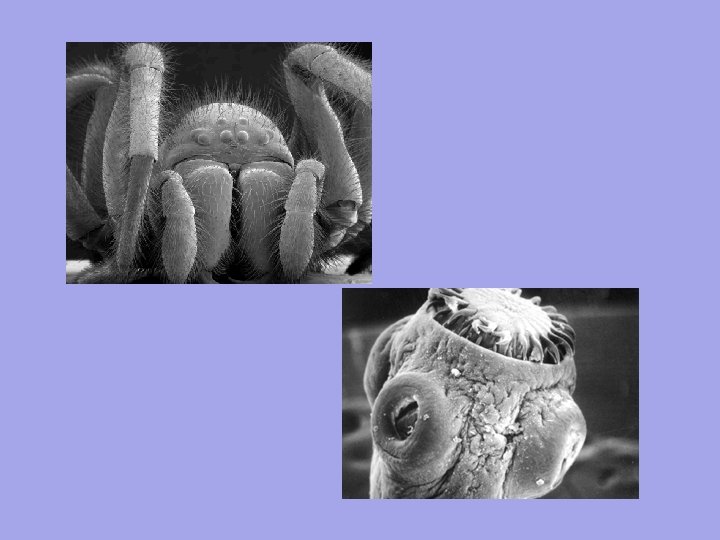
4. Scanning electron microscope (SEM) – electron beams are passed back and forth over the specimen to give a detailed 3 -D image. You also get fine details of the surface of the specimen. It is also expensive, and requires special training to prepare specimens for viewing (specimens are coated in a thin layer of gold and viewed in a vacuumed chamber. Magnification in the 50, 000 – 100, 000 range. Cat Flea
5. Microdissection instruments – delicate tools used under a dissecting microscope to manipulate living cells. Ex. Removal of a nucleus from one cell and placed in another cell.
6. Electrophoresis - technique used to separate substances based on their size and electrical charge. The substances are placed in an electrical field and the substances move at their own characteristic rate. Ex. DNA has a negative charge, move according to size towards the positive pole.
7. Chromatography - technique that separates different substances from each other based on their chemical or physical properties.
8. Ultracentrifuge 3 particles of different densities Time Ultracentrifuge Layers, densest particles on bottom Ultracentrifuge - technique used to separate particles based on their densities. Ultracentrifuge is primarily used for separating cell parts or organelles. Ex. Separate out all of the nuclei from a group of onion cells.
Electron Microscope
TEM Images Chloroplast Golgi Body
SEM Images Cat Flea Bird Feather Hypodermic Needle Moth Antenna
Fly’s Foot Mosquito Head Velcro
Microdissection Tools
Ultracentrifuge
Electrophoresis
Chromatography
http: //www. youtube. com/watch? v= UEKS 4 w 9 bf. Jg http: //www. youtube. com/watch? v= c. E 0 qdqo. BFa 8