The Cell Robert Hooke 1665 British scientist Used

The Cell
 British scientist Used a microscope to study a thin slice of")
Robert Hooke (1665) British scientist Used a microscope to study a thin slice of cork from the bark of an oak tree Named the box-like structures “cells” Saw only dead cells

Hooke’s microscope
 Dutch businessman; his single-lens microscope magnified objects 200 X")
Anton van Leeuwenhoek (1600 s) Dutch businessman; his single-lens microscope magnified objects 200 X and allowed him to see tiny organisms in a drop of water (animalcules)
 Scottish botanist; used newly developed stains; first to describe the cell")
Robert Brown (1831) Scottish botanist; used newly developed stains; first to describe the cell nucleus
 German botanist; concluded that all plants are made up of cells")
Matthias Schleiden (1838) German botanist; concluded that all plants are made up of cells
 German scientist; concluded that all animals are made up of cells")
Theodor Schwann (1839) German scientist; concluded that all animals are made up of cells
 German physician; studied cell reproduction “Where a cell exists, there must")
Rudolf Virchow (1855) German physician; studied cell reproduction “Where a cell exists, there must have been a preexisting cell…”

Cell Theory All living things are made up of cells (organisms are either unicellular or multicellular) Cells are the basic units of structure and function in living things All cells arise from preexisting cells

Microscopes The two most important properties of microscopes are: n n Magnification – how much an image is enlarged Resolution – clarity (sharpness) of the image; ability to show two objects as separate

The Compound Light Microscope Can produce magnifications of 50 X to 500 X Light is passed through the specimen and then through glass lenses The lenses refract (bend) light in such a way that the image of the specimen is magnified

Electron Microscope These scopes focus using a beam of electrons Magnifying powers in excess of 100, 000 X Specimens must be dead or nonliving
 1/10 th the size of")
2 Main Types of Cells Prokaryotic cells Monera (bacteria) 1/10 th the size of eukaryotic cells Lack internal membrane bound organelles Prokaryotes DO have ribosomes

Prokaryotic cell

Eukaryotic cells Found in protists, plants, fungi, and animals have a true nucleus containing chromosomes contain several membrane bound organelles

Parts of the Eukaryotic Cell

Cell Organelles “little organs” Specialized parts of the cell which carry out specific life functions
 Membrane Separates cell from surrounding environment Controls the movement of molecules")
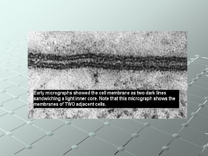
A. Plasma (Cell) Membrane Separates cell from surrounding environment Controls the movement of molecules into or out of the cell selectively permeable (semi-permeable) Visible with the compound microscope
 Flexible structure with freely moving pieces 2)")
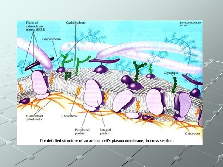
Fluid Mosaic Model of Membrane Structure 1) Flexible structure with freely moving pieces 2) Composed mainly of lipids and proteins http: //home. earthlink. net/~shalpine/anim/ Life/memb. htm http: //telstar. ote. cmu. edu/Hughes/tutorial/ cellmembranes/
 Lipid bilayer (phospholipid molecules + cholesterol) polar heads: hydrophilic (water-loving); in contact with")
3) Lipid bilayer (phospholipid molecules + cholesterol) polar heads: hydrophilic (water-loving); in contact with cytoplasm/extracellular fluid nonpolar tails: hydrophobic (water-hating); interior of membrane

phospholipid

Phospholipid Bilayer
 Proteins extend through the membrane or are found on the outer/inner surface Integral")
4) Proteins extend through the membrane or are found on the outer/inner surface Integral proteins: embedded in the lipid layer; involved in transport across the cell membrane Peripheral proteins: attach to cytoskeleton in the cytoplasm; allows cell to pull membrane in towards center of cell
 Other proteins act as receptors for hormones or chemicals, are involved in cell-cell")
5) Other proteins act as receptors for hormones or chemicals, are involved in cell-cell communication, or join one cell to another

http: //www. bio. davidson. edu/people/macam pbell/111/memb-swf/membranes. swf
 Region between nucleus & plasma membrane 2) Cytosol (cytoplasm) = fluid")
B. Cytoplasm 1) Region between nucleus & plasma membrane 2) Cytosol (cytoplasm) = fluid 3) Suspends organelles 4) Site of biochemical processes/reactions 5) Supported by a cytoskeleton: fibers that controls movement of cell or its internal parts

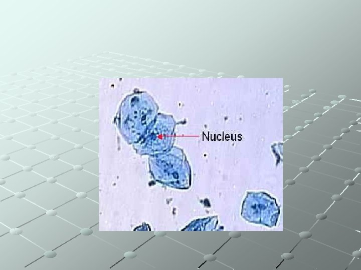
C. Nucleus Contains chromosomes Averages 5 m in diameter Visible under the compound light microscope Enclosed by a nuclear envelope (membrane) perforated by pores that regulates the entrance of exit of molecules

D. Nucleolus “site of ribosome synthesis” Located within the nucleus stains darker than the nucleus because it is more dense

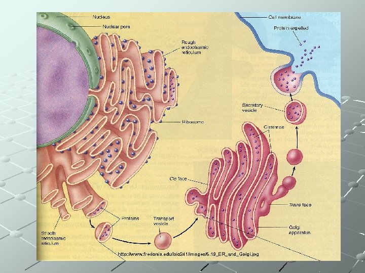
E. Ribosomes “site of protein synthesis” free ribosomes are suspended in the cytoplasm: make proteins that will remain dissolved in the cytosol other ribosomes are attached to the outside of an organelle called the endoplasmic reticulum (ER): make proteins that will be incorporated into membranes or be secreted by the cell

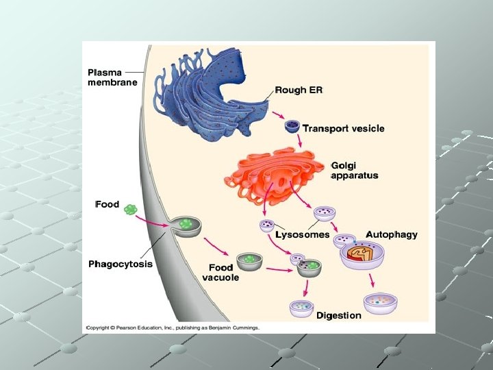
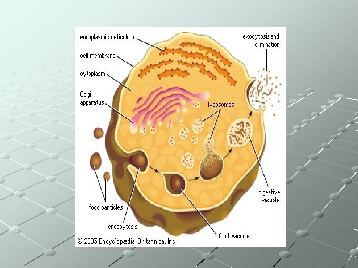
The Endomembrane System Includes the endoplasmic reticulum, Golgi apparatus, lysosomes and vacuoles

F. Endoplasmic Reticulum Network of channels for carrying substances from one part of the cell to another Usually surrounds the cell nucleus
 Rough ER is dotted with ribosomes")
There are two types of endoplasmic reticulum (ER) Rough ER is dotted with ribosomes

Smooth ER lacks ribosomes is involved in synthesis of lipids, metabolism of carbohydrates, and detoxification of drugs and other poisons

G. Golgi apparatus

Stacks of membranes forming flattened sacs Process, package, store cell products to be secreted (transported out of the cell) ribosomes ►RER ►SER ►Golgi ►cell membrane ►release from cell (make proteins) apparatus http: //www. johnkyrk. com/golgi. Alone. html http: //student. ccbcmd. edu/~gkaiser/biotutori als/eustruct/endomembanim. html
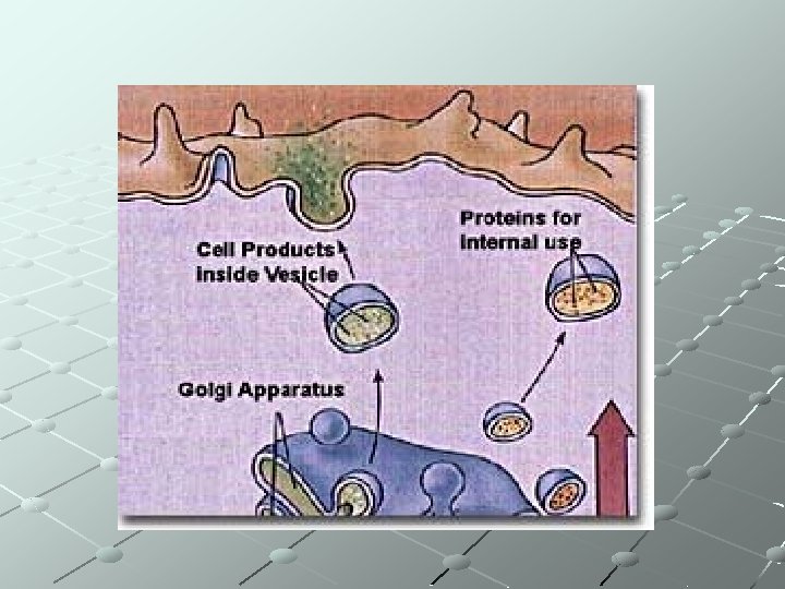

Exocytosis – release of a substance outside of the cell membrane by the fusion of a vacuole

H. Lysosomes “breakdown body” Sac of digestive enzymes Digest organic molecules, worn-out cell structures, harmful bacteria Lysosomes are found in all eukaryotic cells, but are most numerous in diseasefighting cells, such as white blood cells.

I. Vacuoles Variety of functions Food vacuoles Contractile vacuoles in freshwater protists pump out excess water Plant cells have a large central vacuole for water and nutrient storage
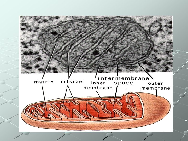
 “powerhouse of the cell” Site of cellular respiration: glucose + oxygen")
J. Mitochondria (Mitochondrion) “powerhouse of the cell” Site of cellular respiration: glucose + oxygen is converted to energy in the form of ATP Each cell contains between 300 -800 mitochondria depending on activity level Shaped like a slipper or peanut Has a double membrane Inner membrane has many foldings = cristae

Although most of a cell's DNA is contained in the cell nucleus, mitochondria have their own DNA

According to the endosymbiotic theory, mitochondria are descended from independent prokaryotes that were engulfed by other cells but not digested The mitochondria gave the cell that engulfed it a selective advantage over other cells

K. Chloroplasts Found in plants, algae Contain chlorophyll, a green pigment that absorbs light to start photosynthesis

Chloroplasts contain their own genome – DNA Chloroplasts are also part of the endosymbiotic theory

L. Centrioles Small bundle of microtubules Found in pairs near cell nucleus Involved in cell division Found in animal cells but not plant cells

M. Cell Wall Found in plant cells only A thick layer of cellulose material that forms fibers which wrap around the cell Provides protection & structure for the cell; prevents expansion

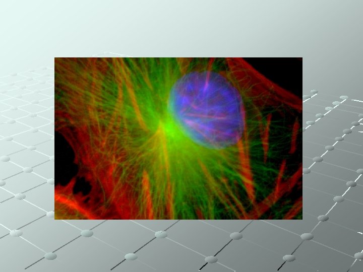
N. Cytoskeleton A network of fibers extending throughout the cytoplasm Microtubules: straight hollow tubes of protein Functions: mechanical support, maintains shape, allows for movement

http: //www. cellsalive. com/cells/cell_model. htm
- Slides: 57