1 VISION II 2 Photoreceptors Rods and cones


 �Consist of: 1) Outer segment (facing choroid) �Detects light stimulus.")


 theory �There are 3 types of cones. Each one is maximally sensitive")

 and affecting males")


 - Concerned with the appreciation of visual")

- Slides: 26
1
. . VISION -II 2
Photoreceptors (Rods and cones) �Consist of: 1) Outer segment (facing choroid) �Detects light stimulus. It contains visual pigment (iodopsin in cones & rhodopsin in rods) that forms 40 % of this segment. 2) Inner segment �Contains metabolic machinery of cell 3) Nucleus 4) Synaptic terminal (facing bipolar cells) �Transmits signals generated in photoreceptor on light stimulation to dendrites of bipolar cells. 4
5
Rods Cones 120 millions / retina 6 millions / retina At the periphery At Center (Fovea centralis) Rhodopsin pigment Photopsin (3 types). More convergence (200 rods : 1 bipolar) One to one connection More sensitive to light (even to 1 photon). Less sensitive to light. Less accurate (less visual acuity). More accurate (Clear vision) More functioning at night (Scotopic vision) More functioning in day (Photobic vision) Vision in shades of grey Color perception (3 types for Red, Green & Blue)
7
Photopigments �Undergo chemical alterations when activated by light �Consists of 2 components: 1 - Opsin �Protein that is integral part of disc plasma membrane 2 - Retinal �Derivative of vitamin A �Light-Absorbing part of photopigment 8
Photopigments 1 - Rod pigment: �Rhodopsin that Absorbs all visible wavelengths 2 - Cone pigments (color pigments): �Respond v. A selectively to various wavelengths of light. single photon of light can activate a rod whereas several hundred photons are required to activate a cone 9
PHOTORECEPTORS RESPONSE TO LIGHT PHOTORECEPTOR POTENTIAL
Chemical response to light
phototransduction 12
13
14
DARK ADAPTATION v If a person has been in brightly lighted surroundings for a long time and then moves to a dark area, the retina slowly becomes more sensitive to light (a decline in visual threshold). v. Due to regeneration of photosensitive pigments in both rods and cones. Retinal and opsins are converted back into photosensitive pigments (It is nearly maximal in about 20 minutes). v. Regeneration of rhodopsin in rods is completed in 20 minutes. Rods play the major role in dark adaptation.
LIGHT ADAPTATION �It occurs when we shift suddenly from a dark to a bright light place. Immediately, light seems very intense and annoying with reflex closure of eye lids. But within 5 min, normal sensation returns again. �Then the eyes adapt to increased illumination & the visual threshold rises. There is a decline in sensitivity of retina to light. �Due to breakdown of photosensitive pigments in both rods and cones. The photosensitive chemicals are reduced to retinal & opsins (bleaching).
COLOUR VISION �It is the sense of discrimination of various wavelengths of visible spectrum (400 - 700 nm). �Each color has a specific wave length. �Shortest wave length is that of violet color. �Longest wave length is that of red color. �Color vision is mainly the function of Cones (highly developed in fovea centralis).
Trichromatic (=Young-Helmholtz) theory �There are 3 types of cones. Each one is maximally sensitive to one of the 3 primary colors. 1 st cone contains pigment maximally sensitive to blue violet (440 nm). 2 nd cone contains pigment maximally sensitive to green of spectrum (535 nm). 3 rd cone contains pigment maximally sensitive to red of spectrum (570 nm). �Any colored object stimulates the 3 cones but in an unequal manner. Number of impulses arising from these cones to visual cortex differ in frequency according to the color. This difference gives the specific sensation by that color. 18
v Equal stimulation of all 3 cones gives sensation of white. v Color perception is a retinal function. v Color translation is a cortical function (area 17).
COLOR BLINDNESS Causes: • Most commonly hereditary (recessive sex - linked) and affecting males (8%) more than females (0. 4 %). • Very rarely acquired e. g. post encephalitis. • Achromatopsia = color blindness due to lesion in V 8 in visual cortex. Types: According to Young - Helmholtz theory, Color blindness is classified to: 1 -Trichromate: Patient has 3 cones but one of them is weak. �Protanomaly: weakness in red color perception. �Deuteranomaly: weakness in green color perception. �Triatanomaly: weakness in blue color perception.
2 - Dichromate: One cone is totally absent & other two are present. �Protanopia: if cone of red color is absent. �Deuteranopia: if cone of green color is absent. �Tritanopia: if cone of blue color is absent. 3 - Monchromate: �Patient has only one cone system and matches the different colors as various degrees of grey color (cone monochromatism). �Or Patient has no cone system at all color & day blindness (rod monochromatism). Test of color vision: �Ishihara's chart test: Colored printed figures and each figure is printed by different colored small circles on a background of colored circles. 21
v. Red green color blindness is most common. Ishehara Chart 22
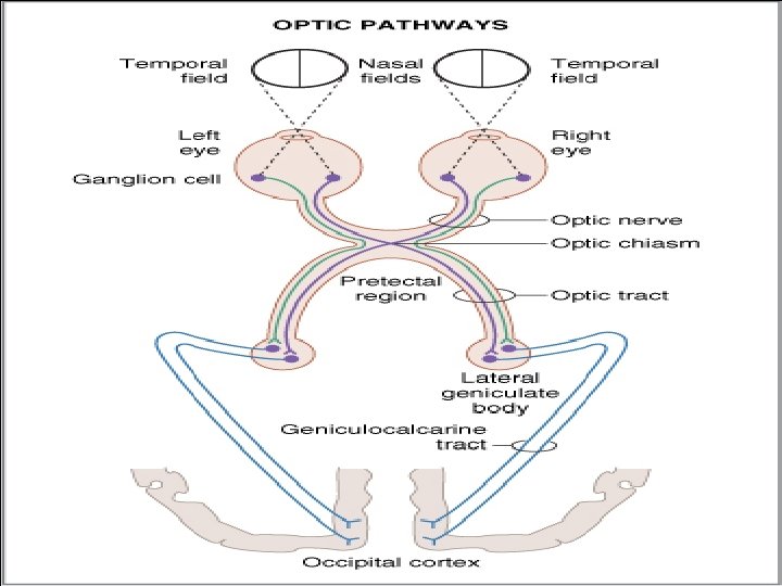
Left eye blind Bitemporal hemianopia Heteronymous Homonymous hemianopia with macular sparring
VISUAL CORTEX Area 17 (=Primary visual area) - Concerned with the appreciation of visual sensations. Area 18 (=Secondary visual area/visual association area) - Concerned with correlation and integration of visual sensations. Area 19 (=Occipital eye field) - Concerned with movement of the eye. v In each visual cortex, both the eyes are represented in alternating columns of cells called Ocular Dominance Columns. v Thus corresponding regions of the retina lie side by side close to each other.
26