1 2 Ultrastructure of cells Essential idea Eukaryotes
- Slides: 37
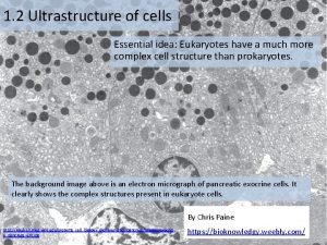
1. 2 Ultrastructure of cells • Essential idea: Eukaryotes have a much more complex cell structure than prokaryotes
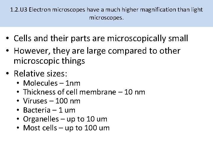
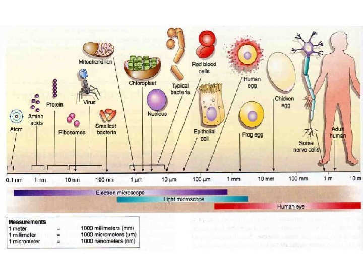
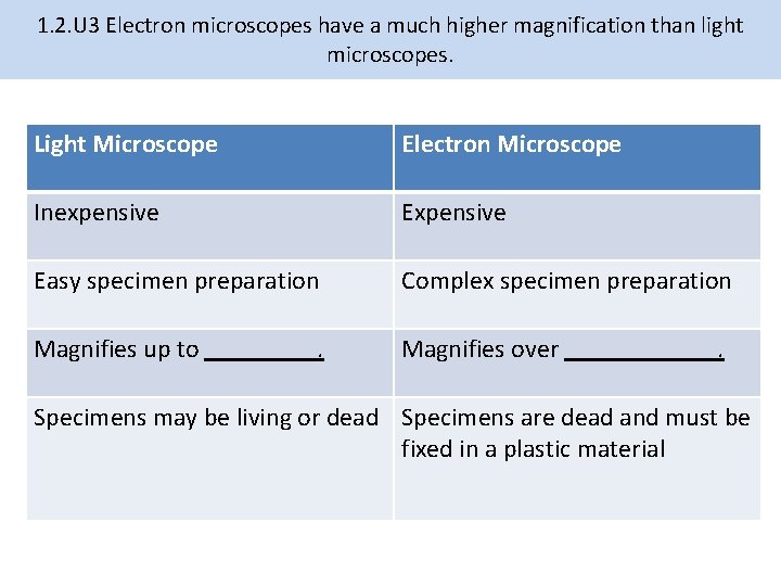
1. 2. U 3 Electron microscopes have a much higher magnification than light microscopes. • Cells and their parts are microscopically small • However, they are large compared to other microscopic things • Relative sizes: • • • Molecules – 1 nm Thickness of cell membrane – 10 nm Viruses – 100 nm Bacteria – 1 um Organelles – up to 10 um Most cells – up to 100 um
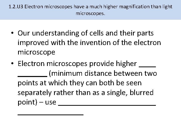
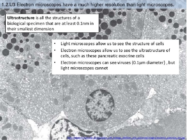
1. 2. U 3 Electron microscopes have a much higher magnification than light microscopes. • Our understanding of cells and their parts improved with the invention of the electron microscope • Electron microscopes provide higher (minimum distance between two points at which they can both be seen separately rather than as a single, blurred point) – use
1. 2. U 3 Electron microscopes have a much higher magnification than light microscopes. Light Microscope Electron Microscope Inexpensive Easy specimen preparation Complex specimen preparation Magnifies up to Magnifies over . . Specimens may be living or dead Specimens are dead and must be fixed in a plastic material
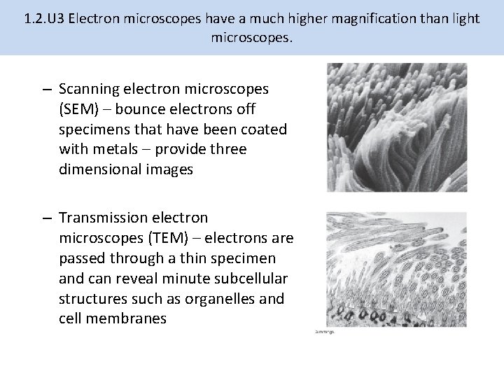
1. 2. U 3 Electron microscopes have a much higher magnification than light microscopes. – Scanning electron microscopes (SEM) – bounce electrons off specimens that have been coated with metals – provide three dimensional images – Transmission electron microscopes (TEM) – electrons are passed through a thin specimen and can reveal minute subcellular structures such as organelles and cell membranes
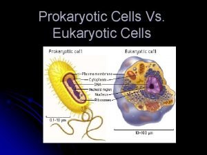
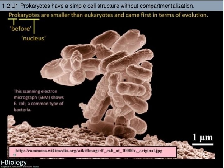
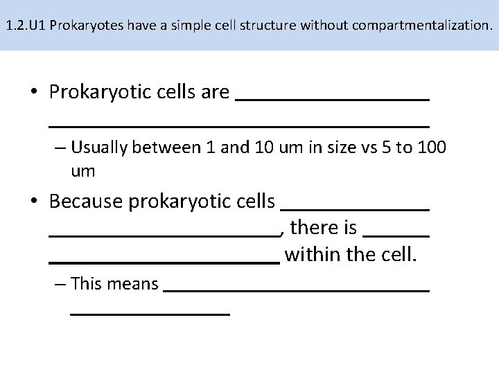
1. 2. U 1 Prokaryotes have a simple cell structure without compartmentalization. • Prokaryotic cells are – Usually between 1 and 10 um in size vs 5 to 100 um • Because prokaryotic cells – This means , there is within the cell.
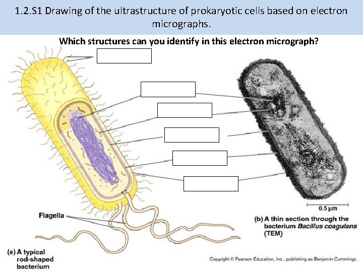
1. 2. S 1 Drawing of the ultrastructure of prokaryotic cells based on electron micrographs. Which structures can you identify in this electron micrograph?
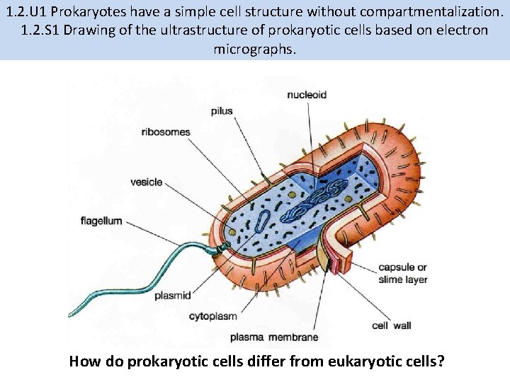
1. 2. U 1 Prokaryotes have a simple cell structure without compartmentalization. 1. 2. S 1 Drawing of the ultrastructure of prokaryotic cells based on electron micrographs. How do prokaryotic cells differ from eukaryotic cells?
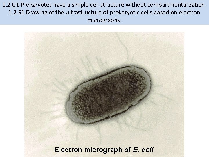
1. 2. U 1 Prokaryotes have a simple cell structure without compartmentalization. 1. 2. S 1 Drawing of the ultrastructure of prokaryotic cells based on electron micrographs. Electron micrograph of E. coli
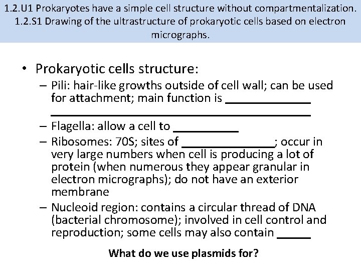
1. 2. U 1 Prokaryotes have a simple cell structure without compartmentalization. 1. 2. S 1 Drawing of the ultrastructure of prokaryotic cells based on electron micrographs. • Prokaryotic cells structure: – Cell wall: protects and maintains the shape of the cell; most prokaryotic cells have cell walls made of (carb-protein complex) – Plasma membrane: lies and it’s composition is similar to eukaryotic cells; controls transport of material in/out; plays a role in binary fission – Cytoplasm: site of the chemical reactions of life
1. 2. U 1 Prokaryotes have a simple cell structure without compartmentalization. 1. 2. S 1 Drawing of the ultrastructure of prokaryotic cells based on electron micrographs. • Prokaryotic cells structure: – Pili: hair-like growths outside of cell wall; can be used for attachment; main function is – Flagella: allow a cell to – Ribosomes: 70 S; sites of ; occur in very large numbers when cell is producing a lot of protein (when numerous they appear granular in electron micrographs); do not have an exterior membrane – Nucleoid region: contains a circular thread of DNA (bacterial chromosome); involved in cell control and reproduction; some cells may also contain What do we use plasmids for?
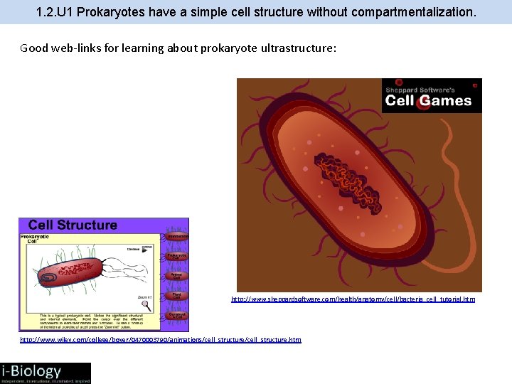
1. 2. U 1 Prokaryotes have a simple cell structure without compartmentalization. Good web-links for learning about prokaryote ultrastructure: http: //www. sheppardsoftware. com/health/anatomy/cell/bacteria_cell_tutorial. htm http: //www. wiley. com/college/boyer/0470003790/animations/cell_structure. htm
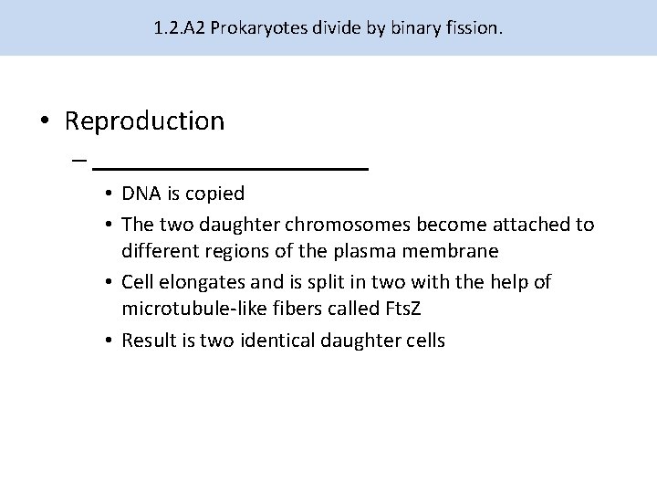
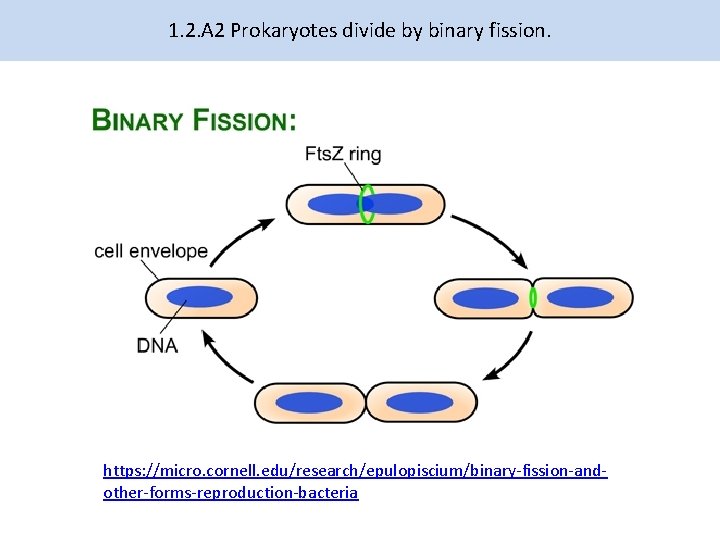
1. 2. A 2 Prokaryotes divide by binary fission. • Reproduction – • DNA is copied • The two daughter chromosomes become attached to different regions of the plasma membrane • Cell elongates and is split in two with the help of microtubule-like fibers called Fts. Z • Result is two identical daughter cells
1. 2. A 2 Prokaryotes divide by binary fission. https: //micro. cornell. edu/research/epulopiscium/binary-fission-andother-forms-reproduction-bacteria
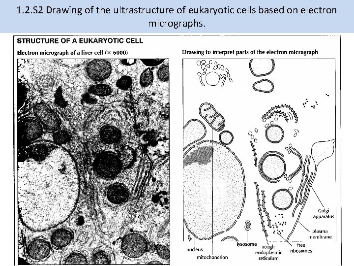
1. 2. S 2 Drawing of the ultrastructure of eukaryotic cells based on electron micrographs.
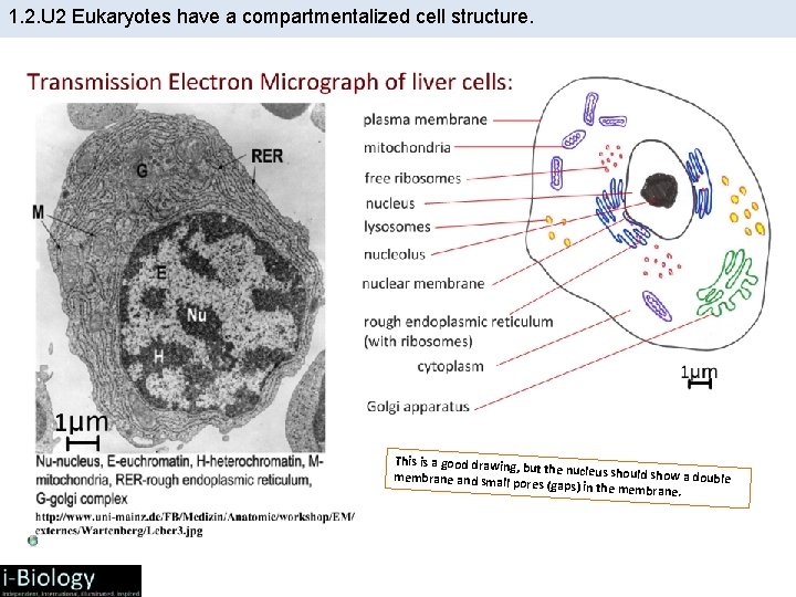
1. 2. U 2 Eukaryotes have a compartmentalized cell structure. This is a good drawing, but the nucleus should show a double membrane and small po res (gaps) in the membr ane.
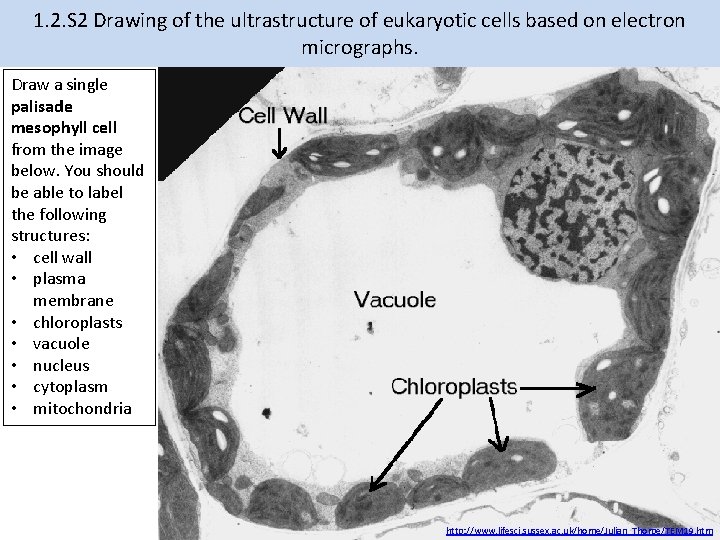
1. 2. S 2 Drawing of the ultrastructure of eukaryotic cells based on electron micrographs. Draw a single palisade mesophyll cell from the image below. You should be able to label the following structures: • cell wall • plasma membrane • chloroplasts • vacuole • nucleus • cytoplasm • mitochondria http: //www. lifesci. sussex. ac. uk/home/Julian_Thorpe/TEM 19. htm
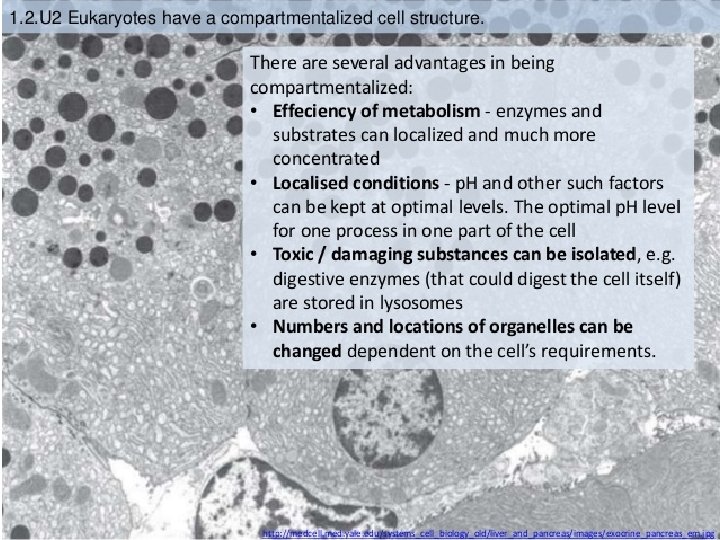
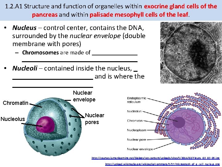
1. 2. A 1 Structure and function of organelles within exocrine gland cells of the pancreas and within palisade mesophyll cells of the leaf. • Nucleus – control center, contains the DNA, surrounded by the nuclear envelope (double membrane with pores) – Chromosomes are made of • Nucleoli – contained inside the nucleus, and is where the Chromatin Nucleolus Nuclear envelope Nuclear pores http: //courses. lumenlearning. net/biology/wp-content/uploads/sites/5/2014/02/Figure_03_03_05. jpg http: //upload. wikimedia. org/wikipedia/commons/5/57/Micrograph_of_a_cell_nucleus. png
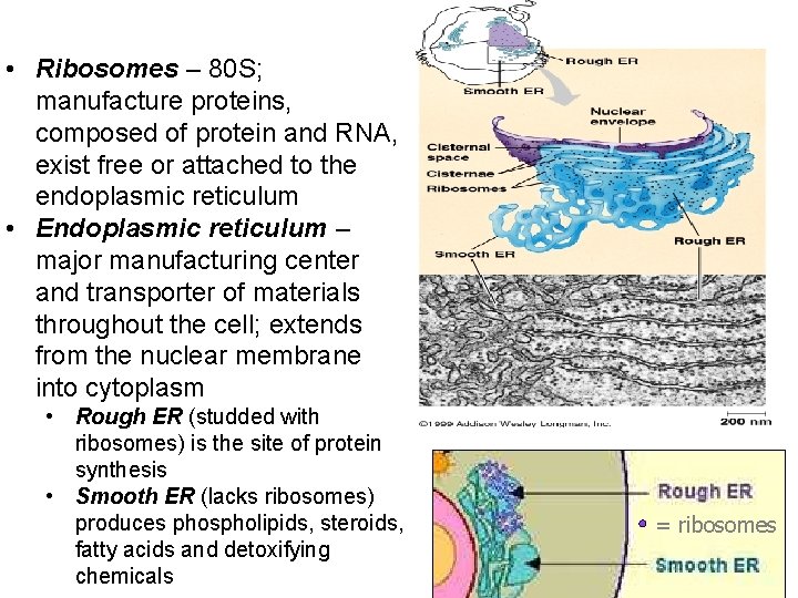
• Ribosomes – 80 S; manufacture proteins, composed of protein and RNA, exist free or attached to the endoplasmic reticulum • Endoplasmic reticulum – major manufacturing center and transporter of materials throughout the cell; extends from the nuclear membrane into cytoplasm • Rough ER (studded with ribosomes) is the site of protein synthesis • Smooth ER (lacks ribosomes) produces phospholipids, steroids, fatty acids and detoxifying chemicals = ribosomes
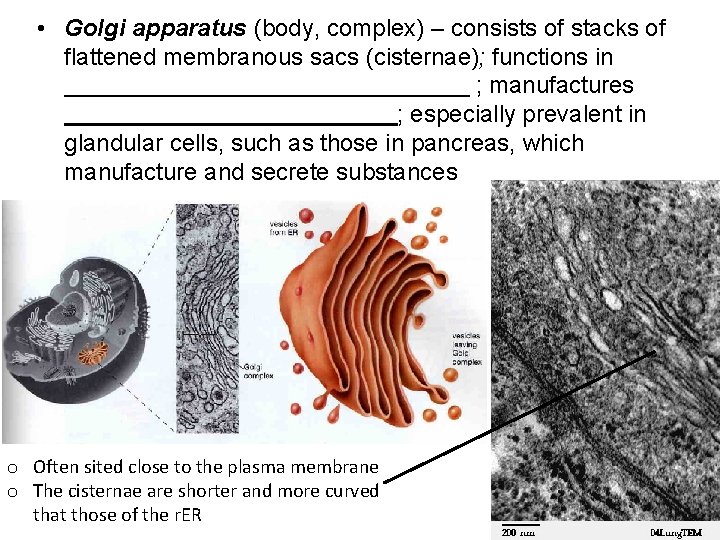
• Golgi apparatus (body, complex) – consists of stacks of flattened membranous sacs (cisternae); functions in ; manufactures ; especially prevalent in glandular cells, such as those in pancreas, which manufacture and secrete substances o Often sited close to the plasma membrane o The cisternae are shorter and more curved that those of the r. ER
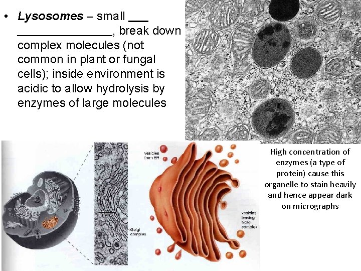
• Lysosomes – small , break down complex molecules (not common in plant or fungal cells); inside environment is acidic to allow hydrolysis by enzymes of large molecules High concentration of enzymes (a type of http: //1. bp. blogspot. com/-WGspb. Etlkls/Tg. CUbtp 7_-I/AAAAAJw/So. Ip 2 v. Xz. H 4 E/s 320/Figure+2 -26. bmp protein) cause this organelle to stain heavily and hence appear dark on micrographs
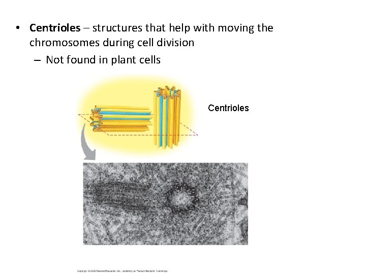
• Centrioles – structures that help with moving the chromosomes during cell division – Not found in plant cells Centrioles
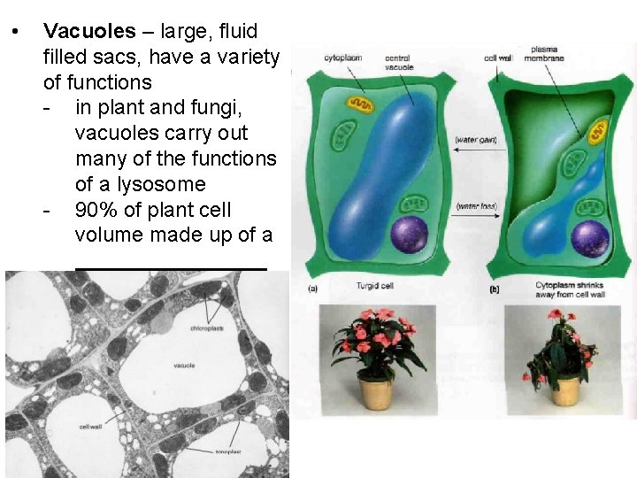
• Vacuoles – large, fluid filled sacs, have a variety of functions - in plant and fungi, vacuoles carry out many of the functions of a lysosome - 90% of plant cell volume made up of a
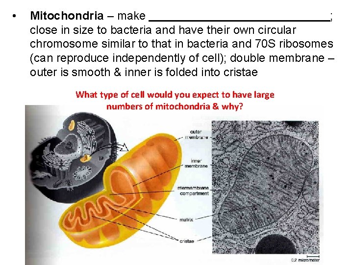
• Mitochondria – make ; close in size to bacteria and have their own circular chromosome similar to that in bacteria and 70 S ribosomes (can reproduce independently of cell); double membrane – outer is smooth & inner is folded into cristae What type of cell would you expect to have large numbers of mitochondria & why?
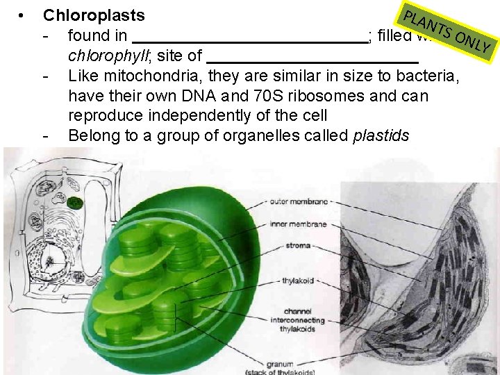
• PLA Chloroplasts NTS - found in ; filled with ONL Y chlorophyll; site of - Like mitochondria, they are similar in size to bacteria, have their own DNA and 70 S ribosomes and can reproduce independently of the cell - Belong to a group of organelles called plastids
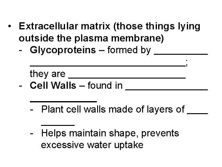
• Extracellular matrix (those things lying outside the plasma membrane) - Glycoproteins – formed by ; they are - Cell Walls – found in - Plant cell walls made of layers of - Helps maintain shape, prevents excessive water uptake
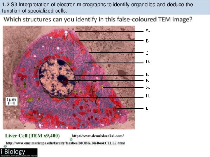
A. B. C. D. E. F. G. H. I.
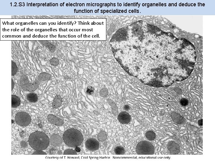
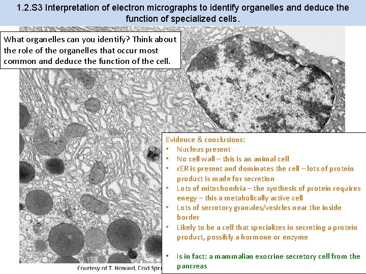
1. 2. S 3 Interpretation of electron micrographs to identify organelles and deduce the function of specialized cells. What organelles can you identify? Think about the role of the organelles that occur most common and deduce the function of the cell.
1. 2. S 3 Interpretation of electron micrographs to identify organelles and deduce the function of specialized cells. What organelles can you identify? Think about the role of the organelles that occur most common and deduce the function of the cell. Evidence & conclusions: • Nucleus present • No cell wall – this is an animal cell • r. ER is present and dominates the cell – lots of protein product is made for secretion • Lots of mitochondria – the synthesis of protein requires enegy – this a metabolically active cell • Lots of secretory granules/vesicles near the inside border • Likely to be a cell that specializes in secreting a protein product, possibly a hormone or enzyme • Is in fact: a mammalian exocrine secretory cell from the pancreas
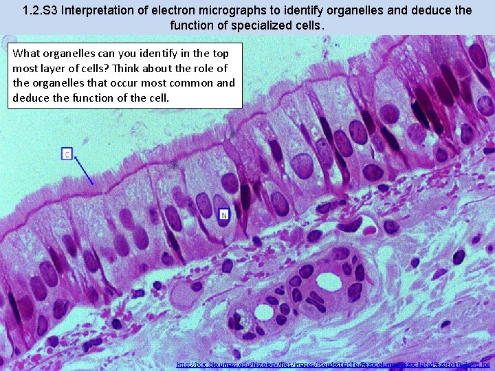
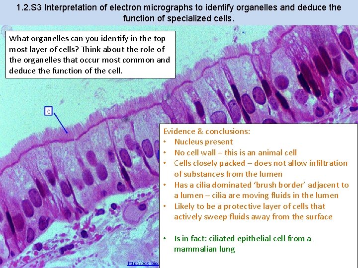
1. 2. S 3 Interpretation of electron micrographs to identify organelles and deduce the function of specialized cells. What organelles can you identify in the top most layer of cells? Think about the role of the organelles that occur most common and deduce the function of the cell. http: //bcrc. bio. umass. edu/histology/files/images/Pseudostratified%20 Columnar%20 Ciliated%20 Epithelium 1. jpg
1. 2. S 3 Interpretation of electron micrographs to identify organelles and deduce the function of specialized cells. What organelles can you identify in the top most layer of cells? Think about the role of the organelles that occur most common and deduce the function of the cell. Evidence & conclusions: • Nucleus present • No cell wall – this is an animal cell • Cells closely packed – does not allow infiltration of substances from the lumen • Has a cilia dominated ‘brush border’ adjacent to a lumen – cilia are moving fluids in the lumen • Likely to be a protective layer of cells that actively sweep fluids away from the surface • Is in fact: ciliated epithelial cell from a mammalian lung http: //bcrc. bio. umass. edu/histology/files/images/Pseudostratified%20 Columnar%20 Ciliated%20 Epithelium 1. jpg
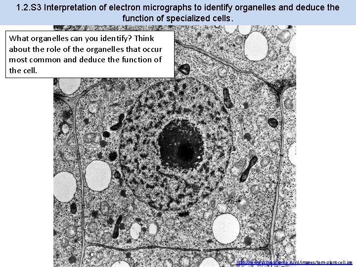
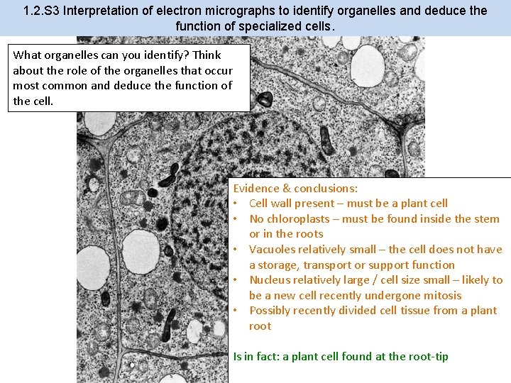
1. 2. S 3 Interpretation of electron micrographs to identify organelles and deduce the function of specialized cells. What organelles can you identify? Think about the role of the organelles that occur most common and deduce the function of the cell. http: //www. vcbio. science. ru. nl/images/tem-plant-cell. jpg
1. 2. S 3 Interpretation of electron micrographs to identify organelles and deduce the function of specialized cells. What organelles can you identify? Think about the role of the organelles that occur most common and deduce the function of the cell. Evidence & conclusions: • Cell wall present – must be a plant cell • No chloroplasts – must be found inside the stem or in the roots • Vacuoles relatively small – the cell does not have a storage, transport or support function • Nucleus relatively large / cell size small – likely to be a new cell recently undergone mitosis • Possibly recently divided cell tissue from a plant root Is in fact: a plant cell found at the root-tip http: //www. vcbio. science. ru. nl/images/tem-plant-cell. jpg