University College London 2018 Retreatment dose prescriptions for


% plots. Existing in vivo data above critical no recovery line")


% • Output parameter")
 Each curve shows BED")


 show greatest change in photon")

![α/β=2 Gy: Central Nervous System [Jones B, Acta Oncol 2017, supplementary section] Dose (Gy)](https://slidetodoc.com/presentation_image_h/0b10304d408e069520719d22fa11c9a0/image-18.jpg "α/β=2 Gy: Central Nervous System [Jones B, Acta Oncol 2017, supplementary section] Dose (Gy)")

 with depth and delivery systems (pre-scattered versus scanned")


- Slides: 25
University College London 2018 Re-treatment dose prescriptions for proton therapy in the spinal cord/CNS structures for scanned and scattered beams. Bleddyn Jones MD FRCR Gray Laboratory, CRUK-MRC Oxford Oncology, University of Oxford & Oxford Univ. Hospitals
Estimations of re-treatment dose fractionation schedules - references Changes in the retreatment radiation tolerance of the spinal cord with time after the initial treatment. Int J Radiation Biology 2018 , Jun; 94(6): 515 -531. TE Woolley, J Belmonte-Beitia, GF Calvo, JW Hopewell, EA Gaffney and B Jones. Based on two earlier articles: Jones B & Grant W. Retreatment of Central Nervous System tumours. Clinical Oncology, 26, 407 -418, 2014. Jones B & Hopewell JH. Alternative models for estimating the radiotherapy retreatment dose for the spinal cord. Int J Radiat Biol. 2014 Sep; 90(9): 731 -41. Many clinical reviews of re-treatment usefulness e. g. Re-irradiation in the Brain: Primary Gliomas. Ho ALK, Jena R. Clin Oncol (R Coll Radiol). 2018 Feb; 30(2): 124 -136
Introduction • Re-treatment results can sometimes be as good as first line chemotherapy! • Particle therapy may be particularly suited for retreatments ……. . due to reduced irradiated volume, either as first or second treatment. • Retreatment may refer not only to tumour recurrences but to tumours arising in a previously irradiated anatomical site, e. g. pelvis, thorax, head and neck. Jones B. Personal View: The potential advantages of charged particle radiotherapy using protons or light ions. Clinical Oncology [Royal College of Radiologists], 20, 555 -563, 2008.
Evidence for time dependent ‘Recovery’ in CNS • Many experiments in small animals…rats, mice, with short retreatment time interval possibilities • One data set in primates (K. Ang et al 2001) • Human evidence from radiotherapy
Paravertebral Sarcoma Reduction in breast, lung cancer induction risk, cardiac sudden death and breathlessness on exertion with protons; but if RBE incorrect and/or Bragg peaks misplaced there could be paralysis (spinal cord) and reduced tumour control IMProton. T IM X-ray RT MGH Boston
Dose-related incidence of radiation myelopathy in the Rhesus monkey: single and a repeated course irradiation of Ang et al 2001 (J Hopewell graphic) Ang et al. , 2001
Biological Effective Dose (BED/BEDtol)% plots. Existing in vivo data above critical no recovery line Rat expts Primates 1, 2&3 years
Human data sets ( black points: Wong et al - myelitis; grey points Nieder et al – No myelitis, All data in agreement with model)
Human and rhesus monkey data from Ang and Hopewell Green data = human, Bluedata = monkey Green curve is conservative interpretation of human (a 10% reduction)
Introducing greater degrees of ‘conservatism’, for patients where tolerance is reduced (surgery chemotherapy, extremes of age, vasculopathies).
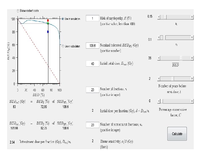
The GUI • Input parameter………BED 1% is the (Given BED/Tolerance BED)% • Output parameter is BED 2%, which is (allowable BED/Tolerance BED)% Graphical User Interface (GUI) can be downloaded to facilitate estimates of allowable dose per fraction and number of fractions for the retreatment. This should be regarded as a boundary value.
For a myelitis risk of 0. 1% (1 in 1000) Each curve shows BED 2(%) increasing with time between treatments for 4, 5 and 6 months followed by 1, 2 and 3 years
Tennis court ‘boundary’ limits…. the model gives an estimate of the boundary, within which it is safe to proceed. The lines change with circumstances
Relative Biological Effect – the ratio of ISOEFFECTIVE doses: The conventional radiation – if / is small (for late tissue effects) this dose will change considerably with dose per fraction The particle radiation – less sensitive to dose per fraction with increasing LET
Late reacting tissues (e. g. CNS, / =2 Gy) show greatest change in photon dose with dose per fraction. This inevitably influences RBE numerator dose, so these tissues have largest RBE`s at low dose per fraction, with sensitivity to dose per fraction Late Tumour prescribed doses Spinal cord max. permitted dose Curve is LQ model isoeffect using / =2 Gy Acute
Some modelled RBE and dose fractionation estimates using methods in Jones B, 2015: Cancers (Basel), but with control LET=0. 22 ke. V. m-1 For / =2 Gy Conventional Tolerance 50 Gy in 25# Conventional Tolerance 60 Gy in 30#
α/β=2 Gy: Central Nervous System [Jones B, Acta Oncol 2017, supplementary section] Dose (Gy) LET=1 d=1. 25 d=1. 5 d=1. 8 d=2. 5 d=3 d=5 d=10 d=12. 5 LET=1. 25 LET=1. 75 LET=2. 0 LET=4. 0 LET=8. 0 1. 12 1. 15 1. 18 1. 21 1. 42 1. 80 (1. 08, 1. 11) (1. 08, 1. 14) (1. 13, 1. 18) (1. 16, 1. 21) (1. 18, 1. 24) (1. 37, 1. 48) (1. 7, 1. 9) 1. 09 1. 11 1. 14 1. 17 1. 19 1. 38 1. 72 (1. 07, 1. 10) (1. 10, 1. 13) (1. 12, 1. 16) (1. 14, 1. 19) (1. 16, 1. 22) (1. 33, 1. 44) (1. 63, 1. 82) 1. 08 1. 10 1. 13 1. 15 1. 17 1. 35 1. 66 (1. 07, 1. 09) (1. 09, 1. 12) (1. 11, 1. 15) (1. 13, 1. 17) (1. 15, 1. 20) (1. 30, 1. 40) (1. 57, 1. 75) 1. 07 1. 10 1. 12 1. 14 1. 16 1. 33 1. 62 (1. 06, 1. 09) (1. 08, 1. 11) (1. 10, 1. 14) (1. 12, 1. 16) (1. 14, 1. 19) (1. 28, 1. 38) (1. 53, 1. 71) 1. 06 1. 08 1. 10 1. 12 1. 14 1. 29 1. 54 (1. 05, 1. 08) (1. 07, 1. 10) (1. 09, 1. 12) (1. 10, 1. 15) (1. 12, 1. 17) (1. 24, 1. 34) (1. 46, 1. 64) 1. 06 1. 07 1. 09 1. 11 1. 13 1. 25 1. 48 (1. 05, 1. 07) (1. 06, 1. 09) (1. 07, 1. 11) (1. 09, 1. 13) (1. 10, 1. 15) (1. 21, 1. 31) (1. 41, 1. 58) 1. 04 1. 05 1. 06 1. 08 1. 09 1. 18 1. 35 (1. 03, 1. 05) (1. 04, 1. 07) (1. 05, 10. 8) (1. 06, 1. 10) (1. 07, 1. 11) (1. 14, 1. 23) (1. 28, 1. 44) 1. 02 1. 03 1. 04 1. 05 1. 11 1. 22 (1. 01, 1. 03) (1. 02, 10. 5) (1. 03, 1. 06) (1. 03, 1. 07) (1. 04, 1. 08) (1. 08, 1. 12) (1. 15, 1. 31) 1. 02 1. 03 1. 04 1. 05 1. 10 1. 19 (1. 01, 1. 03) (1. 02, 1. 04) (1. 02, 1. 05) (1. 02, 1. 06) (1. 03, 1. 07) (1. 06, 1. 15) (1. 12, 1. 28)
RBE changes with depth appear to depend on beam delivery method: passive scattering or scanned beams Actively Scanned pencil beams: Data of Britten et al (Radiation Research 2013), Bloomington USA Passively scattered beams: Data of Megnin-Chanet (Calugaru et al Int J Radiat Oncol Biol & Physics, 2011), Orsay, Paris. Both used two different cell lines for targets at 4 and 20 cm depth, given same dose and LET profile
Variation in RBE (Relative Biological Effectiveness) with depth and delivery systems (pre-scattered versus scanned pencil beams). Modelled Bloomington USA and Orsay, Paris, results. Working Hypothesis : inter-track distances are stable for scanned beams, but increase with depth for pre-scattered beams due to ‘inverse square law’ effects. This will change the averaged LET per voxel of interest. LET ‘Density’ = LET Fluence (Energy/distance N/Area) or Total Energy per unit volume.
Grassburger, Trofimov, Lomax and Pagganetti: IJROBP 2011, 80: 1559 -1566 35% of prescribed dose in optic chiasm, but LET 7. 5 ke. V. m-1
BED with dose sparing + LET
Some re-treatment examples First treatment: Photons to 47. 5 Gy in 30 fractions; with no adverse features Second treatment (Protons), 18 months later, with two different LET possibilities using 1. 6 Gy protons/# (physical dose) (a) LET= 1. 5 ke. V. m-1 RBE=1. 14 N=23 fractions Total Dose 36. 8 Gy (b) LET= 5 ke. V. m-1 RBE=1. 47 N=16 fractions Total Dose 25. 6 Gy Caveat: For ‘generic’ RBE= 1. 1 N=24 #, Tot. Dose=38. 4 Gy But if LET actually=5 then BED=122 Gy [2], which far exceeds tolerance of 100 Gy [2] High Risk
Two proton therapy courses, 2 years apart, no adverse histories First: N=30, d=1. 3 Gy (physical dose) If LET=3, RBE=1. 32, BED=95. 7 Gy [2], equiv. photon dose=1. 72 Gy If LET=1. 5, RBE=1. 15, BED=78. 38 Gy [2], equiv. photon dose=1. 5 Gy Note for LET>3. 5 this would have exceeded tolerance If second course also treated in 30 fractions: Re-treatment schedules: max permissible doses are: If LET=3, N= 29# of 1. 3 Gy If LET=1. 5, N=35 # , so 30# of 1. 3 Gy permissible. Caveat: If RBE=1. 1, then N=38#; with 30# near tolerance limit for LET=3, so for actual LET>3 there is high risk
In principle, the following approach can be used in these difficult clinical situations • Estimate first course BED: • If protons – use LET and dose per fraction RBE. • Use RBE to convert proton dose to equivalent photon dose which can be used in the retreatment GUI • Use ‘conservative factor’ as appropriate for medical history…. . 5 -20% reduction in tolerance BED. • The estimated BED allowed for re-treatment is used with the intended proton dose per fraction, modified by the RBE according to the operative LET, to provide a max permissible number of fractions. • The clinician must finally decide if a lower number of fractions is used.